Contagious Ecthyma (Sore Mouth, Orf, Contagious Pustular Dermatitis, Scabby Mouth)
Bradford P. Smith
Definition and Etiology
Contagious ecthyma (CE) (sore mouth, orf, contagious pustular dermatitis, scabby mouth) is a common disease of sheep and goats that is transmissible to humans and has a worldwide distribution.
The disease has a short 2- to 14-day incubation period and tends to be more severe in goats; Boer and Boer crossbred goats are most likely to have severe generalized persistent infections with proliferative dermatitis, chronic pneumonia, arthritis, and lymphadenopathy.1,2 A good review of human lesions was published in 2005.3 The colloquial name sore mouth describes the most common presentation of the disease in sheep and goats; the name orf (possibly from the old Norse term hrufa, meaning a crust or scab) is more commonly used for the disease in all species in England and for human disease in the United States. The agent is a DNA poxvirus of the parapoxvirus subgroup, which includes several closely related parapoxviruses; these include pseudocowpox (another cause of orflike “milker's nodules” in humans), bovine papularstomatitis (BPS), parapoxvirus of red deer in New Zealand, squirrel parapoxvirus, and parapoxvirus of gray seals. Biological and genetic differences exist among strains of CE virus, and so cross-protection may be limited; vaccines must target the field strain. Wild ruminants (e.g., reindeer, musk ox) and camelids are also affected. A study demonstrated that the sheep orf virus does not cause lesions in camels, and the camel orf virus does not affect sheep, so it is likely that no infection is transmitted between camels and sheep or goats. Other species that can be affected include various wild ruminants, dogs, and cats.4 The virus is epitheliotropic, usually creating proliferative lesions in the skin of the lips, nostrils, oral mucosa, teats, and occasionally the vulva.Clinical Signs and Differential Diagnosis
The most common presentation is of a young animal with crusting, proliferative lesions of the mucocutaneous junctions of the mouth and nose (Fig. 32.88). Proliferative lesions may be seen on the gums (Fig. 32.89). Older immunologically naive animals may be affected, and lesions may occur at the coronary band, on the tongue, interdigitally, on the conjunctiva of the eye, on the external genitalia, or on the udder or teats, with the last site affected especially in does or ewes nursing affected kids or lambs. The disease progresses through papular, vesicular, and pustular stages, which are rarely seen, before the characteristic presentation of proliferative, coalescing, scabbed lesions
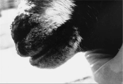
FIG. 32.88 Typical scabby lesions of contagious ecthyma (sore mouth, orf) on the lips of a young goat. Lesions tend to be proliferative rather than ulcerative.
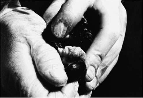
FIG. 32.89 Typical proliferative lesions of contagious ecthyma (sore mouth, orf) on the gums of a young sheep.
appears. In one report, the characteristic proliferative scabs appeared on the margins of healing burn wounds. Affected animals may be reluctant to nurse, eat, walk, or be nursed, depending on the location of lesions.
Secondary bacterial infection or myiasis of affected parts may occur. The disease usually runs its course in 3 to 6 weeks, but chronic cases have been reported. Complete healing without scarring is the norm as scabs fall off. Severe infections may result in stunting of growth, particularly in goats. Overwhelming infection has been noted on rare occasions, with extension of lesions into the deeper respiratory or gastrointestinal tracts. Does and ewes with severe udder infection may develop mastitis from secondary bacterial infection. Sheeppox and goatpox occasionally produce lesions similar to those of CE, but they are virulent diseases with systemic signs, including conjunctivitis, pyrexia, anorexia, and rhinitis.
One report from China documented an outbreak in goats consisting of mixed infections with goatpox, orf, and Mycoplasma capricolum, subspecies cap- ripneumoniae, which together resulted in a 60% mortality rate.5Animals with bluetongue may have a crusted mouth and nose and eye lesions in the convalescent phase, but bluetongue is a disease with more evidence of oral erosive rather than proliferative lesions and systemic signs, including pyrexia, reluctance to move, lesions on the tongue, and conjunctivitis. Bluetongue has a seasonal incidence (late summer, early fall) that coincides with the activity of its insect vector. CE may occur at any time but typically occurs in spring in the lamb or kid crop. Ulcerative dermatosis (lip and leg ulcer) is an uncommon disease caused by a virus similar to that of CE, but the lesions are crusted ulcers, to be distinguished from the crusted proliferations of CE. The author has observed a series of cases of what appeared to be mild CE of long duration (several years in one individual) in a family of Nubian goats; this was confirmed by an immunofluorescence assay. Smith and coworkers6 reported on five sheep, 4 months to 3 years of age, that developed severe refractory distal limb lesions resembling warts. Two also had lesions on the head. The sheep were from three different flocks; one was a Hampshire and four were Suffolk. Lesions were confirmed as orf by electron microscopy and immunohistochemistry.6 A report from Texas described severe multifocal persistent orf lesions in 16 Boer or Boer crossbred goat kids 2 to 5 months of age, from two different farms. Some of the kids had been vaccinated with live-virus vaccine at 1 day of age, others at 2 weeks of age. Lesions were found on lips, nose, ears, body, legs, and feet.7
Laboratory Diagnosis
The diagnosis usually is made in the field by recognition of the typical lesions in a naive flock or in a naive group (young lambs or kids) in a disease-endemic flock.
Definitive diagnosis usually involves identifying the distinctive cross-hatched virus particles in typical early lesions with electron microscopy, PCR, immunohistochemistry, or inoculation into known protected or susceptible animals. Minced biopsy tissues have been used as a source of virus that is identified by fluorescent antibodies after it has been growing in embryonic ovine kidney cell cultures. Complement fixation tests to detect antibodies (with patient serum) or antigen (with vesicular fluid or a suspension of scabs) have also been used.Pathophysiology
The disease follows a similar time course in animals and human beings: approximately 6 weeks. Six stages have been described,8 which begin after an incubation period of 3 to 14 days. Each stage lasts approximately 1 week.
1. Maculopapular stage: An erythematous spot becomes elevated. Histological study of this stage reveals vacuolization of cells in the upper third of the epidermis with intracytoplasmic eosinophilic inclusions in the affected cells.
2. Target stage: A red halo of dilated blood vessels and inflammatory cell infiltrates surrounds a white ring of vacuolated epidermal cells with intracytoplasmic and intranuclear inclusions, which surrounds a red center of pyknotic epidermal cells.
3. Acute stage: The lesion is a red, weeping nodule. Microscopic study reveals reticular degeneration of the epidermis with vesicles. The dermis is infiltrated with macrophages and lymphocytes and is denuded of epidermis in places. The hair follicles are distended with pyknotic epidermal cells.
4. Regenerative stage: The nodule is now dry with small black dots (the pyknotic follicle cells, now extruded to the surface) in a thin yellow surface.
5. Papillomatous stage: The surface of the nodule is roughened with papillomas, which microscopic study proves to be fingerlike, downward projections of epidermis through a full thickness of dermis.
6. Regressive stage: The lesion decreases in size and elevation above the surface, the papillomas regress, and several crusts may come off.
Microscopic study reveals that the papillomas and infiltrates regress, leaving normal architecture.Orf virus encodes for a protein that is apparently homologous to mammalian vascular endothelial growth factors. This family of molecules mediates vascular permeability, angiogenesis, and endothelial cell proliferation, which may account for the swollen, proliferative nature of orf lesions.9 Transient fever and lymphadenopathy are occasionally seen in humans. Lesions in humans that are not excised and from which no biopsy sample is taken heal without a scar.
Orf virus encodes a range of immunomodulatory genes that interfere with host antivirus immune and inflammatory effector mechanisms, allowing time for virus replication in epidermal cells.
Epidemiology
The naturally occurring disease is primarily one of sheep, goats, and human beings, but it has also been reported in a variety of wild ruminants. Experimental transmission has been achieved in cattle, rabbits, horses, and monkeys. No naturally occurring clinical cases in these species have been reported. All ages and classes of sheep and goats are affected, and clinically normal sheep can infect naive individuals.10 In herds and flocks in which the disease is endemic, it usually is seen in the lamb or kid crop and on the udders and teats of some of the nursing mothers. Animals that have had a bout of disease are solidly immune for 1 to several years, but the rate of morbidity is high (often 80%) among naive individuals. Humans are infected through contact with affected animals or fomites that have contacted affected animals (including one report of transmission by a pickup truck that had been used to haul sheep).8 Effective disinfectants include hypochlorite, alkalis, glutaraldehyde, and Virkon. Virkon is a multi-purpose disinfectant containing oxone and inorganic buffers and has a wide spectrum of activity against viruses.
Human-to-human transmission can occur. The mortalityrate among animals is low except when young individuals are severely affected and quit nursing or when mothers have severe udder lesions.
Overwhelming infections are rare except in Boer or Boer crossbred goats, but some outbreaks are more severe than others.The virus is quite resistant to many environmental conditions and persists from year to year on infected premises. Dried scabs allow the virus to persist for years, but wet conditions are less hospitable to it. Reports of PI sheep in wet climates point to another possible source of infection.
Treatment, Prevention, and Control
The infection usually is self-limiting and of minor consequence. Young individuals may need to be tube-fed if the lesions are severe enough to preclude suckling. Secondary infections, myiasis, or mastitis may be treated with topical disinfectants, antibiotics, or insecticides as appropriate. The hard crusts should not be removed because removal may delay healing and promote scarring. Also, humans should limit contact with affected animals and should wear gloves when handling them as necessary. Anecdotal reports, each consisting of one case, have claimed good results for treatment of human orf with intralesional corticosteroids, IFN, idoxuridine in DMSO, diethyl ether, or cryotherapy. An immunosuppressed human with a huge orf lesion on the finger was successfully treated with topical cidofovir antiviral cream. Similar good results were obtained in a group of 12 severely affected, bottle-fed lambs with painful intraoral lesions. The proliferative tissues were debrided and cauterized with a portable diathermy unit. The exposed submucosa was then frozen twice with liquid nitrogen spray. Healing occurred by second intention, and the lambs recovered rapidly.
The infection is prevented by keeping a herd or flock virus free by not introducing or contacting infected animals or humans. Lesions often are not apparent on carriers.10 Once established, the disease persists on the premises because of virus in scabs. Vaccination should be undertaken only if the infection is persistent, because the vaccine consists of virulent live virus. Vaccination can also be achieved with dried scabs from the previous year's outbreak because there are distinct antigenic differences among orf viruses11; the scab material is rubbed into scarified skin in an inconspicuous location (inner thigh, under the tail, in the axilla). A localized inflammatory reaction at the site 1 week after vaccination indicates that inoculation was successful. Infected or vaccinated animals should not be allowed contact with unexposed animals until the lesions have healed. Lambs born to immunized ewes may not be protected by colostral antibodies even though their levels of antibody after suckling are high12; this points to the importance of cell-mediated immunity in protection from the disease. Lambs should be vaccinated at 6 to 8 weeks of age and are immune 3 weeks later. Yearly vaccination of the new lamb or kid crop and of new additions to the herd with farm-specific strains of orf should prevent devastating outbreaks on infected farms. At a minimum, it appears that goat isolates should be used for kids and sheep isolates for lambs.11-13