Corynebacterium pseudotuberculosis Infection
Sharon Jane Spier • Monica Aleman
Definition
Corynebacterium pseudotuberculosis infections occur worldwide and cause external and internal caseous lymphadenitis in sheep and goats; cutaneous excoriated granulomas and mastitic, visceral, or mixed infection in cattle; and ulcerative lymphangitis and external and internal abscesses in horses.1-4 Subacute to chronic lymphadenitis and pneumonia have been reported in humans handling infected sheep.5,6 Several zebras in the United States developed severe or multiple internal abscesses and died weeks after being exposed to horses in California.7 There have been reports of the disease in camels, alpacas, and buffalo.8,9
Microbiology
C.
pseudotuberculosis infection is caused by a 2-μm, grampositive, intracellular, nonmotile, pleomorphic, rod-shaped facultative anaerobe.10 C. pseudotuberculosis grows well at 37° C on blood agar in 24 to 48 hours, and it forms small, pinpoint in diameter, whitish, opaque colonies that are surrounded by a weak zone of hemolysis. Because of the high lipid content in the bacterial cell wall, particularly corynomycolic acid, the colonies spatter in a flame and hold their shape when pushed across the surface of an agar plate.11 High lipid content may facilitate survival of the organism in macrophages.12 Two species-specific biotypes of C. pseudotuberculosis have been identified on the basis of differences in nitrate reduction5 and DNA fingerprinting techniques.11,13,14 Strains isolated from small ruminants are nitrate negative, strains from horses are nitrate positive, and both strains have been isolated from cattle.1,5,13,15 From the results of DNA studies, the terms biovar equi for nitrate-positive and biovar ovis for nitrate-negative strains were proposed.11 Molecular studies revealed that there is considerable heterogeneity of the isolates of C. pseudotuberculosis from small ruminants and from horses13,14,16 and concluded that nitrate reduction may not absolutely distinguish between the isolates, as does ribotyping.13 On the basis of genome sequencing, sheep and goats have specific isolates throughout the world, and horses and cattle have distinct groups of isolates depending on geographic locations.13 Natural cross-species transmission between small ruminants and horses has not been reported.C. pseudotuberculosis produces various exotoxins: phospholipase D (PLD), sphingomyelinase, inhibitory factor of staphylococcal β-hemolysin, hemolysis factor, dermanecrotoxins, and mouse lethality toxins.17 Phospholipase D and sphingomyelinase are important in the pathogenesis of the disease because they hydrolyze lysophosphatidylcholine and sphingomyelin, respectively, enabling degradation of the endothelial cell wall and increasing vascular permeability.18 The synergistic activity of sphingomyelinase with the exotoxin of Rhodococcus equi in lysing red blood cells in agar forms the basis for the synergistic hemolysis inhibition (SHI) test. The SHI test measures IgG to PLD exotoxin.19
Molecular characterization of C. pseudotuberculosis isolates causing various clinical forms of disease, including genome sequencing, has been reported. At present, phenotypic and genotypic strain differences have not been linked to clinical disease presentation (internal or external abscesses), suggesting that other environmental or host factors determine the course of disease.20,21
Clinical Signs and Differential Diagnosis
Sheep and Goats
C. pseudotuberculosis causes caseous lymphadenitis (CLA) in sheep and goats worldwide. Caseous lymphadenitis is a major cause of poor production, premature culling, and mortality. Two forms of CLA exist which manifest as external and internal abscesses.22 The infection in small ruminants is primarily characterized by suppuration and necrosis of the large superficial lymph nodes.
External abscesses are found more commonly involving the mandibular, parotid, prefemoral, or prescapular lymph nodes. The exudate present in those abscesses is thick or inspissated and may appear white in sheep and greenish in goats.4 A breed association with the type of CLA cutaneous lesion was observed in an outbreak in a commercial ram stud in Scotland.23 The disease is commonly known as “cheesy gland” in Australia. Differential diagnosis should include abscesses caused by other organisms, trauma, seroma, hematoma, foreign body, injection reaction, and, less commonly, tumors. C. pseudotuberculosis infection represents a major herd health problem, so abscess culture to determine the precise causative agent is important. Mastitis occasionally develops.2Internal abscesses can be found in the lungs, kidneys, and mediastinal, bronchial, mesenteric, and lumbar lymph nodes.22 Chronic weight loss is the most common presenting complaint. Other clinical signs are related to the organ or tissues affected. Other diagnostic procedures may be necessary for the differentiation of internal abscesses as the cause of weight loss. Signs of spinal cord compression by vertebral abscesses have been seen in lambs born in unsanitary conditions.4 Knowledge of the local prevalence can aid diagnosis of the infection when uncommon anatomic locations are affected. The prevalence in large breeding operations in endemic areas is estimated to be between 5% and 10%.
Cattle
C. pseudotuberculosis infection in cattle occurs as a herd problem with sporadic incidence. The most common clinical form affecting cattle is cutaneous excoriated granulomas; other forms are mastitis, visceral, and mixed infections.2 In the most common form, the lesions do not occur as abscesses but as ulcerative, exuding granulomatous lesions as large as 20 cm in diameter, with necrotic areas that are easily surgically removed, leaving granulation tissue underneath.3,4 Lesions are usually located in the lateral exposed areas of the body: face, neck, thorax,
and flanks.
The exudate varies from bloody to thick greenish in color. Lesions heal spontaneously in 2 to 4 weeks and do not appear to cause significant illness or decrease milk production in cattle in California.4 However, monthly milk production was decreased by 6% in Israeli cattle.3 The prevalence of the infection has been reported to be up to 10% in California dairies.4 Morbidity was reported to be up to 35% in Israeli herds.3 Management problems like broken posts or exposed wires traumatize the skin of cattle, allowing penetration of the organism. Young cattle appeared to be less susceptible to the disease than older cattle in Israel.3 Differential diagnoses include trauma, foreign body, and other masses like tumors. Cutaneous lesions and mastitis were seen in 6%, and cutaneous lesions with concurrent visceral involvement in 1.6% of the cases in Israel with the remainder of the cows (92%) exhibiting only the cutaneous form.3 The most affected organ in the visceral form was the lung.3 The infection has been reported in bison from Egypt, resulting in severe emaciation and edema in the ventral areas and flanks.24Camels and Camelids
Caseous lymphadenitis has been reported in old and new world camelids worldwide.25 One study documented five young alpacas (aged 22 days to 14 months old) in North America with caseous lymphadenitis or subcutaneous abscesses that developed during late summer and early fall.9 The alpacas did not appear clinically ill but developed swellings that progressed to abscesses. The abscesses (1 to 3 per alpaca) were located in the submandibular and cervical areas and in one case adjacent to the eye. Abscess excision appeared to be the most effective treatment in those cases. Differential diagnoses include abscesses caused by Streptococcus spp., Corynebacterium spp., and A. pyogenes2 Severe lymphadenitis with internal and external abscesses was reported in camels in Asia and Jordan.8 A study of experimental infection of adult alpacas resulted in abscesses at the inoculation site and renal lymph nodes.27
Horses
Three forms have been described in horses: ulcerative lymphangitis and external and internal abscesses (Figs.
37.1 and 37.2). In a previous study of C- pseudotuberculosis infection in horses from California, ulcerative lymphangitis was diagnosed in 1%, external abscesses in 91%, and internal abscesses in 8% of the cases.28 There appears to be no breed or sex predilection for development of infection. Ulcerative lymphangitis appears as a severe cellulitis, where the lymphatics are affected in one or more limbs, with multiple draining ulcerative lesions. Horses often develop a non-weight-bearing lameness, fever, lethargy, and anorexia. This form of the disease occurs worldwide and often becomes chronic, resulting in limb edema, lameness, weakness, and weight loss.4 The differential diagnosis should include blunt trauma, fracture, foreign body, puncture wounds, nonseptic cellulitis, staphylococcal cellulitis, and other septic cellulitis. Musculoskeletal infection can result in marked lameness (grades 4 to 5 out of 5) and most commonly involves the axillary and triceps areas, followed by the stifle area.29 In addition to diffuse lymphangitis of the limbs, it can also cause 2829osteomyelitis and septic arthritis.28,29
The median age for horses with external abscesses is 5 years (range, 3 months to 28 years).28 Young horses appear to be predisposed to infection; 52% of the cases in a large retrospective study in California were 5 years old or younger.28 Few cases involved foals younger than 6 months of age, suggesting that foals born to mares in endemic areas may be protected for several months by colostral antibodies.28 External abscesses located primarily in the pectoral and ventral abdominal regions are most commonly diagnosed in areas of the western United States (Texas, Oklahoma, New Mexico, Colorado, Nevada, Utah, California, Wyoming, Arizona, Oregon, and Washington);
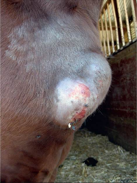
FIG. 37.1 Typical ripe pectoral external abscess in a horse, containing a large pocket of pus, caused by C.
pseudotuberculosis.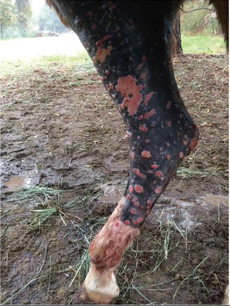
FIG. 37.2 Severe ulcerative lymphangitis in a horse leg caused by C. pseudotuberculosis.
however, disease incidence is increasing and spreading to all regions of the United States, including Hawaii, with reports in Mexico and Western Canada.1,4,20,28,30,31 This clinical form of external infection is commonly known as “pigeon fever” because of the large size of pectoral abscesses, giving the appearance of a pigeon's breast. “Dryland distemper” is another name that reflects its geographical distribution in arid areas. Other common anatomic locations are the prepuce, mammary gland, axilla, inguinal region, limbs, and head.28 Abscesses involving the head include the ears, eyelids, forehead, maxillary, and mandibular regions.28 Severe facial suppurative cellulitis and panniculitis with skin sloughing have also been reported.32 Other less common areas are the thorax, neck, parotid gland, guttural pouches, larynx, flanks, umbilicus, tail, and rectum.28 2833 Central nervous system infection has also been reported.28,33 A large area of edema develops in the area of abscess formation. While the abscess matures, the area becomes hard and painful, and some get large, particularly in the pectoral area. These abscesses typically have a thick capsule measuring up to 10 cm, and can cause severe lameness if located in the axillary or inguinal region.1,28 Maturation can be slow and drainage difficult to establish if the abscess lies deep to muscle. Once drainage is established by spontaneous rupture or lancing, the majority of cases resolve within 10 to 14 days, but many horses can experience a protracted course of disease lasting months with recurring abscesses. Generally, weight loss is not observed in absence of internal infection. Abscesses may contain from 5 to 400 mL of thick, tan, purulent exudate.1 The majority of affected horses present with a single abscess rather than multiple abscesses.28 Fever up to 40° C develops in about 25% of cases. Other signs are nonhealing wounds, lameness, ventral dermatitis, and (less commonly) depression, anorexia, mastitis, and other problems, depending on abscess location.28 In the majority of horses (91.4%), complete recovery occurs with no recurrence of infection in subsequent years, implying a long-lasting immunity. However, 8.6% of equine infections may persist for longer than 1 year or recur as external or internal abscesses.28 In sheep and goats, humoral and cellular immune responses develop following infection, and macrophages acquire the ability to kill the organism.6 The case fatality for horses with external abscesses is low (0.8%).28 The differential diagnosis for external abscess, particularly pectoral, should include trauma, seroma, hematoma, foreign body, or abscess due to a different organism.
In a large study of C. pseudotuberculosis infection in the horse, 8% of 538 horses developed internal abscesses.28 In two different studies, up to 63% of horses with internal abscesses also had a history of, or concurrent, external abscesses.28,34 In a study of 30 horses with internal abscesses, a sex predisposition (70% female) was apparent.34 Mean age is 8 years, with a range of 10 months to 23 years.28,34 The most common clinical signs are anorexia, lethargy, fever (up to 41.1° C), tachycardia, and modest weight loss. Other signs are colic, pale mucous membranes, ventral and/or limb edema, ventral dermatitis, ataxia, hematuria, nasal discharge, and abortion.28 The most commonly affected anatomic location is the liver, followed by mesentery, mediastinum, lungs, kidneys, diaphragm, spleen, pericardium, 12834
blood, and uterus.1,28,34 A postmortem examination was performed on an aborted fetus from a mare with pneumonia and revealed C. pseudotuberculosis abscesses in the liver, lungs, spleen, diaphragm, kidney, and bladder.34 Both single and multiple organ involvement has been documented.34 Bacteremia may also occur. The case fatality for horses with internal abscesses ranges from 30% to 40%.28,34 The differential diagnosis should include other types of abscesses, such as Streptococcus, Actinomyces, Staphylococcus, R. equi in foals, Coccidioides immitis, anaerobes, neoplasia, and other causes of weight loss. The clinical signs and differential diagnosis will depend on the location of the abscess.
Humans
Human infection may result from consumption of unpasteurized infected milk or milk products, continued close contact with infected animals, handling contaminated equipment, and exposure to wounds with exudates.6,35 Human infection has been reported from strains of small ruminants.6 Transmission from horses to humans has not been reported, but precautions should be taken when handling infected horses. Infection in humans occurs as a subacute to chronic lymphadenitis and pneumonia.6
Clinical Pathology and Laboratory Diagnosis
Common findings include anemia of chronic disease and an inflammatory leukogram.28,29,34 Leukocytosis with neutrophilia and increased fibrinogen and serum amyloid A (SAA) are common features of developing bacterial infections, particularly 282934
in the case of internal abscesses. ,, Leukocytosis with neutrophilia was seen in 36% and 76% of horses with external and internal abscesses, respectively.28 Hyperproteinemia due to increased globulins was observed in 38% and 59% of horses with external and internal abscesses, respectively.28 Similarly, infected cattle and small ruminants had increases in white blood cell counts.2,3
Peritoneal fluid from 93% of horses with abdominal abscesses was abnormal.28 The remaining horses with abdominal abscesses and normal peritoneal fluid had abscesses located retroperitone- ally involving the kidneys, without involvement of other abdominal structures. C. pseudotuberculosis was isolated by bacterial culture in 32% of the samples of peritoneal fluid in horses with internal abscesses,28 and infection can also be detected by PCR. Failure to detect the organism from peritoneal fluid does not rule out the disease as infection could be in a retroperitoneal location, sequestered within a thick capsule, or suppressed by local factors or nucleated cells.36
The ELISA test for detection of cell wall antigens appears to have some utility for detection and control of infection in sheep.37-40 The synergistic hemolysis inhibition (SHI) test measures IgG response to the PLD exotoxin in the patient's serum by detecting the highest dilution that will prevent hemolysis of R. equi exotoxin-sensitized bovine red cells when mixed with C. pseudotuberculosis exotoxin of a known concentration.19,41 Increasing titers may be seen with exposure, or active external or internal infection and there exists considerable overlap in values among these groups of horses. In a case control study, SHI test results had greatest utility for determining internal C. pseudotuberculosis infection in horses when there was no evidence of external abscesses.43 A high probability of internal infection with titers greater than or equal to 512 was previously reported, yet a recent case control study of 171 horses revealed higher titers were more indicative of active external or internal disease rather than internal disease specifically. The SHI test was unable to distinguish the occurrence of internal infection when external abscesses were present. Titers increase with chronicity of infection and bacterial culture is the preferred method of diagnosis for external infection. In horses without external abscesses, increased titers greater than 1280 are significantly associated with internal infection. Clinicians are advised against diagnosis of internal infection on the basis of SHI titer; rather, the test should be used in conjunction with other clinical and clinicopathologic evidence of inflammation.
The SHI test can be used in sheep and goats to monitor prevalence and exposure of incoming animals and to detect subclinical infections.4,41 In horses, a low SHI titer does not rule out the disease as horses that are seronegative at the time an external abscess is drained seroconvert later. High SHI titers in horses with internal abscesses probably reflect the chronicity of the disease and the resulting prolonged immune stimulation. In summary, high titers can be seen with both internal and external abscesses and abdominal and thoracic ultrasonography is useful for confirmation of internal infection in suspect cases.28,34,43 Exposed herd mates can also have SHI titers in the absence of clinical infection. Prolonged seropositivity has been observed in horses and goats.28,41,44 Other serodi- agnostic tests used in sheep and goats are tube agglutination, complement fixation, and gel immunodiffusion.17,45
A presumptive diagnosis can be made on the basis of history, local prevalence, time of year, clinical signs, and exudate characteristics.4 For the diagnosis of internal abscesses, the previous features must be considered, as well as the presence of an inflammatory leukogram with elevated fibrinogen, serum chemistry abnormalities, abnormal peritoneal fluid or transtracheal wash, positive blood culture, SHI titer of 512 or higher, and ultrasonographic and/or radiographic evidence of masses.28,34 A definitive diagnosis is established by isolating the organism from abscesses or draining wounds. The organism is readily isolated and grows well in blood agar in 24 to 48 hours, even when contaminant bacteria are present.28
Pathophysiology and Epidemiology
C. pseudotuberculosis is a soil-borne organism that survives for long periods of time, months to years, even in direct sunlight at environmental temperatures.6,46,47 The incidence of infection in horses varies considerably from year to year. External and internal abscesses in horses can present at any time of the year but are more commonly observed during the fall and early winter months, with the highest incidence in September, October, and November.28 However, internal infections are more frequently seen in November through January, 1 to 2 months following the peak number of cases with external abscesses.34 The largest numbers of equine cases have been observed during the dry months of the year, following heavy rainfall, which may result in optimal breeding conditions for insects.1,28,30 The seasonal incidence in horses has been associated with the presence of biting insects like Haematobia irritans (horn fly); this insect's feeding pattern causes ventral midline dermatitis. Insect vectors involved in transmission of disease in horses (e.g., H. irritans, Stomoxys calcitrans, Musca domestica) were identified by detecting the PLD exotoxin gene of C. pseudotuberculosis in an endemic area.48 Disease transmission may occur by direct contact with exudates or contaminated soil or by vector-borne routes.49 Temporal and spatial analysis indicated an incubation period of 3 to 4 weeks in horses.50 There is no breed or sex predilection28; however, a retrospective study indicated a predilection for internal abscesses in females.34 A case-control study in an endemic area revealed young adult horses (prognosis for external abscesses is good. Most resolve in 3 weeks from the day of drainage.4 The prognosis for horses with internal abscesses and ulcerative lymphangitis is guarded but improves if infection is detected early and appropriate long-term therapy is administered. Horses with musculoskeletal infection tend to have a favorable prognosis, but osteomyelitis and septic arthritis can complicate recovery.29
In a study in cattle, skin lesions on individual cows healed (on average) in 23 days after local or parenteral treatment; 17% of severely affected cattle were culled.3 Simple drainage in small ruminants does not usually result in resolution of the disease and can become a potential source of infection. Dilute betadine solutions can be used for abscess lavage. Complete excision of the abscess under general anesthesia may be necessary to keep the abscess from draining and prevent spread of the infection to other animals.4 The treatment of choice in small ruminants is complete surgical removal of affected lymph nodes.
Prevention and Control
Even though C. pseudotuberculosis infection is one of the most commonly diagnosed infectious diseases in California, little is known about its prevention and control. General recommendations to prevent its spread are isolation of infected animals, fly control, good sanitation, careful shearing practices, disinfection of contaminated fomites, and careful disposal of bedding. On small-ruminant farms, morbidity can reach 100%, with depopulation being the most economic option. Because C. pseudotuberculosis can survive in soil and on fomites, the potential for environmental contamination is high.46,49
Immunization trials using whole cells, cell wall toxoids, and bacterin-toxoid combinations have been used to prevent CLA in small ruminants.61-65 These vaccines have been shown to provide a high degree of protection, decreasing the number of infected sheep and number of abscesses per sheep.61-65 C. pseudotuberculosis toxoids are commercially available for sheep and goats (e.g., Caseous D-T [Colorado Serum Co., Denver, Colo.], Glanvac [Rhodes, New South Wales, Australia]). In horses, autogenous bacterin-toxoids have been used for many years with anecdotal success.66,67 A bacterin-toxoid vaccine with a conditional license (Boerhinger Ingelheim, Ingelheim am Rhein, Germany) is available for horses. Vaccinated animals demonstrate increased SHI titers following a 2-dose primary series; however, conferred protection remains to be established.