Diagnosis of Mastitis
Physical examination for changes in the milk or signs of inflammation and culture of the mammary secretion are the most commonly used techniques for identifying clinical or subclinical mastitis.
Various other laboratory tests, especially those relating to cell enumeration, have been investigated as possible indicators of mastitis. Tests currently available other than culture tend to be unsatisfactory in goats, in the author's (MCS) opinion. Monthly somatic cell determinations on all herd members are valuable for keeping track of increased cell counts (and satisfying government regulations), but are rarely worth the expense regarding mastitis control.Clinical Examination
Early indications of clinical mastitis include a decrease in milk production by one gland or a lameness on the affected side as the goat attempts to avoid contact of the hindlimb with the tender half of the udder. Nursing kids may appear to be hungry, and mastitis is associated with increased kid mortality rates (Addo et al. 1980). Visual inspection of the udder from behind and from the sides may reveal asymmetry, whereby the affected gland is swollen (acute) or atrophied (long-term inflammation). Predisposing teat end lesions (wounds, contagious ecthyma, warts; Kapur and Singh 1978) also may be identified. Palpation may disclose the presence of heat, tenderness, and swelling (acute); induration or atrophy (chronic mastitis); or even multiple abscesses. The milk may be watery or contain flakes or clots or may have a normal appearance. Regarding acute mastitis, the goat may be systemically ill, with signs of fever, anorexia, and depression. If the teat is cold and edematous or the secretion red and watery, gangrenous mastitis should be suspected. This severest clinical form is discussed in detail below.
Culture
Culture of an aseptically obtained milk sample is necessary to identify the etiologic agent of bacterial mastitis.
The ideal sampling technique begins with putting on clean disposable gloves, brushing off any loose debris, and discarding several streams of milk. The teats are then predipped in an approved bovine product and the predip is allowed 20 seconds of contact time before being wiped off with an individual paper towel. Finally, the teat end should be carefully disinfected with a cotton ball moistened with 70% ethyl or isopropyl alcohol, before a milk sample is collected in a sterile vial (National Mastitis Council 2017). Refrigerate the samples promptly, or freeze if culture will be delayed more than 48 hours. Freezing results in a minimal decrease in recovery of organisms from goat milk samples (McDougall 2000; Sanchez et al. 2003). Sensitivity testing using routine techniques sometimes assists in selecting antibiotic therapy.Some researchers require positive cultures from two consecutive milk samples from the same gland before a culture result can be interpreted as positive (Contreras et al. 1997a). Two or more consecutive negative culture test results are often presumed to indicate absence of infection.
Practitioners who wish to perform in-house mastitis cultures find that most mastitis isolates other than mycoplasma grow on blood agar at 35-37 °C (95-98.6 °F) and can be identified on the basis of a few simple tests such as colony characteristics and hemolysis on blood agar, Gram stain, and catalase tests. A standard reference book that includes color photographs of culture plates is extremely helpful (National Mastitis Council 2017). A simplified flow chart for identifying common pathogens adapted from this reference is provided in Figure 14.9. An organism is catalase positive if bubbles are produced when a colony is emulsified in 3% hydrogen peroxide. The oxidase test requires special discs and should not be performed on colonies grown on selective media such as MacConkey agar (National Mastitis Council 2017).
Various multipartite culture plates are also available commercially that include selective media for Gramnegative or Gram-positive organisms, staphylococci, or streptococci.
An example is the Bi-plate from the University of Minnesota Laboratory for Udder Health (St. Paul, MN, USA), which includes Factor™ medium (Gram-positive growth, S. aureus shows hemolysis) and MacConkey medium (Gram-negative growth). The same laboratory also offers a Tri-plate, which also contains a selective medium for streptococci.Mycoplasma are more difficult to grow in the laboratory. Mycoplasma mycoides subsp. capri (previously M. mycoides subsp. mycoides large-colony [LC] type) grows on blood agar plates supplemented with ovine blood; “fried egg” colonies 1 mm in diameter and beta hemolysis are detectable after one week (DaMassa et al. 1983). Isolating the other species usually requires special mycoplasma medium and a moist 10% CO2 incubator (National Mastitis Council 2017). Plates are observed under 20-50? magnification for 7-10 days before being termed negative. Most colonies have a fried egg appearance. Exact identification of an isolate is
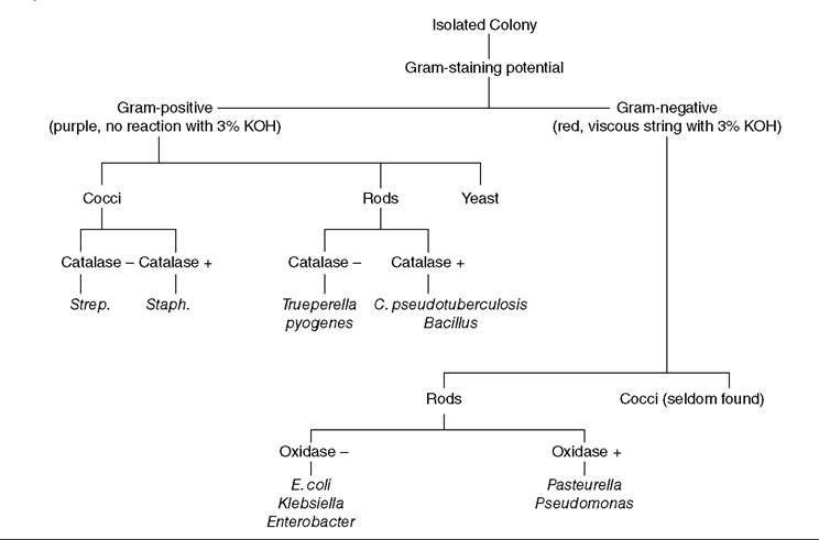
Figure 14.9 Flow chart for identification of bacteria, other than mycoplasma, causing mastitis. - represents negative test result, + represents positive test result.
difficult, and typing is performed by only a few laboratories. Some laboratories offer a mycoplasma PCR.
Cell Enumeration
An increase in the number of somatic cells in milk has been used as an indication of mastitis, including Subclinical mastitis, in cows. Application to dairy goats of tests and regulations developed for cattle frequently has led to panic in the commercial producer, who interprets “high” cell counts as evidence of a serious mastitis problem or who is threatened by the inspector with loss of a milk market. Currenlty the legal limit for somatic cell count (SCC) in goat milk in the United States is 1.5million cells/mL. In a survey of 71 commercial US herds in November and December, only 35% could achieve the previous lower legal limit of 1 million cells/ mL (Droke et al.
1993). A legal limit has not yet been set in the European Union or Canada. Numerous factors affecting cell counts in goat milk have been reviewed (Haenlein 2002; Paape et al. 2007; Jimenez-Granado et al. 2014).Cytoplasmic Particles and Epithelial Cells
In discussions of bovine mastitis, the number of somatic cells per mL of milk is generally assumed to correlate directly with the severity of mastitis or the degree of irritation to the mammary gland. The relationship of SCC to caprine mastitis is limited, unless tests appropriate to caprine milk are used. This is partly because goat milk differs from cow milk due to the presence of cytoplasmic particles and epithelial cells. The caprine mammary gland produces milk by a process called apocrine secretion (Wooding et al. 1970). Portions of the cytoplasm of the epithelial cells are pinched off and appear in the milk as DNA- free particles, similar in size to leukocytes (Dulin et al. 1982). By contrast, ewes produce milk with approximately 10% of the number of cytoplasmic particles found in goat milk (Paape et al. 2001). Also present in variable numbers in goat milk are intact epithelial cells sloughed from acini and ducts.
Interpretation of Reports and Counting Methods
Very careful attention must be paid to the counting technique when comparing or contrasting various reports or when attempting to estimate the prevalence of mastitis in a goat herd. Another inadequacy of much of the literature is in the realm of determining mean SCC. Most older studies have not used a log transformation such as the linear score (also called somatic cell score) developed for bovine SCC, where linear score = 3 + log2 (X/100) for an SCC of X ? 103 cells/mL. Somatic cell scores and equivalent SCC are 1 = 25 000; 2 = 50 000; 3 = 100 000; 4 = 200 000; 5 = 400 000; 6 = 800 000; 7 = 1.6 million; 8 = 3.2 million/mL.
The direct microscopic SCC, using a stain appropriate for goat milk, is the standard against which other counting methods should be judged.
Direct Microscopic Somatic Cell Count
The confirmatory test for somatic cell numbers in bovine milk is usually a direct microscopic examination of 0.01 mL of milk on a slide within an area of 1 cm2. The Levovitz- Weber modification of the Newman-Lampert stain is commonly used to stain somatic cells for counting (Schalm et al. 1971). This stain is inappropriate for goat milk, because staining is similar for cytoplasmic particles and cells (Dulin et al. 1982). A research technician might be able to recognize the difference between particles and cells, but overcounting would be expected in a commercial laboratory.
Currently, the stain preferred for determining SCC in goat milk in the United States is the pyronin Y-methyl green stain (Dulin et al. 1982; Paape et al. 2001), often referred to simply as the green stain. Methyl green is specific for DNA and pyronin Y is specific for RNA. Chromosomes, then, stain blue-lavender while cytoplasmic particles and the cytoplasm of epithelial cells stain red. Neutrophils do not contain pyronin Y-positive material (Paape et al. 1963). Unfortunately, this staining is difficult to do and the reagents are potentially toxic to laboratory workers. Neutrophils are the predominant cell type in goat milk from both infected and uninfected glands, whereas the macrophage is the predominant cell in milk from uninfected sheep and cows (Sierra et al. 1999; Paape et al. 2001).
Leukocytes and epithelial cells in goat milk have also been differentiated by a modified Wright's stain technique (Hinckley and Williams 1981). Others have reported that many leukocytes are masked by the background in smears prepared with Wright's stain (Paape et al. 1963).
California Mastitis Test and Teepol Test
The California Mastitis Test (CMT) is a simple, semiquan- titative test for determining the number of nucleated cells (both neutrophils and epithelial cells) in milk (Schalm and Noorlander 1957). Equal quantities (typically 2 mL) of milk and the commercial reagent (Nasco, Fort Atkinson, WI, USA and others), which contains 3% alkyl-arylsulfonate and bromcresol purple as a pH level indicator, are combined in the cup on a white paddle and swirled.
Different scoring schemes are used in different parts of the world. As described originally, there is no change with a negative reaction, while a T (trace) reaction consists of a slight thickening of the milk. Scores of 1, 2, and 3 are given with increasing gel formation and deepening of color. A score of 3 means that a gel has formed that pulls away from the edge and collects in the center of the cup. It is potentially confusing that some workers designate the same five categories as scores of 1-5 (Scandinavian system).The reaction detects the presence of DNA, which is released when the detergent ruptures somatic cells. The approximate numbers of leukocytes for each score for goat milk (Schalm et al. 1971) are indicated in Table 14.1. In a Norwegian study, the total numbers of leukocytes and
Table 14.1 California Mastitis Test (CMT) scores on goat milk.
| CMT score | Scand. system | Samples | Neutrophil leukocytes/mL | |
| Range | Median | |||
| 0 | 1 | 46 | 0-480000 | 60 000 |
| Trace | 2 | 43 | 0-640 000 | 270000 |
| 1 | 3 | 29 | 240000-1440000 | bgcolor=white>660 000|
| 2 | 4 | 16 | 1 080000-5850000 | 2400000 |
| 3 | 5 | 6 | >10000000 | - |
Source: Schalm et al. (1971) / Lee & Febiger.
epithelial cells were 690, 800, 820, 1230, and 4520 ? 103∕mL for the five CMT scores from 1966 goat milk samples (Pettersen 1981). A French study of midlactation samples that categorized any reaction greater than a trace as positive concluded that a positive CMT result had a sensitivity of 88% and a specificity of 93% for detecting SCC by Fossomatic methods greater than 750 000 cells/mL (Perrin et al. 1997). It is generally believed that scores of T or 1 (up to 1 million cells/mL) are usually no reason for concern.
In France, the equivalent to the CMT is called the Teepol test, after a European brand of neutral liquid detergent. The commercial detergent is diluted 1 : 10 and then used as the reagent (Lefrileux 2002). It reacts with both epithelial cells and neutrophils. Test results with goat milk have been graded and interpreted as follows (Roguinsky et al. 1971):
1) No or fine precipitation (as many as 500 000 cells/ mL): normal.
2) Granular precipitate (200 000 to 1 million cells): mild irritation, as by improper milking.
3) Filamentous precipitate (500 000 to 2 million cells/mL): weakly pathogenic organism such as non-hemolytic staphylococcus.
4) Viscous precipitate (more than 1.5 million cells/mL): suggests presence of S. aureus.
The CMT is more useful for ruling out than for diagnosing mastitis in goats (Contreras et al. 1996). In a Norwegian study of 1161 milk samples, 331 of 422 samples with CMT values of 1-3 in the American system yielded no bacterial growth; 732 of 739 with a negative or trace CMT score yielded no growth (Nesbakken 1978b). High scores at the end of lactation (Maisi 1990a) or in systemically ill goats with drastically reduced milk production occur in the absence of mastitis. A sick goat with a negative or trace CMT reaction is probably not sick because of mastitis. If there is a marked difference between the scores of two halves of a goat's udder, mastitis is very likely. The usefulness of the CMT (or any other test) for diagnosing subclinical mastitis depends on the prevalence of mastitis in a herd. In a well-managed herd, the predictive value of a positive test is unacceptably low (Hueston et al. 1986).
Wisconsin Mastitis Test
The Wisconsin Mastitis Test (WMT) uses diluted CMT reagent. It is more objective than the CMT because the viscosity of the milk-reagent mixture is estimated from the volume remaining in a special tube after draining through a standard-sized hole for 15 seconds (Schalm et al. 1971). The WMT is considered to be DNA specific. Results obtained using standard conversion factors for cow milk are similar to counts obtained by the Fossomatic method (Dulin et al. 1982).
Fossomatic and DeLaval Cell Counters
The Fossomatic™ method (FOSS, Hillerod, Denmark) of determining SCC is an automated fluorescent technique that uses a dye that specifically binds to the DNA of cell nuclei. With Fossomatic equipment currently used by dairy herd improvement associations in the United States, both epithelial cells and leukocytes are counted, but counts are not confounded by cytoplasmic particles. There is a good correlation between Fossomatic counts and those obtained by direct microscopic exam using the pyronin Y-methyl green stain (Droke et al. 1993).
Test temperature (40 °C versus 60 °C; 104 °F versus 140 °F), use of bronopol preservative, and sample storage time (one to four days) have little effect on SCC results (Sierra et al. 2006). Counts are slightly lower when azidiol is used as the preservative (Sanchez et al. 2005). If the Fossomatic machine is calibrated according to the manufacturer's directions using goat milk standards, the SCC is 27% lower than when cow standards are used (Zeng 1996).
The DeLaval counter (DeLaval International, Tumba, Sweden) is a newer device that is portable and can be used on the farm. A single-use cassette is employed for each milk sample and the technique is DNA specific. Fluorescence of cell nuclei is detected as a digital image. There is a high correlation (95%) with cell counts obtained with the more laborious direct microscopic method using the pyronin Y-methyl green stain on goat milk (Berry and Broughan 2007). Early, mid, and late-lactation goats were included in this study. In another Swedish study, the DeLaval counter results correlated well with CMT results and with infection status as determined by culture (Persson and Olofsson 2011).
Coulter Counter
The Coulter counter enumerates particles as milk flows past an electronic eye. Because cytoplasmic particles are similar in size to leukocytes, they too are counted in goat milk. Certain counters with channels that permit categorizing cells by cell diameter may improve differentiation of mastitic from non-mastitic samples (Smith and Roguinsky 1977). Coulter counter cell counts tend to be approximately double the counts in goat milk determined by Fossomatic equipment (Poutrel and Lerondelle 1983; Lerondelle 1984).
One study involving 483 half samples from goats in Scotland found that SCC by Coulter counter is neither specific nor sensitive for diagnosing caprine mastitis. Approximately one-third of non-infected samples or those yielding non-aureus (previously coagulase-negative) staphylococci gave counts more than 2 million, whereas 73% of samples yielding S. aureus had SCC more than 2 million (Hunter 1984). The stage of lactation and yield were not specified for these goats. In a longitudinal study in Greece, the SCC by Coulter counter increased as lactation progressed in uninfected goats, varied with the breed of goat, and was higher is goats infected with S. aureus than in animals with non-aureus staphylococci, which in turn were higher than the count from goats with no infection (Boscos et al. 1996), demonstrating the same relationships seen by others with Fossomatic counts.
Cell Counts in the Course of a Normal Lactation
The number of cells and distribution of cell types are not constant throughout lactation. Epithelial cells are most numerous in late lactation. In a study that did not distinguish stage of lactation and did count cytoplasmic particles, epithelial cells accounted for 5.6% of the total cells plus particles (Sierra et al. 1999). Macrophages are also increased in late lactation and may have a foamy cytoplasm due to phagocytized fat globules. They are extremely difficult to differentiate from epithelial cells. In the fall, when many goats in the herd are “stressed” by estrus, the percentage of neutrophils in the milk increases (Atherton 1992), as does the overall SCC (Haenlein 2002). An association has also been proposed between herd events such as vaccination or nutritional problems leading to acidosis and increased cell counts (Lerondelle et al. 1992). Cytoplasmic particle numbers change little with stage of lactation (Dulin et al. 1983).
The proportion of polymorphonuclear cells in the milk increases as days in milk increases (Rota et al. 1993). A differential count of cells from uninfected goat milk in late lactation has revealed that approximately 80% of the cells are polymorphonuclear cells. This is due to the presence of chemotactic factors different from those found in mastitic milk (Manlongat et al. 1998). These cells may participate in the involution of the udder and protect from new infections. In one study, increased SCC was clearly linked to advancing lactation and decreased production. Goats producing less than 454 g of milk per day all had counts more than 5 million/mL (using a DNA-specific method; Perez and Schultz 1979). Other studies have also shown SCC above 1 million/mL in uninfected goats in late lactation (Zeng and Escobar 1995). In a seasonal dairy goat herd, then, where most animals are in late lactation simultaneously, cell counts determined by whatever method often exceed regulatory standards for cow milk even with low prevalence of mastitis. It has been proposed that the regulatory threshold should be adjusted according to herd average days in milk (Haenlein 2002), but implementation might be easier if month of the year were considered instead, in regions where seasonal breeding is the norm. The cell count may also vary with time of day, as one study found higher SCC for afternoon than morning milk samples (Randy et al. 1988).
The arithmetic mean cell count by Coulter counter at drying off is approximately three times the cell count in midlactation in halves infected with non-aureus staphylococci or in halves with negative bacterial cultures. Thus, in non-infected halves, the Coulter counter cell count mean in one study was 1.54 ? 106 cells/mL (n = 1061) in lactation versus 4.31 ? 106 cells/mL (n = 617) at drying off (Lerondelle and Poutrel 1984).
Cell Counts in Mastitis
It is very difficult to establish a threshold cell count for the diagnosis of mastitis. Many authors have attempted to do so, but their results cannot be combined because different techniques for cell enumeration were used. Marked herd differences in SCC of milk from uninfected goats have been reported in several studies. Also, the specificity of a given threshold as an indicator of mastitis is greatly decreased at or near drying off (Lerondelle and Poutrel 1984). In other words, goats with a high SCC in midlactation are more likely to be infected than are late-lactation goats with elevated counts. A threshold of 1 million cells/mL has been proposed for detecting major pathogens in early and midlactation (Poutrel and Lerondelle 1983).
The presence of more than 3 million cells/mL in one or both of the first two monthly tests in lactation has been used in France to diagnose infection with S. aureus, with a sensitivity of 82% and a specificity of 95% (Baudry et al. 1999). In another French study of 5905 samples from 1060 goats in eight herds, the geometric mean cell count per mL (by Fossomatic) was 272 000 for uninfected glands, 932 000 for non-aureus (coagulase-negative) staphylococci, and 2 443 000 for glands infected with major pathogens (Poutrel et al. 1996). In an eight-year survey of herds in Rhode Island and Connecticut, 2911 milk samples were evaluated. Of these, 466 had SCC above the 1 ? 106 threshold but were culture negative, representing 44% of all samples classified as mastitic by SCC (White and Hinckley 1999). A marked difference in the cell count (by whatever test) between halves is a very good indicator of infection in the gland with the higher cell count.
Whatever the cell-counting technique and threshold used, it is also very important to realize that the prevalence of infection affects the predictive value of a positive test. Thus, using sensitivity and specificity determined in a Fossomatic study at 40 days of lactation, the predictive value of an SCC above 1 million was 0.21 at an infection prevalence of 5%, 0.57 at a prevalence of 20%, and 0.84 at a prevalence of 50%. The corresponding predictive values with a 3 million threshold were 0.39, 0.76, and 0.93 (McDougall et al. 2001). One study concluded that approximately 90% of the difference in goat SCCs cannot be explained by infection, and that a better test for milk quality is needed (Wilson et al. 1995).
Most (but not all) goats with subclinical S. aureus infection show an elevated cell count (Lerondelle and Poutrel 1984). The nucleated cell count in the milk from a half chronically infected with S. aureus can fluctuate widely from week to week (Nesbakken 1978a). The SCC in the half infected with S. aureus is higher than the SCC of the goat's other gland if it is uninfected (Moroni et al. 2005b). A decrease from 10 million to 1 million per mL cannot be used as evidence of elimination of the infection.
Most studies show increased SCC in goats infected with non-aureus staphylococci compared with non-infected herd mates, while others show no or minimal difference (Sheldrake et al. 1981; Hunter 1984; Manser 1986; Paape et al. 2001; Moroni et al. 2005a; Schaeren and Maurer 2006). In one study, the proportion of cells that were neutrophils was increased (approximately 75% compared with approximately 50%) in milk from goats with non- aureus staphylococci when compared with milk from goats with negative culture test results (Dulin et al. 1983). It has been proposed that strain differences in patho - genicity are responsible for variation in inflammatory response in different herds. Contreras et al. (1999) reported that Staphylococcus epidermidis was associated with a higher SCC than other non- aureus staphylococci in the same herd. Hemolytic strains of coagulase- negative staphylococci induce a higher SCC response than nonhemolytic strains (Bergonier et al. 2003). However, one recent study failed to find a difference in SCC according to species of non-aureus, coagulase-negative staphylococci (Leitner et al. 2004b).
Infections with coliforms or other bacteria producing endotoxin can result in increased nucleated cell counts, and specifically neutrophils, in goats as in cattle. Several workers have demonstrated this by infusing endotoxin into the udder (Dhondt et al. 1977; Jarman and Caruolo 1984). Various species of Mycoplasma have also been associated with increased leukocyte counts in goat milk (Prasad et al. 1985) and bulk tank SCC was increased (1 176 000 cells/mL vs. 875 000 cells/mL) in herds with Mycoplasma infections (Contreras et al. 2008).
Some researchers feel that CAE virus infection leads to higher cell counts in goats and accounts for part of the difference between SCCs of “normal” goats and cows. French workers have noted an increase in the proportion of mononuclear cells in milk from goats with interstitial mastitis caused by CAE (Lerondelle 1988; Lerondelle et al. 1989, 1992). In one study, goats serologically positive for CAE had increased cell counts, but also had more sub- clinical infections with staphylococci than did CAEnegative herd mates (Smith and Cutlip 1988). A similar study from Italy found significantly higher SSC and lower protein and lactose in seropositive doelings, but did not determine bacteriologic status (Turin et al. 2005). A Norwegian study of 1799 goats from 66 herds found increased SCC but no statistical differences in production of milk, fat, protein, or lactose in seropositive goats (Nord and Adnoy 1997). Several studies of goats free of bacterial infection have reported increased SCC in milk of CAEpositive animals (Ryan et al. 1993; Sanchez et al. 2001). Some researchers have found no effect of CAE status on SCC (Luengo et al. 2004).
The feeding of avocado leaves (Persea americana) of the Guatemalan but not Mexican variety to goats has caused a marked drop in milk production, udder edema, grossly curdled milk, and SCCs that were markedly increased (mean more than 7 million/mL; technique not reported but probably Fossomatic; Craigmill et al. 1984, 1989). There is an apparent injury to the microcirculation of the gland followed by coagulative and lytic necrosis of acinar epithelium (Craigmill et al. 1992). Doses of 20 g fresh leaves/kg bodyweight cause mastitis, while higher doses cause a cardiomyopathy.
Finally, some intramammary infusion products cause a marked increase in SCC; swelling and tenderness of the udder and flakes or clots in the milk occur when given to healthy goats. Oxytetracyline increased the SCC by an average of 42 times 12 hours after infusion, erythromycin by an average of 23 times, and penicillin and cephapirin by 6 times pretreatment cell counts (Ziv 1984). In a more recent study using bovine mastitis infusion tubes available in South Africa, after three consecutive milkings a cefurox- ime product caused no irritation, whereas an ampicillin/ cloxicillin product caused a significant (but unspecified) increase in SCC, and the irritation caused by a cephalexin/ neomycin/prednisolone treatment was less marked (Karzis et al. 2007).
Cell Counts and Cheese Yield
Numerous studies with cow milk have shown a small decrease in cheese yield from milk with high SCC. It is believed that proteolytic enzymes from neutrophils break down milk solids into smaller fragments that are then lost in the whey. Lipolysis, which results in off flavor and decreased yield, also increases, apparently caused by increased susceptibility of the milk fat to lipolysis when mastitis is present (Murphy et al. 1989). Preliminary work with goat milk has failed to show any abnormal protein composition related to increased neutrophil production (Atherton 1992). However, a goat study that compared the milk of uninfected halves with halves infected with non- aureus (coagulase-negative) staphylococci demonstrated higher SCCs, lower lactose concentration, and decreased cheese curd yield from the infected glands (Silanikove et al. 2005). Another study found that cheese yield increased in late lactation and was negatively correlated with CMT, but that there was no correlation with SCC by Fossomatic (Galina et al. 1996). Addition of somatic cells to raw or pasteurized goat milk has been shown to increase lipolysis, and thus potentially alter the flavor of the cheese (Sanchez-Macias et al. 2013).
Other Tests Correlated with Mastitis
Various constituents and properties of normal goat milk have been reviewed by Jenness (1980). The chloride content of goat milk is greater than in cow milk, with means in various studies ranging from 121 to 204 mg/100 mL.
Bacterial infections in the udder alter cell wall permeability and permit an increased flow of sodium and chloride into the milk. Lactose and potassium concentrations decrease (Linzell and Peaker 1972), but these substances have not been used to diagnose caprine mastitis.
Electrical Conductivity
Subclinical infections by minor pathogens that do not damage mammary epithelium may be of little or no concern to udder health or milk production. With this in mind, researchers have tried to use electrical conductivity of the milk as an indicator of the severity of a mastitis infection (Linzell and Peaker 1975; Fernando et al. 1982; Sheldrake et al. 1983; Norberg et al. 2004). A convincing increase in accuracy of detecting bovine mastitis, relative to somatic cell counting, has not been demonstrated.
Preliminary work has not shown electrical conductivity to be useful in screening for subclinical mastitis in goats. One group failed to find a correlation between SCC (Fossomatic) and electrical conductivity (Park and Nuti 1985; Park 1991). They noted little variation between conductivity of foremilk and strippings of the same goat, but demonstrated a negative correlation between electrical conductivity and butterfat percentage. Another study showed no correlation of electrical conductivity with SCC, a positive correlation with butterfat, and an increase in electrical conductivity as lactation progressed (Das and Singh 2000). Another study showed that electrical conductivity of goat milk was a poor predictor of infection status and less accurate than SCC (McDougall et al. 2001). Although electrical conductivity does not seem to be very useful as a screening test for caprine mastitis, it may be of value when determined daily in an automated system, as infection does increase the conductivity of the milk (Diaz et al. 2012).
NAGase
N-acetyl-β-D-glucosaminidase (NAGase) has received attention as a possible marker for inflammation in both bovine and caprine milk. Because this enzyme is present in the cytoplasm of epithelial cells of the mammary gland and in sloughed-off cytoplasmic particles, and because NAGase is increased in the colostrum and late-lactation milk of goats free of bacterial mastitis (Maisi 1990a), it suffers from the same problems relative to interpretation as do cellcounting techniques.
Several studies have confirmed that NAGase is elevated in milk from halves with major pathogens and with non-aureus staphylococci (Timms and Schultz 1985; Maisi and Riipinen 1988, 1991; Vihan 1989; Leitner et al. 2004a, 2004b). Increased NAGase has also been demonstrated in milk from CAE-positive goats free of bacterial infection, when compared with CAE-negative, bacteria-negative goats (Ryan et al. 1993). The test has been reported to be less sensitive than CMT for detecting infection and there is no significant difference between NAGase levels in the infected and uninfected halves of the same goat according to one study (Maisi 1990b). Others have concluded that NAGase is superior to cell-counting techniques for diagnosing subclinical mastitis in goats (Vihan 1989, 1996).
Cathelicidin
Cathelicidins are a family of small peptide proteins involved in the innate immune response of epithelial and mucosal tissues (Zanetti 2005). They exhibit both direct antimicrobial activity as well as chemotactic and regulatory functions. Cathelidicins are produced by mammary epithelial cells in response to microbial pathogens (Cubeddu et al. 2017), but are mainly stored preformed in intracellular granules in polymophonuclear neutrophils (PMNs) (Borregaard et al. 2007). When PMNs are recruited into the mammary gland in response to infection by many different etiologic agents, they release massive amounts of cathelicidin. Thus the cathelicidin concentration increases significantly in the milk of ruminants, including cattle (Addis et al. 2016b), sheep (Addis et al. 2016a), and goats (Tedde et al. 2019) with mastitis, and the proteins, measured by enzyme-linked immunosorbent assay (ELISA), might be used as a diagnostic marker for mastitis. Work in this field is just beginning. In a preliminary study with goats over the course of lactation, cathelicidin correlated with SCCs, but did not increase as much in late lactation (Tedde et al. 2019). The assay had good sensitivity for detecting intramammary infections (mainly non-aureus staphylococci in the study herd of Alpine goats in Italy), but had poor specificity at the diagnostic thresholds that were used. Goat cathelicidins have also been studied as possible novel therapeutics for human infections with resistant microorganisms (Panteleev et al. 2018).
Total Bacteria Counts
Total bacteria counts in raw goat milk are often determined as an indication of milk quality. Although goats with bacterial mastitis will indeed have higher bacterial counts than uninfected goats, counts also increase with factors such as poor cleaning of the teats, inadequate sanitation of milking equipment and the bulk tank, improper cooling of the milk, or prolonged storage of the milk. Because there are now automated systems for rapidly counting bacteria, such as the Bactoscan™ (FOSS), a practitioner may be asked to evaluate both the udder health and the milk-handling protocols. It should be noted that the Bactoscan flow cytometry system gives an appreciably higher value for the regulatory limit of 50 000 colony-forming units (321 000 individual bacteria count) with goat milk than with cow milk (Ramsahoi et al. 2011).