Diagnosis of UrinaryTract Disease by Presenting Sign
Abdominal Distension
Rupture of the urinary bladder with passage of urine into the abdominal cavity secondary to obstructive urolithiasis is the most common cause of abdominal distension related to the urinary system.
In rare cases, the ureter may rupture instead of or in addition to the bladder. Either condition is more likely observed in males than in females. The contour of abdominal distension is bilateral and ventral. A similar “pot-bellied” appearance may be seen in normal Pygmy goats, obstructions of the forestomachs, gastrointestinal parasitism, infectious peritonitis, and reproductive conditions leading to distension of the uterus.There are two case reports of young goats presenting with abdominal distension in which congenital polycystic disease affecting the kidneys and liver was identified at necropsy. One was a 1-month-old Pygmy goat that presented with a history of progressive abdominal distension from birth and, subsequently, hematuria (Newman et al. 2000). The second case report involved a 3-week-old Nubian kid presenting with abdominal distention, which at necropsy also had cystic lesions in the pancreas in addition to the kidneys and liver (Krotec et al. 1996).
Subcutaneous Swelling
Rupture of the urethra secondary to obstructive urolithiasis leads to subcutaneous pooling of urine in either the perineal or preputial region. Leakage of urine into the perineal region can also result from trauma to the urethra at the urethral recess, from careless or forceful attempts at bladder catheterization.
Focal swelling on the ventral aspect of the prepuce also is seen in cases of congenital urethral diverticulum in young males. This infrequent lesion consists of single or multiple outpocketings of the penile urethra, sometimes in association with a bipartite or split scrotum. Urine is not free in the subcutis, but does pool in the diverticulum.
Contrast urethrography can be used to confirm the diagnosis and successful surgical correction of the defect has been reported (Gahlot et al. 1982; Fuller et al. 1992; Chaudhari and Vishwasrao 2009).Hypospadias, a more extreme form of congenital urethral defect, is seen in young phenotypically male goats (Eaton 1943). These animals may be genetically female intersexes, as discussed in Chapter 13. In hypospadias, the urethra remains open on the ventral surface of the penis and is visible externally on the preputial midline. Urine may seep constantly from the opening. Phenotypically male goats showing this condition are probably infertile. In ulcerative posthitis, preputial swelling may result from accumulation of urine in the preputial space, because inflammation and scabbing of the preputial orifice do not permit extension of the penis or passage of urine.
Balanoposthitis can occur in venereal caprine herpesvirus infection, causing edematous swelling of the prepuce in bucks.
Hypoproteinemia from any cause may cause subcutaneous ventral edema. The prepuce may be involved in this swelling.
Abnormal-Appearing Vulva and Vagina
Hyperemia and swelling occur normally during estrus, and a clear to cloudy mucous discharge may be observed. The normal discharge of late estrus is white and tenacious and full of neutrophils. It is easily misinterpreted as pus by the inexperienced observer.
The intersex condition is common in goats and much variation in the structure of the external genitalia can be observed in affected female phenotypes. A bulbous or projecting vulva and an enlarged or protruding clitoris are indicative of the intersex condition. Additional internal abnormalities in the genitourinary organs may accompany these external signs.
Caprine herpes vulvovaginitis produces vulvar edema and erythema and a cloudy to gray or yellow vulvar discharge, in addition to vesicular lesions and erosions, as discussed later in this chapter.
Granular vulvovaginitis can result from Mycoplasma spp.
infections, as discussed further in Chapter 13. Ulcerative vulvitis with purulent vulvar discharge caused by Trueperella (Arcanobacterium) pyogenes and Staphylococcus spp. also occurs in goats.Abnormal Appearance of Urine or Abnormal Urinalysis
Normal urine is clear and pale to dark yellow. Cloudy urine suggests inflammation associated with pyelonephritis, cystitis, or possibly vulvovaginitis. When inflammation is severe, the urine may also contain visible clumps of inflammatory cells and debris.
Pink to red urine usually indicates either hematuria or hemoglobinuria, though phenothiazine-based drugs can also turn the urine pink. Hematuria can occur in association with obstructive urolithiasis, pyelonephritis, cystitis, or, as in a case known to the author (DMS), by an infiltrative carcinoma at the neck of the urinary bladder. It has also been reported in goats with nephritis caused by consumption of broomweed (G. microcephala) during drought in the southwestern United States (Mathews 1936). Bloody urine may be found in the bladder in goats dead of anthrax. There is a case report of two kid goats with red urine from excessive water consumption leading to hypotonicity, hemolysis, and hemoglobinuria (Middleton et al. 1997). Additional causes of hemoglobinuria secondary to hemolytic anemia are discussed in Chapter 7.
Brown urine results from myoglobinuria. In goats this may be seen in nutritional muscular dystrophy and in certain plant poisonings resulting in muscle necrosis. Brownish yellow urine can occur with bilirubinuria. Causes of bilirubinuria are poorly documented in goats.
Proteinuria caused by proteins other than myoglobin and hemoglobin does not alter the color of urine, but often accompanies inflammatory conditions of the urogenital tract. Postpartum does may also have protein in the urine as a contaminant from normal discharge of lochia. Bacterial endotoxemia can result in proteinuria.
Persistent, elevated proteinuria, in conjunction with prolonged weight loss, is a hallmark of renal amyloidosis, which is the deposition of fibrillar amyloid protein in the kidney, mainly in the glomeruli.
The condition is not common in small ruminants, but when it occurs it is most likely associated with the AA form of amyloid, which derives from serum amyloid A (SAA), an acute-phase protein involved in cholesterol transport but also as a chemoattractant in the inflammatory process. Chronic inflammation leads to increased concentrations of SAA and the cleavage of certain isoforms of SAA, the fragments of which tend to form fibrillar aggregates (amyloid) that are deposited systemically, but mainly in the kidney, liver, and spleen (Menusa et al. 2003). In goats, amyloidosis has been most frequently seen in hyperimmunized animals that are used for commercial antibody production, and thereby subjected to chronic stimulation of the immune system (Gezon et al. 1988). In addition, sporadic cases of amyloidosis involving the kidney and/or liver have been reported in relation to other chronic diseases of goats, including caseous lymphadenitis (Tham and Bunn 1992), caprine arthritis encephalitis (Crawford et al. 1980), contagious agalactia (Menusa et al. 2003), and chronic arthritis, putatively due to Erysipelothrix rhusiopathiae (Wessels 2003).Marked ketonuria may be seen in pregnant does and is virtually diagnostic for pregnancy toxemia in the non-lactating pregnant doe. It can also occur in lactational ketosis.
Glucosuria is recorded in enterotoxemia caused by Clostridium perfringens type D. Glucose may occur in the urine of goats stressed by other serious disease problems, including convulsions from any cause. Iatrogenic causes of glucosuria include xylazine and parenterally administered dextrose. Goats on aspirin therapy or those grazing Salix spp. may have false-positive glucose tests caused by salicylates in urine (Wilkinson 1969).
Crystalluria can be observed in conjunction with or before clinical obstructive urolithiasis, or after consumption of ethylene glycol or plants high in oxalates.
Casts in the urine reflect tubular damage in the kidney, usually because of poor renal perfusion, toxins, or drugs.
Increased white cells, red cells, and epithelial cells in the sediment indicate inflammation. Urine of sexually active bucks can normally contain sperm.Anuria, Oliguria, or Polyuria
Male goats with obstructive urolithiasis may produce no urine, but dysuria is more common. Instances of anuric renal failure are poorly documented in goats. In most documented cases of toxic nephropathy, affected goats were oliguric initially and later polyuric. Oak poisoning is a common cause of nephrosis with polyuria in cattle and sheep. Goats are considered highly resistant to oak poisoning, though there is one documented case in a goat herd (Katiyar 1981).
Dysuria, Pollakiuria, and Stranguria
Abnormal behavior related to urination in goats is most commonly associated with obstructive urolithiasis in males. The signs are described later in this chapter.
In ulcerative posthitis cases, scabbing over of the preputial orifice can also lead to dysuria in males. “Hair rings,” or accumulations of loosely matted hairs encircling the penis behind the glans, have been recorded in feral goats in Australia (Figure 12.5) (Tarigan et al. 1990). These also can be associated with dysuria.
In females, abnormal urination occurs with cystitis and may also occur in cases of vulvovaginitis. Stranguria was observed in a doe with obstructive uropathy from trauma and adhesions of the urinary tract secondary to dystocia (Morin and Badertscher 1990). Pollakiuria, or frequent urination due to external pressure of an enlarged uterus on the urinary bladder, can be associated with hydrometra, mucometra, or pyometra in does and has also been reported in a case of uterine enlargement due to neoplasia (Pfister et al. 2007). Pollakiuria may be noted in does with reproductive tract tumors infiltrating the uterus or the cervix, as described in Chapter 13.
Malignant enzootic dysuria has been reported in grazing goats, sheep, and especially cattle in Morocco. The condition is considered to be most likely caused by consumption of leaves, buds, or acorns of Quercus suber, the cork tree, common in the Mediterranean countries.
In addition to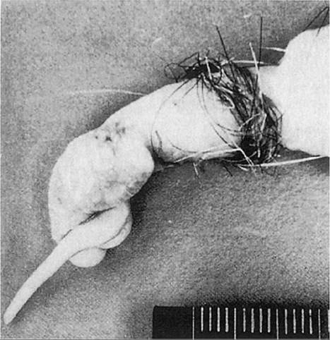
Figure 12.5 Hair ring encircling the penis of a goat. This can lead to dysuria. Source:Tarigan et al. 1990 / John Wiley & Sons, Inc.
dysuria, affected animals show poor body condition, hypothermia, muzzle lesions, purulent nasal discharge, and recurring keratitis and conjunctivitis. Death can occur in two to four weeks, but more prolonged cases also occur (Mahin and Chadli 1982).
There is a single case report of dysuria and pyuria in an 8-year-old male, pseudohermaphrodite goat. The dead goat was presented for necropsy and determined to have a metastatic urothelial carcinoma, with masses and nodules at the base of the urinary bladder and in the proximal urethra, kidney, liver, spleen, heart, lung, and adrenal gland (Mamom 2012).
Uremia
Uremia is a systemic, toxic condition associated with failure to remove the products of protein metabolism from the body via the urinary tract. The origins of uremia can be prerenal, renal, or postrenal. Elevated concentrations of urea nitrogen in the blood or plasma are the main laboratory indicator of uremia, also referred to as azotemia. Prerenal uremia is associated mainly with dehydration or poor renal perfusion, which may result from a variety of causes unrelated to the urinary system.
In cattle, very high levels of BUN can occur in the absence of primary renal disease (Divers et al. 1982). Therefore, marked azotemia must be interpreted cautiously. Experimental evidence suggests that this is also true in goats. Induction of pyloric stenosis by ligation resulted in serum urea levels of 353 mg/dL with no evidence of impaired renal function. The increase correlated with a decrease in effective renal blood flow (Jorna 1978).
Uremia of renal origin is associated with decreased renal function or outright kidney failure. Naturally occurring, clinical cases in goats are not widely documented or reported, though they undoubtedly occur. Experiments involving partial or total nephrectomy in goats provide indications of the clinical manifestations of uremia of renal origin in the species.
Experimentally, five of nine goats subjected to subtotal nephrectomy survived for 52 weeks without clinical signs, while four died within eight days. All goats with total bilateral nephrectomy were terminally ill within eight days. Clinical signs included decreased appetite, rumen atony, depression, weakness, excess salivation, wiry pulse and jerky respiration, subnormal temperature, recumbency, convulsions, and coma. The degree of azotemia was not measured (Vyas et al. 1978).
Hypocalcemia, hyperphosphatemia, and hypermagnesemia have been recorded in toxic nephrosis in the goat. Hyperkalemia is not a consistent finding. In experimental lead poisoning, hypokalemia was observed along with hypocalcemia and hyperphosphatemia (Gouda et al. 1985). There is one reported case of renal (uremic) encephalopathy in a 2-year-old male goat in which spongiform lesions of the brain were observed. The cause of the underlying nephrosis was not determined, but the brain lesions were considered to be secondary to the uremia (Radi et al. 2005).
In goats, most reported clinical cases of uremia are post- renal in origin and are associated with obstructive urolithiasis. The condition is discussed in more detail later in the chapter.