Specific Diseases of the Urinary System Infectious Diseases
Caprine Herpesvirus Vulvovaginitis and Balanoposthitis
In the goat, an alphaherpesvirus, caprine herpesvirus-1 (CpHV-1), has been identified as the cause of a venereally transmitted vulvovaginitis and balanoposthitis.
The same agent has been implicated in epizootics of fatal viremia involving kids 1-2 weeks of age and abortion in does. It has also been isolated in conjunction with Mannheimia (Pasteurella) haemolytica from an outbreak of severe pneumonia of goats. CpHV-1 infection should be viewed as an emerging disease of growing importance in goats, especially in Mediterranean countries. The respiratory form, the viremic kid form, and the abortion form are discussed further in Chapters 9, 10, and 13, respectively.Etiology
CpHV-1 is an icosahedral, double-stranded, linear DNA virus measuring 135 nm in diameter. It is sensitive to lipid solvents and trypsin and is inactivated at a pH value of 3 and at a temperature of 50 °C (122 °F). It can be cultivated on a variety of cell culture types and is cytopathic (Berrios and McKercher 1975). The biologic and physicochemical properties of the virus have been described (Engels et al. 1983).
In early reports of disease, CpHV-1 was sometimes referred to as bovine herpesvirus type 6, but it is now recognized as a distinct entity, CpHV-1. Still, CpHV-1 belongs to an important group of alphaherpesviruses pathogenic to ruminants and closely related to each other. This group includes bovine herpesvirus-1 (BoHV-1), the cause of infectious bovine rhinotracheitis and infectious pustular vulvovaginitis in cattle; bovine herpesvirus-5 (BoHV-5), which causes bovine encephalitis; bubaline herpesvirus-1, associated with subclinical genital infections in water buffalo; cervid herpesvirus-1, which causes an ocular syndrome in red deer; cervid herpesvirus-2, associated with subclinical genital infections in reindeer; and elk herpesvirus-1, associated with subclinical genital infections in elk.
The relatedness of these viruses has practical significance relative to disease diagnosis and control. Crossspecies infections occur, though rarely. This has been investigated under experimental conditions, as reviewed by Thiry et al. (2006a). Because of the antigenic similarity of these alphaherpesviruses, most serologic tests of antibody detection cannot distinguish reliably among them, as discussed further below in the clinical pathology section. Also, if multiple infections occur, recombination between these related viruses is theoretically possible, though it has not yet been confirmed between CpHV-1 and BoHV-1 (Meurens et al. 2004). Of greatest concern is that goats and other ruminants may serve as reservoirs for BoHV-1, and thereby confound national and international efforts to eradicate infectious bovine rhinotracheitis in cattle. This underscores the need for diagnostic tests that can reliably discriminate among the alphaherpesviruses. The molecular and epidemiologic relationships of the alphaherpesviruses have been reviewed (Thiry et al. 2006a).
Goats have been experimentally infected with BoHV-1, resulting in mild clinical signs, high levels of BoHV-1 excretion for several days during primary infection, an antibody response, and establishment of latent infection in the trigeminal ganglia of the challenged goats (Six et al. 2001). These latent infections could be reactivated by treatment of the goats with high doses of dexamethasone.
Conversely, calves experimentally challenged with CpHV-1 also became infected. They did not manifest any clinical signs, but they did excrete CpHV-1 virus and produce an antibody response. Latent infection in the trigeminal ganglia was established as demonstrated by polymerase chain reaction (PCR), but could not be reactivated (Six et al. 2001).
Naturally occurring infections of goats with BoHV-1 have also been reported. In 1972, it was reported that the virus was isolated from nasal and ocular swabs from two goats in Maryland with fever and signs of respiratory disease (Mohanty et al.
1972). The BoHV-1 virus was also isolated from four goats in Washington state, one with clinical signs of vulvovaginitis, two with pneumonia, and one with a wart-like lesion. All these goats had commingled with cattle. The isolates were found to be more closely related to bovine vaccine strains of BoHV-1 than to CpHV-1 on the basis of restriction endonuclease analysis (REA) (Whetstone and Evermann 1988).A gammaherpesvirus, known as caprine herpesvirus-2, has been isolated from goats and characterized (Li et al. 2001). It has been shown to cause malignant catarrhal fever in whitetail deer (Odocoileus virginianus) (Li et al. 2003). In a subsequent report, a putative case of malignant catarrhal fever in a Pygmy goat was described (Twomey et al. 2006). The goat, which was kept with sheep, had a multisystemic necrotizing vasculitis and the gammaherpesvirus ovine herpesvirus-2 (OvHV-2) was identified in the goat's tissues by PCR. The gammaherpesviruses are biologically distinct from the alphaherpesviruses and are not considered further here.
Epidemiology
The isolation and identification of CpHV-1 in association with naturally occurring disease was first reported from California in 1974 in cases of severe systemic infection of goat kids (Saito et al. 1974). Vulvovaginitis caused by CpHV-1 was first observed in Saanen does in New Zealand in 1981 and then in Australia in 1986 (Horner et al. 1982; Grewal and Wells 1986). Posthitis in males was identified in New Zealand in 1982, in Australia in 1984 (Tarigan et al. 1987), and in bucks in California from herds experiencing abortions due to CpHV-1 (Uzal et al. 2004). Typical of herpesviruses, CpHV-1 infection demonstrates latency and recrudescence. In one New Zealand goat herd, the genital form of the disease was reported to reoccur a year after the first outbreak (Horner 1982).
Transmission of the genital disease is presumed to be venereal, but bucks need not be infected or have active lesions to spread the disease from infected to non-infected does mechanically.
Increases in clinical prevalence or seroconversion can occur after the onset of the breeding season, suggesting either venereal transmission of new infections or recrudescence of latent infections secondary to stress associated with estrus and/or breeding activity. Clinical signs of vulvovaginitis in does have been reported to appear within 11 days of introduction of teaser bucks (Horner et al. 1982).There is growing evidence of widespread CpHV-1 infection in world goat populations. Serologic evidence of CpHV-1 infection has been reported from Norway, northern Ireland, Spain, Italy, Turkey, Greece, France, and Syria (Kao et al. 1985; Thiry et al. 2006a, 2008). In Greece, neutralizing antibodies were identified in 52.6% of 795 goats from various locations in the country (Koptopoulos et al. 1988). The Mediterranean region in general has a high seroprevalence of CpHV-1 infection in goats, and clinical disease results in notable economic loss for goat producers in the region. The infection has been confirmed in goats in France by virus isolation (Sauvet et al. 2016).
Pathogenesis
Genital infections in goats have been established experimentally by intranasal and intravaginal inoculation, but the pathogenesis appears to vary with the route of introduction. With intranasal challenge, virus first replicates locally, producing epithelial lesions in the nasal cavity. This is followed by viremia, as evidenced by the presence of virus in the buffy coat, and then infection of the genital tract, where virus produces characteristic lesions in the external genitalia and is shed in high titer (Tempesta et al. 1999a). The viremia associated with intranasal inoculation has also been demonstrated experimentally by recovery of virus or its detection by PCR assay from the dead fetuses of pregnant does (Tempesta et al. 2004). Case reports of natural infections indicate that abortion in does due to CpHV-1 as well as the fatal syndrome observed in 1-2-week-old kids are also the result of viremia, as aborted fetuses (Williams et al.
1997; Chenier et al. 2004; Uzal et al. 2004) and dead kids (Roperto et al. 2000) demonstrate the presence of CpHV-1 virus or viral DNA in multiple organs.When goats were experimentally infected intravaginally, successful infection was manifested by genital lesions and recovery of virus from vaginal swabs for five to seven days post infection. However, no virus was identified from ocular, nasal, or rectal swabs or buffy coat, suggesting that genital infections can be established without an associated viremia (Tempesta et al. 2000).
As is characteristic of herpesviruses in other species, CpHV-1 results in latent infections with the capacity for recrudescence. The presence of virus in the sacral ganglia of latently infected goats has been confirmed by PCR assay (Tempesta et al. 1999b). Experimentally, recrudescence with viral shedding has been induced by repeated administration of high doses of dexamethasone over several days (Buonavoglia et al. 1996). In natural infections, recrudescence occurs in infected does in association with estrus, but primarily in does with low neutralizing antibody titers (Tempesta et al. 1998, 2005). Because recrudescence is associated with viral shedding, this suggests that the condition could spread rapidly through goat herds during the breeding season. Venereally infected bucks can shed the virus from the prepuce for 24 days following infection and therefore can play a significant role in spreading the infection during the breeding season (Camero et al. 2015).
CpHV-1 was isolated from the lungs of goats in a naturally occurring outbreak of severe, fatal pneumonia in New Zealand in 1986. However, M. (Pasteurella) haemolytica was isolated from affected lungs in addition to CpHV-1 (Buddle et al. 1990a). Experimental follow-up challenge studies were undertaken to clarify the etiologic role of CpHV-1 in these cases. Challenge with CpHV-1 alone did not produce pneumonia, while challenge with M. haemolytica alone or M. haemolytica with CpHV-1 did (Buddle et al.
1990b). CpHV-1 readily proliferated in the upper respiratory tract and lungs of the challenged goats, but its role in the pathogenesis of pneumonia remains unclear.Clinical Findings
All goats of breeding age are susceptible to the genital form of disease. Clinical cases occur most frequently in epidemic form shortly after the onset of the breeding season. This reflects the presumption that hormonal changes associated with the onset of estrus trigger recrudescence of virus shedding in latently infected does, which may then be spread to susceptible does by breeding bucks.
The initial signs of vulvovaginitis in does are vulvar edema and hyperemia, with possibly a slight amount of blood-stained discharge. No systemic signs of disease are observed. Over the next several days, a more copious yellow to gray discharge may develop, and multiple, focal, shallow erosions appear on the vulvar and vaginal mucosa. These erosions may be covered with yellow to red-brown necrotic scabs. The vulva is painful to the touch and dysuria is possible, though not often reported. Lesions usually heal spontaneously within two weeks. Reproductive performance and conception rates are not affected, though animals that experienced the disease may have subsequent recurrences of lesions due to recrudescence of latent infections, usually at the next breeding season.
Males used in breeding may or may not show signs. When present, signs are indicative of balanoposthitis and include preputial edema and hyperemia of the penis, with pain on palpation and bloody or purulent discharge. Crusts may be noted as well as focal, punctate, epithelial erosions on the penis, and more frequently the prepuce (Figure 12.6).
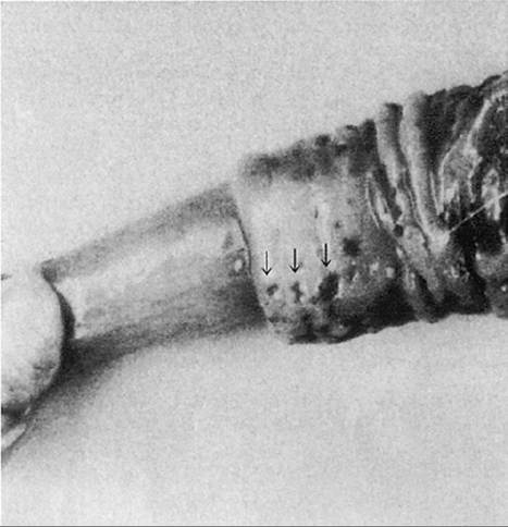
Figure 12.6 Erosive lesions of the penis (arrows) in balanoposthitis caused by caprine herpesvirus. Source: Tarigan et al. 1987 / John Wiley & Sons, Inc.
Fever may be present early in the course of infection. Lesions may resolve by 10 days post infection, but affected bucks may continue to shed virus for up to 24 days (Camero et al. 2015).
Clinical Pathology and Necropsy
The virus can be isolated from vaginal or preputial swabs of acute lesions for confirmation of CpHV-1 infection. Rising titers to CpHV-1 can be detected in both clinical and subclinical infections immediately after an outbreak of vulvovaginitis and balanoposthitis. Acute and convalescent serum sampling should be initiated early in the course of disease, because titers may begin to decline again by three weeks after onset of disease. Serum neutralization titers in infected goats range from 1 : 4 to greater than 1 : 256.
While a rise in titer using serum neutralization or enzyme-linked immunosorbent assay (ELISA) techniques is certainly indicative of CpHV-1 infection, definitive diagnosis of CpHV-1 infection by serologic methods is difficult because of the common cross-reactivity of the alphaherpesviruses and the fact that cross-species infection can also occur, particularly with regard to BoHV-1. A blocking ELISA based on the B glycoprotein of BoHV-1 has been reported to be a good screening test for tentative identification of CpHV-1 in goat herds (Keuser et al. 2004). Confirmation can be achieved by following up with double or cross-seroneutralization tests using BoHV-1 and CpHV-1 antigens and finding higher titers to the CpHV-1 antigen (Thiry et al. 2008; Sauvet et al. 2016).
Definitive diagnosis depends on virus isolation with precise characterization of the virus or detection of CpHV-1 viral DNA in tissues by appropriate means. Current techniques used for detection or characterization of the CpHV-1 virus include REA, real-time polymerase chain reaction (RT-PCR; Elia et al. 2008), and an immunofluorescence assay able to differentiate five related alphaherpesviruses in infected cells (Thiry et al. 2006a).
Necropsy diagnosis of the genital form of CpHV-1 infection is rarely required or undertaken. However, necropsy is most valuable in the diagnosis of other forms of the disease, notably abortion and death of kids 1-2 weeks of age. Virus isolation and viral DNA identification techniques should be conducted on tissues of aborted fetuses and dead kids.
Diagnosis
Normal estrus produces some degree of hyperemia and edema of the vulva, and should not be confused with the early signs of CpHV-1 infection. Granular vulvovaginitis caused by various Mycoplasma spp. must be differentiated from CpHV-1 infection. Granular vulvovaginitis is reported most often from India and Nigeria (Singh et al. 1975; Singh and Rajya 1977; Tiwana et al. 1984; Chima et al. 1986). The condition is discussed further in Chapter 13. An ulcerative vulvitis with copious purulent vaginal discharge has been reported in Nigerian goats. Trueperella (Arcanobacterium) pyogenes and Staphylococcus spp. were isolated (Ihemelandu 1972).
Treatment
There is no practical, specific treatment for CpHV-1 infection. However, there is a report of the use of the human antiviral drug cidofovir in goats. The drug, administered intravaginally, inhibited onset of local vaginal lesions in does and reduced viral shedding (Tempesta et al. 2007b). Does and bucks with the genital form usually heal spontaneously within two weeks, though recurrent lesions are possible. Risk of secondary bacterial infections may be controlled by administration of prophylactic antibiotics.
Control
Efforts to eradicate genital CpHV-1 infection from infected flocks have been proposed based on the assumption that the disease is venereally transmitted. The evidence for this was that kids born to seropositive does and left to nurse on them did not acquire infection by passage through the birth canal, nursing, or close contact. Therefore, it was proposed that gradual elimination of infection from known infected flocks could be achieved according to the following recommendations (Horner 1988):
• Kids should be separated from older animals before they become sexually active and maintained as a separate group.
• Only seronegative bucks or teasers should be used for mating in this separate kid group.
• Buck kids tested before 4 months of age may have maternal antibody present and should be retested at a later age.
• At the same time, the mature, source herd should be serotested regularly and positive animals culled at detection.
• No new animals should be introduced into the herd that have not first been found seronegative.
Since those recommendations were made, it has been demonstrated, at least experimentally, that goats infected with CpHV-1 by the intranasal route can develop vulvovaginitis and shed virus from the genital tract (Tempesta et al. 1999a). This suggests that a better understanding of the transmission of CpHV-1 under field conditions may be needed to formulate management interventions for control of CpHV-1 in infected herds.
Nevertheless, successful eradication of CpHV-1 has been reported from a dairy goat herd used for biopharmaceutical production (Pollock et al. 2019). Eradication, achieved over a period of 10 years, involved annual serologic testing for the virus using a serum neutralization test and the culling of positive animals in association with implementation of strict biosecurity measures, maintenance of a closed herd, and separation of kids from adults at birth.
Currently, there are no commercial vaccines available for controlling CpHV-1 infection. As the commercial market for a CpHV-1 vaccine is considered to be limited, there is interest in the potential for use of commercial bovine herpesvirus vaccines, which may offer some cross-protection. In that regard, a commercially available, live, attenuated, glycoprotein E-negative, BoHV-1 virus vaccine has been reported to be safe for use in goats and demonstrated partial efficacy against subsequent challenge with CpHV-1 (Thiry et al. 2006b, 2007). However, experimental efforts to produce a CpHV-1-specific vaccine are ongoing. A β- propiolactone inactivated CpHV-1 vaccine given either subcutaneously or intravaginally protected does from clinical signs of vulvovaginitis when they were subsequently challenged intravaginally with CpHV-1 (Tempesta et al. 2001; Camero et al. 2007). A recombinant vaccine using a non-pathogenic bovine herpesvirus 4 (BoHV-4) as a vector and expressing the immunodominant CpHV-1 glycoprotein D has been developed and shown to be protective against CpHV-1-induced genital disease in vaccinated, experimentally challenged goats (Donofrio et al. 2013). An inactivated CpHV-1 vaccine has been developed for intra- vaginal administration mixed with LTK63, an enzymatically inactive mutant of heat-labile enterotoxin of Escherichia coli that is known to act as a potent mucosal adjuvant, able to elicit both strongly protective antibody response, including IgA and persistent T-helper type 1 response. Vaccinated animals displayed high levels of secretory IgA and were significantly protected after challenge with the virulent CpHV-1 strain (Tempesta et al. 2007a). Another inactivated CpHV-1 vaccine mixed with the adjuvant MF59™ also was able to confer effective protection against vaginal CpHV-1 challenge in goats (Marinaro et al. 2012). MF59 is a detergent-stabilized oilin-water emulsion consisting of small drops of oil surrounded by a monolayer of non-ionic detergents, and is known to promote both cell-mediated and humoral immunity.
Ulcerative Posthitis
Though principally a disease of sheep, this infectious, inflammatory condition of the prepuce and penis is sometimes seen in castrated male goats as well. The condition is also known as enzootic posthitis, sheath rot, or pizzle rot.
Etiology and Pathogenesis
The disease is associated with the Gram-positive diphtheroid bacterium C. renale, which is capable of hydrolyzing urea. The organism is present in the prepuce of infected animals and is believed to be transmitted venereally or by insects. However, the presence of the organism in the sheath alone is not sufficient to produce clinical disease. The feeding of a high-protein diet increases urine urea content and thereby provides a substrate for the production of large quantities of ammonia by C. renale. The ammonia is believed to irritate the preputial mucosa and skin around the preputial orifice. The condition is more common in wethers than in intact males, probably because of the hypoplastic nature of the penis in castrated males. In wethers, the preputial and penile mucosa do not completely separate and urination frequently occurs without exteriorization of the penis from the prepuce. This promotes urine pooling in the preputial space and allows more complete degradation of urea by C. renale. Another contributing factor may be excessive hair or wool at the preputial orifice, causing the area to remain moist with urine and allowing prolonged bacterial activity on urea substrate, and may explain why the condition is more common in Angora wethers, which are long-haired.
Epizootiology
The disease in goats has been reported only from the United States, though the potential for it to occur elsewhere exists, because it is seen in sheep in Australia, the United Kingdom, and Spain (Loste et al. 2005). While the disease is known to be venereally transmitted by rams to ewes in some flocks, there are no reports of the condition affecting does.
The principal report of posthitis in goats comes from south central Texas, where Angora wethers may be kept to advanced ages for mohair production (Shelton and Livingston 1975). The condition was observed in a large flock of wethers on rangeland containing guajillo (Acacia angustissima). Guajillo, a leguminous shrub, has a protein content in the range of 22-27% and is readily consumed by goats. Morbidity was estimated at 10% and no mortality was directly attributable to the problem.
The author (DMS) has observed ulcerative posthitis in wethers in a different context. Affected goats were castrated males of dairy breeds kept to advanced ages for the purpose of commercial antibody production. These animals were maintained on a commercial dairy ration containing 16% protein. Morbidity was low and the condition was not severe, but obvious swelling and inflammation were evident around the preputial orifice.
Clinical Findings
The condition may be mild to severe. In the mild form, signs are limited to a swelling of the prepuce, readily noted in short-haired dairy wethers or detected in long-haired Angora wethers at shearing time. In severe cases, swelling and inflammation interfere with normal urination and signs of straining are present. These animals may kick at the abdomen, show a stiff gait or an arched back, and may lie down and rise repeatedly. Examination of the ventral abdomen reveals scabs or ulcers on the skin dorsal to and surrounding the prepuce and preputial orifice. The orifice itself may be distorted, reduced in diameter, or completely scabbed over. The sheath may be filled with urine, and exudate may be expressed by applying pressure to the prepuce. Fistulous tracts may be present. The preputial cavity and the penis may show severe ulceration and possibly maggot infestation associated with fly strike. Animals with complete occlusion of the preputial orifice may die of uremia.
Clinical Pathology and Necropsy
Swabs of the preputial cavity should allow isolation of C. renale. Animals with extensive ulceration inside the prepuce and on the penis may die. Necropsy demonstrates extensive exudation within the preputial cavity and ulcers and scar tissue on the mucosa. The urethral process may be necrotic and ulcers be present on the glans penis.
Diagnosis
On rare occasions, goats may show scabby lesions of contagious ecthyma on the prepuce, but similar lesions also likely would be found elsewhere on the face and body. Ulcerative dermatosis, a viral disease of sheep, is not reported in goats. Ulcerative posthitis may appear similar to balanoposthitis due to caprine herpesvirus 1, but in herds with that virus female goats would likely be showing vulvovaginitis as well. Obstructive disease of the gastrointestinal tract and obstructive urolithiasis must be considered in the differential diagnosis of goats with signs of straining or abdominal discomfort.
Treatment
Affected animals should be separated from the herd or flock and the high-protein component of their diet withdrawn. Clipping hair from around the preputial area may improve the rate of healing. Reestablishing patency of the urethral orifice is the first therapeutic goal in animals unable to urinate. Lesions should be debrided and topical treatment begun. C. renale is usually sensitive to penicillins, ampicillins, and cephalosporins. As these antibiotics are excreted in urine, systemic treatment will also provide local therapy. Intramammary infusion tubes designed for mastitis treatment are convenient vehicles for administering these antibiotics into the preputial cavity, though care should be taken not to transmit infection to unaffected herd mates. Repeated daily treatment with ophthalmic ointment containing bacitracin, neomycin, and prednisolone applied topically to the prepuce has also been reported to be effective as an ancillary treatment in sheep (Loste et al. 2005). In severe exudative cases, opening the prepuce surgically with scissors along the ventrum is indicated for salvage. Except in the most severe cases, the prognosis for recovery is good.
Control
Dietary protein should be limited to a level consistent with the nutritional requirement of the animals. This may be problematic under range conditions. Preputial hair should be kept clipped to reduce urine accumulation. Animals with external lesions identified at shearing time should be treated topically with antiseptics or antibiotics to control lesions and reduce spread in the flock. Implantation of wethers with 70-100 mg of testosterone at three-month intervals has been effective in controlling ulcerative posthitis, but this method has not been evaluated in goats (Kimberling 1988). Castration using the so-called short scrotum technique has also been suggested, because it renders wethers infertile but allows the urethra and penis to mature under the influence of testosterone. In this technique, both testes are pushed up into the inguinal canal or inguinal region and a rubber ring is applied around the neck of the scrotum below the testes. There are animal welfare concerns associated with the use of this technique (Molony et al. 2002) and it does not invariably result in sterility.