DIAGNOSTIC CONSIDERATIONS
Optimal management of patients with acute intestinal disease hinges on a specific diagnosis if possible. This is particularly true for patients with enteritis caused by an infectious agent that may have a specific treatment (Box 6-2).
Although initial treatment in most cases varies little (fluid administration, correction of volume and electrolyte disturbances), specific treatment is preferred to more general, nonspecific measures such as indiscriminate administration of antibiotics, motility modifiers, adsorbents, and intestinal protectants. For these reasons, diagnostic tests should be done early, before administering treatments that may interfere with test results.Physical examination allows immediate assessment of hydration status, severity of volume depletion, and initial fluid replacement needs. Careful abdominal palpation may reveal evidence of intestinal obstruction.Viral gastroenteritis is more likely in young and in febrile patients, especially those without adequate vaccinations. Rectal examination may often provide early evidence of impending diarrhea and allow assessment of stool characteristics (presence of blood or mucus, odor,
Infectious Disease Differential Diagnoses for Vomiting and Diarrhea in Dogs and Cats
| Viruses Astrovirus (S) Canine and feline coronaviruses (S) Canine parvovirus (S) Canine distemper virus (S) Feline leukemia virus-associated lymphoma (S, M) Feline immunodeficiency virus (S) Feline panleukopenia (S) Rotavirus (S) | Helminths Ancylostoma/Uncinaria (S,M,L) Dirofilaria immitis (V only; mainly cats) Ollulanus tricuspis (V only; cats only) Physaloptera (V only) Strongyloides spp. (S) Toxascaris (V mainly) Toxocara spp. (V mainly) Trichuris vulpis (dog, L) |
| Bacteria Bacterial cholangiohepatitis (S) Bacterial overgrowth (S) Bacterial peritonitis (S) Campylobacter jejuni (S,M,L) Clostridium perfringens (M, L) Enterotoxigenic E. coli (S, M) Helicobacter spp. (V) Salmonella spp. (S, M, L) | Protozoans Balantidium coli (ciliate; L) Cryptosporidium spp. (coccidian; S) Giardia spp. (flagellate; S) Isospora spp. (coccidian; L) Entamoeba (amoeba; L) Pentatrichomonas (flagellate; L) Miscellaneous Prototheca (algae; L) Histoplasma (fungal; L) Neorickettsia spp. (rickettsia; S) |
S, Small bowel; L, large bowel; M, mixed; V,vomiting.
color, consistency). Occasionally parasites may be noted.
Due to the high prevalence of parasitic and infectious diseases associated with acute diarrhea, fecal testing is mandatory. To adequately assess patients for infectious causes of diarrhea, a direct smear, fecal flotation, fecal cytologic study, and Cryptosporidium parvum screening test should be performed.
Direct Smear. Liquid feces or feces that contains large quantities of mucus should be microscopically examined immediately for the presence of protozoal trophozoites, including those of Giardia spp., Pentatrichomonas hominis, Balantidium coli, and Entamoeba histolytica. A direct saline smear can be made to potentiate observation of these motile organisms. The amount of mucus and feces required to cover the head of a pin is mixed thoroughly with one drop of 0.9% NaCl. Following application of a coverslip, the smear is evaluated for motile organisms by examining it under ? 100 magnification. Use of fresh feces gives the highest yield of positive results.
Fecal Flotation. Cysts, oocysts, and ova in feces can be concentrated to increase sensitivity of detection. Most ova, oocysts, and cysts are easily identified after zinc sulfate centrifugal flotation. This procedure is considered by many to be optimal for the demonstration of protozoan cysts, in particular Giardia spp., and so is a good choice for a routine flotation technique in practice (Figure 6-1). Sugar centrifugation can be used for routine parasite evaluation and may be superior to many techniques for the demonstration of oocysts
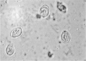
FIGURE 6-1 Giardia cysts.
of Toxoplasma gondii and C. parvum. Giardia cysts are distorted by sugar centrifugation but can still be easily identified. Fecal sedimentation will recover most cysts and ova but will also contain debris. This technique is superior to flotation procedures for the documentation of fluke eggs. Some parasites such as Trichuris spp. and Giardia spp. are shed intermittently and so can be occult. Performing two to three fecal flotations over 5 to 7 days will increase sensitivity of detection of these parasites. Feces should be refrigerated, not frozen, until assayed for parasites. If a fecal sample is to be sent to a diagnostic laboratory for further analysis and will not be evaluated for parasites within 48 hours, it should be preserved. Polyvinyl alcohol, Merthiolate-iodine-formalin, and 10% formalin preservation can be used. Because of routine availability, 10% formalin is commonly used; add 1 part feces to 9 parts formalin and mix well.
Stained Smear. A thin smear of feces should be made from all dogs or cats with large or small bowel diarrhea. Material should be collected by rectal swab if possible to increase chances of finding white blood cells. A cotton swab is gently introduced 3 to 4 cm through the anus into the terminal rectum, directed to the wall of the rectum, and gently rotated several times. Placing a drop of 0.9% NaCl on the cotton swab will facilitate passage through the anus of cats but not adversely affect cell structure. The cotton swab is gently rolled on a microscope slide multiple times to give areas with varying thickness. Following air drying, a slide should be stained with Diff-Quik or Wright's or Giemsa stain. The slide should be examined for white blood cells and bacteria morphologically consistent with Campylobacter jejuni or Clostridium perfringens (Figure 6-2). Presence of neutrophils on rectal cytologic examination can suggest inflammation induced by Salmonella spp., Ca. jejuni, or C. perfringens; fecal culture is indicated in these cases (see the following section).
Histoplasma capsulatum or Prototheca may be observed in the cytoplasm of mononuclear cells. Methylene blue in acetate buffer (pH 3.6) stains trophozoites of the enteric protozoans. Iodine stains and acid methyl green are also used for the demonstration of protozoans. Acid-fast staining of a fecal smear is one of the C. parvum screening procedures that should be performed in dogs or cats with diarrhea. C. parvum is the only enteric organism of approximately 4 to 6 μm in diameter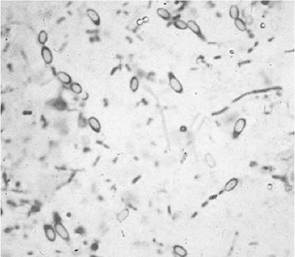
Figure 6-2 Clostridium perfringens spore-forming rods.
that will stain pink to red with acid-fast stain (Figure 6-3). Alternatively, antigen testing is available for this organism (see the following section). conjunction with results from fecal examination techniques. C. perfringens enterotoxin can be detected in both healthy and diseased animals, and so positive test results do not confirm disease due to this organism (see bacterial diseases section).
Culture. Culture of feces for Salmonella spp., Campylobacter spp., and Cl. perfringens is occasionally indicated in small animal practice. Approximately 2 to 3 g of fresh feces should be submitted to the laboratory immediately for optimal results; however, Salmonella and Campylobacter are usually viable in refrigerated fecal specimens for 3 to 7 days. The laboratory should be notified of the suspected pathogen so appropriate culture media can be used. If a delay in sample submission is expected or to increase yield of positive results, a transport medium such as Cary-Blair[‡‡‡] [§§§] medium should be used.
Immunologic Techniques. Parvovirus,
C. parvum, Giardia spp., and C. perfringens enterotoxin detection procedures are available as point-of- care tests for use with feces. Parvovirus assays detect both vaccine and field strains of canine parvovirus, and so results should be interpreted cautiously (see the viral diseases section). Recently, human C. parvum and Giardia spp. antigen assays have been applied to feces from small animals.* Minimal sensitivity and specificity results are currently available. For example, it is not known if the assays detect the canine and feline genetic variants of C. parvum. If used, results of these assays should be interpreted in
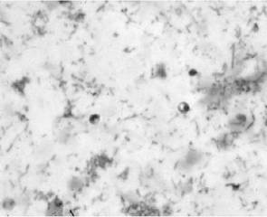
Figure 6-3 Cryptosporidium parvum oocysts.
Electron Microscopy. Electron microscopy can be used to detect viral particles in feces of dogs and cats with GI signs of disease. Approximately 1 to 3 g of feces without fixative should be transported to the laboratory* by overnight mail on cold packs for performance of this assay.