Ecology
Sporothrix schenckii s. str., the classical etiological agent of sporotrichosis, is a saprophyte widely distributed in nature. It is associated with decaying organic matter in soil, water, and rotten bark (Lacaz et al.
2002). Ideal conditions for fungal development in the environment include temperatures between 25 and 28 °C and relative humidity between 92 and 100% (Kenyon et al. 1984; Findlay et al. 1984; Conti Diaz 1989). From propagules in the environment, Sporothrix is able to cause disease in humans and other animals such as cats, dogs (Schubach et al. 2001; Govender et al. 2015), rats, mice, pigs, camels, chimpanzees, armadillos, cattle (Rodrigues et al. 2013a,b), donkeys, and horses (Crothers et al. 2009). Due to those climatic conditions, sporotrichosis is common in temperate and subtropical regions, being rarer in arid, semiarid, and cold zones (Dixon et al. 1991). Brazilian Sporothrix brasiliensis is mainly cat-transmitted, whereas the mainly East Asian species S. globosa practically never infects cats. It might be expected that the habitat determinants of these two species are fundamentally different, but as yet no data are available.Few ecological studies have evaluated the natural reservoirs and the factors that affect the population growth of Sporothrix in nature (Mackinnon et al. 1969; Criseo and Romeo 2010; Rodrigues et al. 2014a). Studies on physiological characteristics of S. schenckii allow development of strategies for selective isolation (Fernandes et al. 2009b). Ghosh et al. (2002) reported that the mycelial phase tolerates osmotic pressure of up to 20% glycerol. The yeast form is slightly more tolerant to osmotic pressure, tolerating 6-7% salt concentration, but all are inhibited at 8% salt. Sporothrix species grow well between pH 3.0 and 11.5, but few can tolerate pH 12.5 (Ghosh et al. 2002; Fernandes et al.
2009b).Sporothrix species exhibit thermal dimorphism, growing in the mycelial phase at 25 °C but as yeast at 37 °C (Howard 1961). In addition to temperature, pH seems to have an important role in the transformation of S. schenckii. At pH 4.0-5.0 at 25 °C in rich medium, only hyphae develop; however, yeast cells grow in the same medium at pH 6.5-8.0 and at 35 °C (Rodriguez-Del Valle et al. 1983).
At room temperature, colonies are initially white to cream and, over time, usually become dark, with colors ranging from brown to black. Some isolates may take months to exhibit these characteristics, and some may never become black (Rippon 1988) (Fig. 10.4). Microscopically, the hyphae are thin and septate, and the diameter does not exceed 3 μm; ovoid conidia are 2-3 x 3-6 μm in size and are arranged sympodially, in the shape of a daisy at the conidiophore apex. Sessile brown conidia are inserted along undifferentiated hyphae (Fig. 10.4).
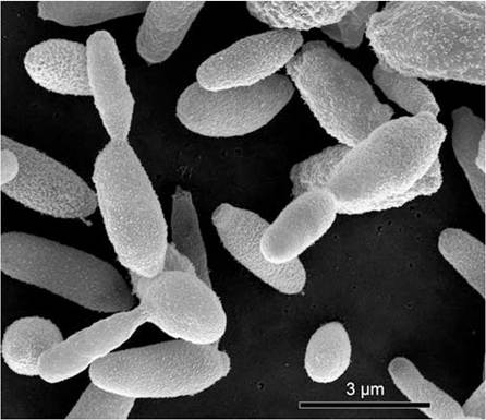
At 37 °C the colony morphology changes from pasty white to grayish yellow, resembling a bacterial colony (Fig. 10.4). Mother yeast cells are spherical and daughter cells are oval to cigar-shaped, budding on a narrow base. Yeast cell size ranges from 1-3 to 3-10 μm; multiple buds may be observed, but a single bud is typical (Howard 1961; Rodrigues et al. 2015d) (Fig. 10.5).
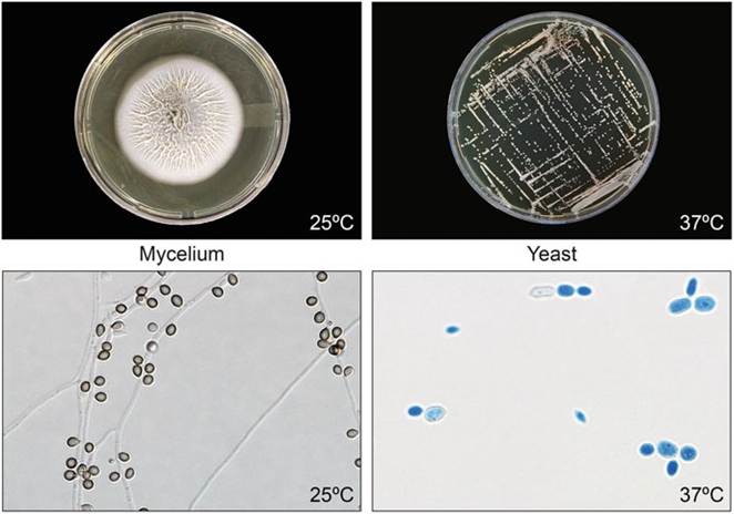
Fig. 10.4 Thermal dimorphism in Sporothrix spp. is a morphophysiological adaptation for infection of warm-blooded hosts. The saprophytic phase occurs at room temperature (25 oC). When grown on Sabouraud agar, the colonies are at first white to cream and leathery with a grooved surface. Microscopically (25 °C), many formations of sessile dematiaceous conidia along undifferentiated hyphae are present. When the isolates are grown at 37 °C in brain heart infusion (BHI) agar, the yeast grows, forming small, creamy colonies. Microscopically (37 °C) yeast cells are spherical to oval, commonly known as cigar-shaped. Petri dish: 9 cm in diameter
Fig. 10.5 Morphology of Sporothrix brasiliensis yeast cells. The parasitic phase is represented by oval or spherical cells (1-3 μm to 3-10 μm), sometimes elongated and with buds
10.4