ESOPHAGITIS
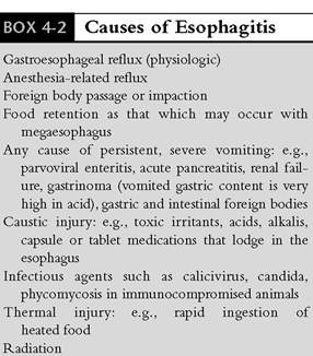
Inflammatory diseases of the esophagus occur more commonly than they are recognized. The major causes of esophagitis are listed in Box 4-2.
Inflammatory changes can range from mild mucosal inflammation that may or may not be grossly evident, to moderate to severe ulceration and transmural involvement.Any disorder that causes acute or chronic frequent vomiting can potentially cause esophagitis.
This especially includes causes of severe vomiting, such as intestinal foreign bodies, gastric foreign bodies, acute pancreatitis, parvoviral enteritis, and gastrinoma. Dogs with parvoviral enteritis that are debilitated and recumbent are especially at risk. Vomited fluid that is retained in the esophagus is not cleared adequately in weak and recumbent patients. As a result the esophageal mucosa is bathed with gastric acid and activated enzymes that will cause mucosal injury.Other causes of esophagitis include esophageal foreign bodies (extent of injury depends on the size, texture, and duration of lodgment) and chemical and thermal injuries (e.g., ingestion of hot food). The latter two factors are uncommon causes of esophagitis. Chemical injury may result from ingestion of toxic chemicals or from failure of the esophagus to transport capsule or tablet medication completely to the stomach. It is not infrequent in humans for medications taken without water to become lodged somewhere in the esophagus. When certain medications dissolve there, mucosal irritation results. Medication associated esophagitis from caustic compounds such as NSAIDs and doxycycline have been associated with esophagitis or even stricture formation. Owing to anatomic and physiologic differences between the canine esophagus and the human esophagus (canine esophageal muscle is entirely striated; the human esophagus is mostly smooth muscle), this irritation is somewhat less likely to be a problem in dogs than in humans.
However, it is likely to be more common in cats than in dogs. There have been reports of severe esophagitis occurring in patients secondary to injury from ingested capsule medication (e.g., doxycycline, chloramphenicol). There is also potential for a stricture to develop secondary to esophagitis. In fact, doxycycline-induced esophagitis with esophageal stricture formation has been reported in cats.Gastroesophageal Reflux Disease
A study was reported recently on normal cats in which the passage of tablets and capsules when given alone (dry swallow) and when followed by a water bolus (wet swallow) was evaluated. This investigation was undertaken as a result of experience with cats developing esophagitis and esophageal strictures subsequent to doxycycline tablet administration. Thirty healthy cats of various ages were used in this study. Each cat was given a 20-mg barium tablet and a 190-mg (size 4) capsule both as a dry and wet swallow. A wet swallow consisted of immediately following administration with 6.0 ml of water orally via syringe. Fluoroscopy was used to evaluate tablet or capsule passage at 30, 60, 90, 120, 180, 240, and 300 seconds following administration. Dry swallows and wet swallows were evaluated. Successful passage was defined as complete passage into the stomach at a given time interval.
The percentage of dry tablet swallows that successfully passed into the stomach was 0.0% at 30 and 60 seconds, 6.7% at 90 seconds, 13.3% at 120 seconds, 26.7% at 180 and 240 seconds, and 36.7% at 300 seconds. Wet tablet swallows successfully passed 90.0% of the time at 30 seconds, 93.3% at 60 seconds, and 100.0% of the time thereafter. The percentage of dry capsule swallows that successfully passed was 16.7% at each time interval. Wet capsule swallows successfully passed 96.7% of the time at 30 seconds and 100% of the time thereafter. For each time interval, wet swallows achieved significantly greater percentage passage into the stomach when compared with dry swallows.
The results of this study show that tablets or capsules given as a dry swallow in cats have prolonged retention in the esophagus. A water bolus following tablet or capsule administration results in significantly faster passage through the water bolus for cats receiving oral tablets or capsules to prevent possible medication associated esophagitis. It is therefore now recommended that cats that receive oral tablet or capsule medications without food be given approximately 6 ml of water immediately after the medication to promote rapid clearing from the esophagus and transit to the stomach. It is also probably a good idea to do the same in dogs whenever medications are not given in food.
Gastroesophageal reflux is the most common cause of esophagitis in animals and humans. This disorder is discussed in detail in this section. The term reflux refers to movement of gastric or duodenal contents into the esophagus without associated eructation or vomiting. As such this can be a “silent” disease. Reflux esophagitis is a disorder in which esophageal inflammation of variable degree occurs as a result of mucosal contact with gastric or duodenal fluid or ingesta. In many cases the inflammation may not be visible grossly. A variety of factors can contribute to its development in individual patients. It can be a particularly difficult diagnosis to make without special instrumentation (e.g., pH probe monitoring, endoscopy) because in many patients clinical signs are quite subtle. History and recognition of suggestive clinical signs constitute the basis for performing diagnostic procedures or instituting empirical therapy. Because significant discomfort can result from reflux episodes, it is important that reflux esophagitis be diagnosed and treated in a timely manner.
Gastroesophageal reflux disease in humans
Gastroesophageal reflux disease (GERD) is among the most common GI disorders that affect people. It is estimated that up to 50% of adults in the United States experience heartburn type of symptoms at least once a month.
GERD is a very difficult diagnosis to establish in animals, largely because our patients are not able to describe for us the fact that they are experiencing the symptoms of this disorder and there are no hallmark signs, and it can also be difficult to diagnose definitively in humans.There is still no single test that can uniformly detect GERD. Of all the GI disorders that affect humans, the symptom pattern for GERD is among the most specific of any GI disorder. However, there are now well-recognized supra- esophageal and extraesophageal reflux symptoms (e.g., laryngeal problems, cough) that will not be diagnosed with just a standard history. The tests used in human medicine to investigate for GERD are endoscopy, looking for esophagitis or other GERD complications, and 24-hour pH probe recording. Barium swallow is also performed in some cases, mostly to screen for other upper GI disorders. None of these tests is always diagnostic, however (e.g., it is known that 30% to 70% of patients undergoing endoscopy for GERD symptoms may have a grossly normal examination), and some gastroenterologists therefore use parallel tests to determine the presence of GERD. More recently another diagnostic approach in humans that has received attention is a therapeutic trial of high dose proton pump inhibitor (PPI) therapy. The basis for this test centers on the ability of the high dose PPI therapy to completely inhibit gastric acid secretion. If symptoms do not resolve on high- dose PPI therapy, they are not likely caused by GERD. There are sensitivity and specificity issues with this test as well, however.Veterinarians should recognize that GERD is very common in people and that it likely occurs in animals much more commonly than we are able to recognize. Therefore we should maintain a high index of suspicion for this disorder when presented with patients that may be exhibiting any of the potential signs of esophagitis.
Pathophysiology
Normal LES function is essential to the prevention of gastroesophageal reflux and esophagitis.
The LES is located at the GEJ and is a zone of high resting pressure that acts to prevent reflux of gastric contents into the esophagus. In response to esophageal peristaltic contractions, the LES undergoes a phase of initial relaxation that is followed by postdeglutition contraction. Initial relaxation begins when an esophageal peristaltic contraction is in the proximal esophagus. Postdeglutition contractions prevent reflux of a food bolus following its passage into the stomach.Reflux of small amounts of fluid is considered a normal physiologic phenomenon in both animals and humans. Functional defense mechanisms prevent esophageal mucosal damage when these minor reflux episodes occur. These defenses include acid clearance by means of one or two esophageal peristaltic sequences that empty all or most of the acid from the esophagus,local mucosal protective factors, and neutralization of any post- peristaltic residual acid by bicarbonate-rich saliva. It has been shown in humans that some individuals can experience significant reflux episodes without developing demonstrable esophageal mucosal changes. Although clinical signs of reflux may be experienced (heartburn, indigestion, dyspepsia), significant sequelae such as esophagitis, esophageal stricture, and chest pain often never develop.
Although the relationships and factors responsible for individual variations in response to reflux are unknown, a number of factors are probably involved in determining how significant a problem reflux episodes will be in an individual. These include volume and frequency of reflux, the duration of contact between the refluxate and the esophageal mucosa, character of the refluxed material, competency of esophageal clearing mechanisms, and gastric emptying patterns. It has been estimated that up to 7% of the general human population has symptoms of heartburn daily, and a much larger percentage experiences these symptoms monthly (approximately 50% as previously stated). Many humans never seek medical attention for what they consider a minor, normal physiologic event.
The frequency in animals is unknown, since signs of mild reflux are extremely difficult to detect.Manometric measurements of the LES have shown that a decrease in resting pressures is the major factor in the pathogenesis of gastroesophageal reflux. Reflux occurs primarily by one of three different mechanisms: transient complete relaxation of the LES, transient increase in intraabdominal pressure, or spontaneous free reflux associated with a low resting pressure of the LES. Human studies have shown that in normal individuals, reflux episodes are almost always caused by transient sphincter relaxation. The predominant reflux mechanism in reflux esophagitis patients varies, although transient LES relaxation seems to be most common. This transient relaxation mechanism may explain why some reflux esophagitis patients have resting LES pressure values that overlap those of normal controls.
Transient changes in intraabdominal pressure may intermittently overcome a hypotensive LES; however, complete sphincter relaxation alone does not guarantee that significant reflux will occur. Factors that may influence reflux in this situation include body position, intragastric volume, intra- gastric pressure, and relaxation of the diaphragmatic hiatus. Significant reflux can occur in animals that undergo general anesthesia, especially when there is ingesta or fluid retention in the stomach. Anesthetic agents promote relaxation of the LES, and any procedure that involves positioning the patient with the rear quarters elevated (e.g., tilting the surgery table so that the head and the upper body are below the lower body) can promote gravitational flow of gastric contents to the esophagus. Mild to severe esophagitis may result, and in some cases esophageal stricture formation occurs. Clinical situations in which a reflux episode may be exacerbated must be recognized, and preventive measures must be taken to decrease serious sequelae.
Mechanisms of Esophageal Mucosal Damage
Although both acid and pepsin were implicated in the past as the major injurious agents in reflux disease, it now appears that the importance of acid has been overemphasized and that of pepsin minimized. Animal studies have shown that pepsin, rather than acid, is a major causative agent of erosive esophagitis resulting from reflux of acid gastric contents.
Of the potentially injurious agents in acid gastric contents (e.g., acid, bile salts, pepsin, and trypsin), pepsin produces a mucosal injury consistent with both the macroscopic and the microscopic appearance of reflux esophagitis in symptomatic human patients. Hydrochloric acid (HCl) at physiologic pH values does not appear to break the esophageal squamous mucosal barrier to hydrogen ion back diffusion or to cause esophagitis. Pepsin, however, can cause mucosal permeability changes, resulting in severe hydrogen ion back diffusion. Rabbit esophageal perfusion studies have demonstrated that pepsin causes significantly more esophageal injury than does bile, trypsin, or acid alone. In these studies the extent of injury increased in a dose-dependent manner as pepsin concentration was increased. Pepsin injury was characterized by mucosal erosion and ulceration with submucosal hemorrhage. Acid, bile, and trypsin damage was generally limited to submucosal edema without mucosal disruption.
Excessive alkaline gastroesophageal reflux produces inflammatory changes comparable to those seen with excessive acid gastroesophageal reflux. The alkaline nature of refluxed material alone does not appear to produce mucosal damage. Rather, with alkaline reflux the pancreatic enzyme trypsin has been shown to be the factor that causes the most significant damage. Pepsin causes minimal esophageal changes in the presence of an alkaline environment. Trypsin is present in the gastric contents of patients with decreased pyloric tone and duodenogastric reflux. The pH of the refluxate appears to control which agent will be the most active in causing esophageal damage. Pepsin's optimal pH range for proteolytic activity is 2 to 4.5, and it is the most injurious agent when the reflux- ate is acid. Trypsin's optimal pH range for proteolytic activity is 5 to 8.
The bile salt taurodeoxycholate has been found to protect the esophageal mucosa from the injurious effects of acid and pepsin, but the effect of trypsin in the alkaline medium has been potentiated. Bile salts decrease pepsin's proteolytic activity, and the protective bile salt effect is dose related. The combination of bile, trypsin, and an alkaline refluxate could potentially cause the most severe degree of esophageal injury. Bile salts may play an important role in modulating the injurious effect of acid and pepsin in certain clinical settings. The concentration of injurious agents in the refluxed gastric fluid and the duration of their contact with the esophageal mucosa are the major factors determining the likelihood and severity of mucosal injury.
Etiology
Pharmacologic agents that have been associated with decreased LES pressure and reflux include atropine and other anticholinergic drugs, morphine, meperidine, diazepam, and pentobarbital. Phenothiazine-derivative tranquilizers also can decrease LES pressure. Glycopyrrolate does not cause as significant an effect on the LES as atropine does. For this reason, some clinicians use glycopyrrolate rather than atropine as their standard preanesthesia agent.
Pregnancy in humans is associated with an increased frequency of heartburn, a sensation of chest pain that is due to esophageal pain from mucosal contact with refluxate. This was originally thought to be due to reflux exacerbated by increased gastric pressure from an enlarging uterus. However, it is now recognized that elevated progesterone levels decrease LES pressure, increasing the likelihood of reflux. Reflux esophagitis in humans can also be predisposed by high-fat or spicy foods, chocolate, alcohol, and nicotine.
The most common causes of reflux esophagitis in dogs and cats are general anesthesia and persistent vomiting due to any cause (e.g., pancreatitis, gastric or intestinal foreign body, parvoviral enteritis). Hiatal hernia disorders, neuromuscular disorders (e.g., myasthenia gravis) that interfere with function of the esophagus and LES, delayed gastric emptying, and duo- denogastric reflux are also important, but less common, disorders that are associated with reflux esophagitis episodes.
During anesthesia there is suppression of normal esophageal motility and decreased LES pressure. As a result, acid and other refluxed agents cannot be cleared as quickly as in an awake animal with normal esophageal defenses. Problems tend to occur more commonly in patients that have undergone prolonged surgical procedures; however, it has been shown that reflux can occur between 5 and 15 minutes after induction of anesthesia. Therefore reflux should be considered a possibility in any patient that undergoes anesthesia. In a study involving 100 dogs, it was found that the frequency of gastroesophageal reflux was 25% with regard to the type of surgery, 48% of the cases of reflux appeared during orthopedic surgery, 24% during abdominal surgery, and 28% during other types of surgery (e.g., skin, eyes). Consideration should be given to routinely treating these patients for reflux esophagitis for several days during the immediate postoperative period. It may also be beneficial to pretreat patients scheduled to undergo a prolonged (greater than 1 to 2 hours) surgical procedure with an H2-receptor antagonist before induction of anesthesia (e.g., administer injectable famotidine or ranitidine 1 to 2 hours prior). As the duration of anesthesia increases, the risk of a significant reflux event also increases.
Some clinicians have also instituted a postoperative practice of lavaging the esophagus with saline or warm water after long surgical procedures, so as to dilute and remove offending substances before significant mucosal damage can occur. This can be done either under endoscopic guidance or simply by passing a tube blindly into the esophagus for lavage purposes, while the patient is still intubated. Use of an endoscope offers the advantage of lavage and suction under direct visualization. In addition to the anesthetic agents used, tilting of the surgery table so that the patient's abdomen is elevated relative to the thorax and improper preparation (e.g., incompletely evacuated stomach) also can play a major role in exacerbating reflux. Moderate to severe esophagitis can result in esophageal stricture formation (see later discussion).
Most hiatal hernia patients have some degree of reflux esophagitis. Decreased LES pressure leads to esophageal reflux in most patients with sliding hiatal hernias. Hiatal hernia patients are often presented for evaluation because of clinical signs that suggest a significant degree of esophagitis (e.g., salivation, inappetence, decreased activity, regurgitation). Treatment involves both management of esophagitis and medical management or surgical correction of the hiatal hernia.
Gastric emptying and gastric motility may be reduced in some patients with gastroesophageal reflux. Delayed emptying of liquids or solids would be expected to increase esophageal reflux. However, only a fraction of human patients with reflux esophagitis have delayed gastric emptying. Detailed studies have not been performed in animals, but clinical signs and endoscopic evidence of esophagitis have not been commonly observed in animals with gastric motility disorders. Probably the most important clinical situation regarding animals with gastric motility disorders involves general anesthesia. Every effort must be made to ensure that there is sufficient time for the stomach to empty before anesthetic induction, since the combination of anesthesia and an incompletely evacuated stomach would increase the likelihood of a reflux episode and subsequent development of esophagitis.
Duodenogastric reflux may be damaging for two reasons: It increases gastric volume available for gastroesophageal reflux, and it adds bile and other potentially damaging duodenal fluid components to the gastric contents. Patients that have a chronic intermittent pattern of vomiting bile fluid may have duodenogastric reflux and should be watched carefully for signs of esophagitis.
Diagnosis of Esophagitis
The clinical signs of esophagitis vary considerably, depending on the degree of inflammation present. The clinician must maintain a high index of suspicion because in many cases only subtle clinical signs may be evident. With mild esophagitis there may be increased swallowing motions, salivation, and inappetence. In more severe cases there may be gulping, regurgitation, dysphagia due to pain, total anorexia, and signs that suggest esophageal pain, such as reluctance to move, standing with the head extended, reluctance to lie down, and trembling. Heartburn pain in humans can be quite intense, and it is suspected that a similar situation exists in animals. Esophageal hemorrhage may occur in severe cases.
The immediate past medical history must be reviewed carefully because it may provide important clues regarding both diagnosis and etiology. Signs such as increased attempts at swallowing, salivation, regurgitation, and inappetence that occur within 1 to 4 days of an anesthetic procedure strongly suggest reflux esophagitis. Coughing may indicate aspiration pneumonia. Patients with persistent vomiting should be observed carefully for signs of esophagitis. Severe esophagitis must be identified and treated early, since one of the potential sequelae is stricture formation.
Chronic reflux esophagitis occurs most commonly in patients with hiatal hernia disorders. Clinical signs include hypersalivation, regurgitation, and vomiting, which often are noted shortly after the patient eats. There also may be coughing, dyspnea, and exercise intolerance. Hiatal hernias are most commonly identified in immature patients.
Physical examination is usually unremarkable, but there may be physical evidence of glossitis or pharyngitis related to ingestion of a caustic substance. There may also be sensitivity on palpation of the cervical esophagus in animals with esophageal inflammation in that area.
Radiographic survey and contrast studies are often normal in patients with mild to moderate esophagitis. Survey films may show increased esophageal density in moderate to severe esophagitis. There also may be various degrees of esophageal dilation, since esophageal inflammation may inhibit motility. In fact, megaesophagus can occur secondary to inflammatory disease. Segmental narrowing and irregularity of luminal contour may occasionally be identified on contrast studies. Persistent contrast in the thoracic esophagus or esophageal dilation or both suggest the possibility of gastroesophageal reflux. In hiatal hernia the gastric cardia, the fundus, and the LES will be cranial to the esophageal hiatus. A definitive diagnosis may not always be possible in sliding hiatal hernia on spot films. Fluoroscopy may be needed to confirm the diagnosis.
A definitive diagnosis of esophagitis is most often made by endoscopic visualization of the esophageal mucosa. Variable degrees of mucosal erythema or isolated patches of eroded mucosa may be seen. Mucosal friability may be evidenced by bleeding caused by gentle manipulation with the endoscope tip or biopsy forceps. Fluid pooling in the esophagus or a markedly dilated gastroesophageal junction or both are not diagnostic, but these findings should alert the endoscopist to the possibility of a reflux disorder.
Numerous human studies have reported that 30% to 70% of patients with symptoms suggesting gastroesophageal reflux have an endoscopically normal esophagus. Symptom severity often does not predict the degree of endoscopic abnormality. When esophagitis is suspected in the absence of visible diagnostic changes in the mucosal surface, an esophageal mucosal biopsy specimen should be obtained from an area 2 to 5 cm proximal to the gastroesophageal junction. With proper technique, adequate mucosal biopsy specimens can be obtained with standard flexible forceps. Alternatively, specimens can be obtained with a suction biopsy instrument. Histologic changes appear before significant symptoms and endoscopically observable changes, and they persist after the endoscopic indicators have disappeared in response to therapy. An endoscopically demonstrable hiatal hernia is nearly always associated with reflux esophagitis.
Treatment
Because there are a variety of pathophysiologic mechanisms that contribute to reflux esophagitis, an individualized treatment program for each patient is often necessary. It is important to note that, although the esophagus is physically a very tough and resilient structure, once it is injured it does not always heal very quickly. For inflammatory disorders fairly aggressive combination drug therapy is often required. Treatment may include dietary modification, PPIs, H2-receptor antagonists, GI promotility agents, anti-inflammatory drugs, and mucosal protectant therapy. Single or combination drug therapy may be required, depending on factors that include whether treatment is designed mostly for prevention, duration or severity of mucosal injury, and clinical signs. Most affected dogs and cats are managed with either an H2-receptor antagonist or a PPI. Additionally, high-protein and low-fat diets, a promotility drug, and cytoprotective medication are indicated in some cases.
Mild reflux esophagitis is often asymptomatic and generally resolves without therapy. If clinical signs suggestive of reflux esophagitis occur within several days of an anesthetic procedure, treatment should be instituted, regardless of whether endoscopy is available for definitive diagnosis. Treatment in this situation usually includes an H2- receptor antagonist or a PPI (e.g., omeprazole), the cytoprotective drug sucralfate, and a promotility drug (metoclopramide or cisapride). The duration of therapy will typically be 7 to 14 days. A longer duration will be required if clinical signs persist.
H2-receptor antagonists such as cimetidine (Tagamet), ranitidine (Zantac), and famotidine (Pepcid) are used to decrease gastric acid production, thereby decreasing acid volume available for reflux. H2-receptor antagonists also reduce the volume of gastric juice that is produced. There is no adverse effect on resting or stimulated LES pressure levels. Large multicenter human clinical trials have shown that H2-receptor antagonist therapy results in consistent improvement in symptoms of reflux esophagitis. (However, as stated in the following discussion on PPIs, numerous studies on humans have documented the clinical superiority of PPIs relative to H2-receptor antagonists in both relief of symptoms and healing of esophagitis). Cimetidine (2.5 to 5 mg/lb orally every 6 to 8 hours), ranitidine (1 mg/lb [dog], 1.5 mg/lb [cat] orally every 12 hours), or famotidine (0.25 to 0.5 mg/lb orally every 24 hours, or every 12 hours if there is severe esophagitis) is generally used for 2 to 3 weeks in dogs and cats with acute reflux esophagitis. I prefer to use famotidine because of its long dosage interval and the fact that it is associated with fewer side effects. Another ∏2~receptor antagonist that can be tried is nizatidine (Axid).The dosage is 1.25 to 2.5 mg/lb orally every 24 hours. Ranitidine and nizatidine also have a gastric prokinetic effect. Long-term therapy should be used in hiatal hernia patients with chronic reflux esophagitis if corrective surgery either is not performed or is unsuccessful.
PPIs are drugs that completely inhibit gastric acid secretion in response to all modes of stimulation. PPIs include omeprazole (Prilosec), lansoprazole (Prevacid), esomeprazole (Nexium, the S optical isomer of omeprazole), pantoprazole (Pro- tonix), and rabeprazole (Aciphex). Omeprazole is the PPI that has been used most frequently in animal patients. PPIs decrease acid secretion by inhibiting H+,K+ATPase (commonly called the proton pump), thereby blocking the final, common step in the secretion of gastric acid. PPIs control both basal and meal-stimulated acid secretion. Therefore the acid suppression achieved by a PPI is more complete and longer lasting than can be attained with an H2-receptor antagonist.
In humans, PPIs have now been shown to be superior to H2-receptor antagonists in management of erosive esophagitis. Concurrently it is now also recommended in veterinary medicine that animals with esophagitis are better managed with a PPI than with an H2-receptor antagonist. Although PPIs are more expensive, they produce quicker relief from symptoms in humans and total treatment time is also reduced in some patients. Results from human trials that investigated the use of PPIs in gastroesophageal reflux disease patients with nonerosive disease have demonstrated the superiority of PPI therapy over H2-receptor antagonists. PPIs are now considered the most effective first-line treatment for nonerosive reflux esophagitis. More rapid responses have also been observed in animal patients treated with the PPI omeprazole. Therefore if esophagitis is judged to be greater than mild in degree, it is probably best to choose a PPI over an H2-receptor antagonist as the primary therapy for controlling acid release. The recommended dosage for omeprazole is 0.3 mg/lb once daily.
Prokinetic drug therapy with metoclopramide or cisapride provides several beneficial effects. Promotility drugs increase LES pressure, thereby decreasing reflux, and stimulate more rapid gastric emptying by increasing gastric contractions. They also enhance relaxation of the pylorus for more effective aboral movement of gastric contents and increase distal esophageal contractions. One problem with metoclopramide is that it may cause bothersome side effects such as restlessness, hyperactivity, and occasionally aggressive behavior. In my experience, these side effects are not common in dogs and cats, but owners should always be forewarned of the possibility that they may occur. If side effects occur, they usually will be noted within 1 hour of the first or second dose and subside within 3 to 4 hours. Unfortunately, lowering the dose does not usually alleviate side effects. The dosage of metoclopramide is 0.1 to 0.2 mg/lb (maximum starting dose, 10 mg) two to three times daily 30 to 45 minutes before feeding and at bedtime. Cimetidine or famotidine and metoclopramide are often used concurrently. Occasionally, the side effects of metoclopramide will be increased when it is used with cimetidine.
One significant advantage of cisapride is that, unlike metoclopramide, it is not associated with any significant side effects in animals. I have used cisapride in many patients that have experienced neurologic side effects from metoclopramide. I have observed no adverse reactions to cisapride in any of these patients, even in those whose side effects from metoclopramide included very bizarre behavior changes. The suggested dosage of cisapride is the same as that recommended for metoclopramide (see previous paragraph).
Early studies in humans showed promise for use of cisapride as primary therapy for reflux esophagitis. However, more recent studies have been disappointing. Cisapride provides symptomatic relief in less than half of patients with results comparable to those achieved with standard doses of an H2- receptor antagonist. And cisapride used in combination with an H2-receptor antagonist is less effective for symptomatic relief of esophagitis symptoms in humans than that achieved with a PPI. So cisapride and metoclopramide can play an important adjunctive role in management of reflux esophagitis, but prokinetic agents used alone are not likely to be very successful. Effective acid control is essential.
One of the most important forms of reflux esophagitis therapy involves use of sucralfate (Carafate) to provide an esophageal mucosal cyto- protective effect. Sucralfate is an aluminum salt that has been shown to bind selectively to areas of injured GI tract mucosa and to form a local protective layer that binds pepsin and bile and prevents them from causing further mucosal damage.
Sucralfate cytoprotection against pepsin- induced esophageal lesions has been demonstrated using a liquid preparation in short-term experiments in rabbits. A study in cats demonstrated a protective effect of liquid sucralfate against intermittent, repeated esophageal exposure to acid over a period of days. Sucralfate acts not only by adhering to damaged mucosa but also by enhancing normal mucosal defenses. Based on this information, it is recommended that administration of sucralfate in a liquid form be considered for patients with evidence of esophagitis. Its greatest value may be in treatment of acute reactions in the esophagus and in prevention of further damage. Sucralfate should also be considered for use as a preventive medication in situations in which a significant reflux episode could potentially occur (e.g., emergency surgery in a patient with an incompletely evacuated stomach). The recommended dosage is 1 g per 65 lb given orally every 6 to 8 hours. For treatment of esophageal disorders, a suspension form of sucralfate should be used. Sucralfate is now commercially available in suspension form. Alternatively, sucralfate tablets can be mixed into suspension. Sucralfate tablets readily dissolve in lukewarm water (10 to 15 ml). Once the suspension is thoroughly mixed, it is administered as a gavage.
A short course (several days to 2 weeks) of corticosteroid therapy (e.g., prednisone, 0.25 to 0.5 mg/lb orally every 12 hours) may be indicated in severe reflux esophagitis to minimize fibrosis and possible stricture formation. Corticosteroids are not indicated in mild cases of esophagitis.
Patients with moderate to severe esophagitis should be held nothing by mouth (NPO) for 24 to 72 hours. When food is resumed, a high-protein, low-fat diet is indicated. High-protein diets enhance LES function, whereas high-fat diets interfere with LES function. Patients that have been held NPO are generally started on a gruelconsistency diet for the first several days. Owners of overweight patients that are prone to developing esophagitis (e.g., due to hiatal hernia) should be encouraged to initiate weight reduction measures for their pet.
Animals with evidence of reflux esophagitis following anesthesia should be treated early and aggressively, since there is potential for esophageal stricture formation. Early recognition of esophagitis symptoms in clinical situations that can potentiate development of the disorder is very important. Early treatment often minimizes mucosal injury and in severe cases may help decrease the likelihood of stricture formation. One of the clinical situations in which early recognition is most important involves patients that have undergone general anesthesia, especially, but not exclusively, for a prolonged surgical procedure. Clinicians and nursing personnel should monitor for signs of esophageal reflux (e.g., salivation, pronounced gurgling, and regurgitation of fluid from the mouth or nostrils). Recommended early treatment measures include using low-grade suction attached to a feeding tube that has been passed into the esophagus in an attempt to remove retained fluid before its contents can cause mucosal injury. If there is concern that a significant amount of reflux has occurred, an endotracheal tube can be used to lavage the esophagus. Warm water is instilled as a diluting agent and then suctioned. Care must be taken to ensure that the endotracheal tube is cuffed adequately in order to prevent aspiration. The best method is to use an endoscope for direct visualization, lavage, and suction, following a prolonged anesthetic event. Any retained fluid can be quickly suctioned through the endoscope. The endoscope should be used to examine the stomach as well. Any fluid that is present should be suctioned; otherwise, it might be refluxed to the esophagus during the recovery period. Sucralfate or sucralfate and an H2-receptor antagonist are then administered for 24 to 72 hours. This approach is very effective in helping prevent significant postoperative esophagitis and possibly later formation of esophageal strictures. If signs of esophagitis develop despite this therapy, a PPI such as omeprazole should be used and H2-receptor antagonist therapy is discontinued.
Clinicians are also cautioned to be more attentive to patients that might have esophagitis secondary to frequent or severe vomiting (e.g., caused by GI foreign bodies, parvoviral enteritis, acute pancreatitis, or renal failure). Esophagitis can easily develop in these situations, and it no doubt adds significantly to the discomfort that the patient is already experiencing. In these cases, both sucralfate and an H2-receptor antagonist are used to treat esophagitis. I use famotidine injectable at 0.25 mg/lb intravenously every 12 hours. An antiemetic drug such as chlorpromazine (Thorazine) is injected to help decrease the frequency of vomiting. Sucralfate is given orally, usually 30 to 60 minutes after antiemetic therapy has been administered.
The duration of therapy in patients with reflux esophagitis depends on the cause and degree of inflammation. For moderate to severe esophagitis, 4 to 8 weeks of therapy or more may be required to achieve full healing of the esophagus. For esophagitis related to frequent or severe vomiting (e.g., parvoviral enteritis, pancreatitis, toxic enteritis, linear foreign body), treatment is usually administered for 5 to 7 days, and only longer if clinical signs or endoscopic findings warrant. As discussed later in this chapter, patients with a hiatal hernia may require long-term therapy using either a PPI alone or a PPI with sucralfate. Patients should be monitored carefully for signs that can be associated with esophagitis, and use of endoscopy as both a diagnostic and a monitoring tool should be encouraged.