Gastric glands
The gastric mucosa is composed of pits and glands. Located at the base of the pits are the openings of the gastric glands (Figure 4.2). The gastric mucosa contains three different types of glands that are named based on their localization: cardiac glands are located in the gastric cardia, fundic glands (also called oxyntic glands) in the fundus and corpus, and pyloric glands in the pyloric portion of the stomach.
Each of these glands contains different secretory cells, which differ in their localization within the gastric glands as well as in the type of products they secrete.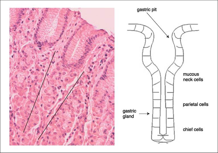
Figure 4.2:
Localization of secretory cells within the gastric gland. This figure shows the idealized localization of the gastric glands on the right and a histopathological view of gastric glands on the left. Please note the orientation of the gastric gland indicated by the two fine lines.
Cardiac glands. Cardiac glands consist mainly of mucussecreting cells.
Fundic glands. Fundic glands are characterized by the presence of mucous neck cells, chief cells, and parietal cells. All these cell types most likely develop from a common progenitor cell.1 The chief cells are located at the base of the fundic gland and secrete different isoforms of pepsinogen, a precursor of the proteolytic enzyme pepsin. In dogs two distinct groups ofpepsinogens have been identified, pepsinogen A and B.2 The parietal cells are located in the upper third of the fundic gland and secrete hydrochloric acid, R-protein, and in dogs, a glycoprotein called intrinsic factor. R-protein and intrinsic factor are crucial in the absorption of cobalamin (vitamin B12) in the small intestine. In contrast to humans, the stomach appears to be a limited source of intrinsic factor in domestic animals.
In the dog, intrinsic factor is almost exclusively produced by the exocrine pancreas and to some extend in the salivary glands, while in the cat intrinsic factor is almost exclusively produced by the exocrine pancreas.3 Mucous neck cells are scattered between the chief cells and parietal cells. In the dog, the mucous neck cells and mucous pit cells of gastric glands also produce gastric lipase.4Ghrelin is synthesized in the epithelial cells lining the fundus of the stomach, with smaller amounts also produced outside the GI tract. Ghrelin is a peptide hormone that stimulates the release of growth hormone from the anterior pituitary. Ghre- lin has a significant effect on appetite and energy balance, and chronic obesity has been reported to be associated with a significant decrease in plasma ghrelin concentration.5
Trefoil factors 1 and 2 (TFF 1 and 2) are peptides expressed in the stomach by the mucous neck cells in the gastric fundus and antrum, and by the pyloric glands. Trefoil factors play an important role during epithelial restitution after mucosal damage and /or during inflammation, and decreased expression of TFFs have been implicated in the development of gastric cancer.6
Pyloric glands. Pyloric glands contain mainly mucus-secreting cells and gastrin-producing endocrine cells (G cells). Gastrin stimulates gastric acid secretion and has an important trophic influence on the gastric mucosa.
4.3.2