Genetic Tests for Large Animals
DANIKA L. BANNASCH, Consulting Editor CARRIE J. FINNO
Genetic testing based on deoxyribonucleic acid (DNA) involves the analysis of an animal's DNA to determine its genotype for an inherited disorder, trait, or anonymous marker.
Genetic testing can be used for positive or negative selection in a population, depending on whether it is being used to identify a disease (negative) or a trait (positive). Genetic testing can also be used for permanent individual identification and parentage determination. Many breed registries require parentage verification to ensure the accuracy of their pedigrees.Using genetic testing results for selection requires an understanding of the mode of inheritance of the disease or trait. Most often, a genetic test will be performed for a recessive disorder to determine if an animal is a carrier. Carriers are asymptomatic but have the potential to produce diseased progeny. Because they have no outward manifestation of disease, a genetic test is extremely valuable for managing their breeding appropriately. Carrier animals can be bred to noncarriers if needed to retain valuable characteristics while not producing diseased offspring. In the case of positive selection for a trait of interest, carrier animals may have higher breeding values because they can produce a trait if bred to other carriers or to animals with the trait. Genetic tests may also be used for dominant disorders if the disease/trait has a late age of onset or if it is inherited in a codominant manner. DNA testing for traits that are controlled by more than one locus (polygenic) may also be used for selection for economically important traits. In these cases, one particular genotype may confer a slight advantage over another and therefore, in a large population, can have a significant effect on production.
Box 52.1 defines key genetic terms; see also Chapter 51.
Individual Identification and Parentage Testing
Researchers use genetic markers distributed along all the chromosomes as tools to identify regions associated with diseases or traits. One type of marker used for individual identification and parentage testing is composed of small nucleotide repeats and is called microsatellite markers or short tandem repeats (STRs). These markers have a feature that makes them extremely useful to geneticists; the markers have been chosen to be “polymorphic” (show differences) between individuals. In other words, individual animals will have different lengths of the nucleotide repeats for each of these markers. The high level of polymorphism of this type of marker makes them useful for “mapping” (identifying the chromosomal location of diseases and traits).
The microsatellite markers are assayed by polymerase chain reaction (PCR) amplification using fluorescent-labeled primers. Primers are short (20 base pairs), single-strand lengths of DNA that are complementary to a specific region of the genome. PCR is the amplification of a section of DNA contained between two primers designed to complement the unique sequence flanking the STR. The PCR products are then resolved by electrophoresis on the basis of their length. Markers with many different alleles are said to be polymorphic and would be a useful marker for individual identification or parentage. Because the markers show differences between individuals, a collection of these markers can be used as a form of identification of an animal. High statistical significance can be obtained with as few as 10 markers, depending on the species and breed. The DNA type of an animal will not change over its lifetime and can therefore be used as a form of permanent identification.
Many purebred registries require parentage verification for registration purposes. To accomplish parentage verification, a DNA sample must be available from both parents, as well as the offspring. DNA samples are taken in the form of hair, blood, or buccal swabs (depending on the species and registry) and submitted at the time registration is requested.
Each animal inherits one copy of each marker from its sire and one copy from its dam, so the markers can also be used to verify parentage. The most useful marker has a high polymorphism rate because that type of marker will be most likely to show differences not only between the sire and the dam but also between the two copies (alleles) of the marker. A set of polymorphic markers (10 to 20) is used to verify parentage to ensure a high probability that the parentage is correct. Table 52.1 shows the allele sizes for a set of markers in a parentage case. For marker A the offspring inherited a 122 allele and a 126 allele. The 122 came from its dam, so the 126 came from the sire. Because sire 1 does not have a 126 allele, it has been excluded. In this example, sire 1 is excluded as the sire of the offspring and sire 2 is verified on the basis of the results for all three markers.Genome Sequence
Whole-genome sequencing and genome assembly of large animal species was completed in the early 2000s (horse September 2007, cow October 2011, sheep February 2010, goat February 2013). The whole-genome sequence of an organism provides the exact base pairs along each chromosome of the individual animal that was sequenced. This “assembly” of DNA sequence then becomes the reference assembly for each organization, and genetic variation is compared with that reference providing a frame of reference for all DNA sequencing. In the horse, a Thoroughbred mare (Twilight) was selected for sequencing because of her low heterozygosity rate (1/1380 base pairs). In 2010, a Quarter Horse mare underwent whole-genome sequencing and was aligned to Twilight.1 In cattle, a single partially inbred Hereford cow was selected to contribute 6? whole-genome shotgun (WGS) reads and another 1.5? came from individual animals of the Holstein, Angus, Jersey, Limousin, Brahman, and Norwegian Red breeds for detection of single nucleotide polymorphisms (SNPs). Following sequencing of each species, over 20,000 protein-coding genes were annotated on the sequences by virtue of previously sequenced cDNAs and also by prediction software that compared known genome
■ BOX 52.1
Definitions of Genetic Terms
Allele One of the variant forms of a gene at a particular locus, or location, on a chromosome.
Base pairs Two bases that form a “rung of the DNA ladder.” A DNA nucleotide is made of a molecule of sugar, a molecule of phosphoric acid, and a molecule called a “base” The bases are the “letters” that spell out the genetic code. In DNA the code letters are A, T, G, and C, which stand for the chemicals adenine, thymine, guanine, and cytosine, respectively.
Genotype Genetic makeup, either at a single locus or at all loci. Genome wide association study A study that uses genomewide markers, typically microsatellites or single nucleotide polymorphisms (SNPs) to determine an association between a phenotype and region of particular chromosome.
Linked Association of genes and markers that lie near each other on a chromosome; linked genes and markers tend to be inherited together.
Locus Place on a chromosome where a specific gene is located; a type of “address” for the gene.
Marker Segment of DNA with an identifiable physical location on a chromosome, the inheritance of which can be followed. A marker can be a gene or some section of DNA with no known function. Also known as a genetic marker.
Microsatellite Repetitive stretches of short sequences of DNA used as genetic markers to track inheritance in families.
Phenotype Observable traits or characteristics of an animal (e.g., coat color, weight, presence or absence of a disease).
Recombination Genetic transmission process by which the combinations of alleles observed at different loci in two parental individuals become shuffled in offspring individuals.
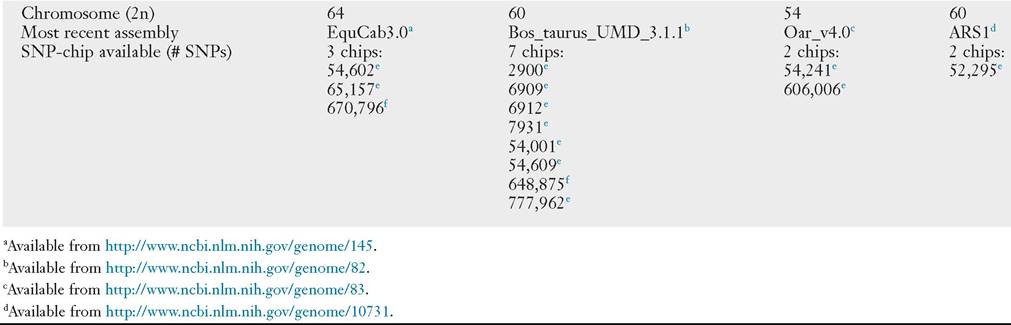
Single nucleotide polymorphism (SNP) Differences in a single base pair of DNA (A, C, T, G) often used as genetic markers for linkage and association studies sequences in other species with newly sequenced genomes. Since these initial assemblies, revisions have occurred within species. These initial and updated annotated genome databases are publicly available (Table 52.2).
With the advent of whole-genome sequencing and assemblies, there has been a rapid expansion in the number of SNPs discovered in the genomes of large animal species.
SNPs are single base pairs of DNA with differences between individuals. They generally have only two alleles and are therefore less informative than the STR markers mentioned earlier. They have distinct advantages, however, when it comes to ease of genotyping. Very large numbers of SNP markers can be genotyped for a fraction of the cost of microsatellite markers. SNPs are now considered the next generation of markers to conduct breed diversity and to map disease-causing traits. The availability of such a tool as an SNP chip facilitates rapid mapping of diseases to specific chromosomal regions and analysis of candidate genes. SNP arrays are currently available on many large animal species (Table 52.2) and are currently being used in mapping studies of various complex disorders. Haplotype structures are being identified across species with targeted resequencing and, most recently, whole-genome next-generation sequencing, of animals from different breeds.In addition to genomic mapping, functional genomic tools have become more readily available and affordable to researchers. More recently, sequencing of the transcriptome, through technologies such as RNA-Seq, have allowed researchers to evaluate gene expression differences among tissues and between animals of different disease states.
Identifying Genetic Mutations
Initial genetic mutations in large animal species were discovered through the use of comparative genomics. Genes involved in a specific disease were targeted because of equivalent diseases in other species, namely humans. In the horse, the genetic mutations for many diseases that have genetic tests currently available, including hyperkalemic periodic paralysis (HYPP)2 and severe combined immunodeficiency (SCID),3 were uncovered by evaluating candidate genes that had been associated with similar diseases in humans (Table 52.3). With the sequencing and annotation of whole genome maps, other diseases were discovered through whole-genome linkage mapping (hereditary equine regional dermal asthenia [HERDA]4), genome-wide association studies with microsatellites (Type I polysaccharide storage myopathy [PSSM]5) and genome-wide association studies using SNP array technology (Lavender foal syndrome6).
A similar theme is evident in cattle. Initial genetic mutations were discovered based on sequencing of candidate genes known to cause similar disease in humans (bovine leukocyte adhesion deficiency [BLAD]7). Later studies used microsatellite markers and performed linkage analysis (complex vertebral malformation8) and, most recently, the use of SNP-based genome-wide association studies has identified recessive defects (congenital muscular dystony types 1 and 29). At the time of publication,
■ TABLE 52.1
Parentage Verification Using Microsatellite Markers
AUele Sizes for 4 Markers
| Individuals | A | B | C | D |
| Dam | 122/124 | 110 | 131/133 | 80/82 |
| Offspring | 122/126 | 110/112 | 133 | 82/86 |
| Sire 1 | 128 | 112/114 | 131 | 84 |
| Sire 2 | 126 | 112/114 | 131 | 86/90 |
■ TABLE 52.2
Map Status and Genomic Resource for Large Animal Species
Horse Cattle Sheep Goat
eIlluminaInc., San Diego, Calif. fAHymetrix, Inc., Santa Clara, Calif.
■ TABLE 52.3
Genetic Tests for Horses
| Disease/Trait | Gene | Mode of Inheritance | Reference |
| Androgen insensitivity syndrome (AIS) | AR | X-linked recessive | 18 |
| Cerebellar abiotrophy (CA) | TOE1/MUTYH | Autosomal recessive | 19 |
| Curly coat | KRT25 | Autosomal dominant | 20 |
| Dwarfism, ACAN related | ACAN | Autosomal incompletely dominant | 21,22 |
| Dwarfism, Friesian | B4GALT7 | Autosomal recessive | 23 |
| Hereditary regional dermal asthenia (HERDA) | PPIB | Autosomal recessive | 4 |
| Junctional epidermolysis bullosa (JEB) | LAMA3 | Autosomal recessive | 24 |
| JEB. | LAMC2 | Autosomal recessive | 25 |
| Foal immunodeficiency syndrome (immunodeficiency | SLC5A3 | Autosomal recessive | 26 |
| of Fell ponies) | |||
| Gaitedness | DMRT3 | Multifactorial | 27 |
| Glycogen storage disease IV (glycogen branching | GBE1 | Autosomal recessive | 28 |
| enzyme deficiency) | |||
| Hoof wall separation disease (HWSD) | SERPINB11 | Autosomal recessive | 29 |
| Hydrocephalus | B3GALNT2 | Autosomal recessive | 30 |
| Immune-mediated myositis | MYH1 | Autosomal semidominant | 31 |
| Incontinentiapigmenti | IKBKG | X-linked semidominant | 32 |
| Lavender foal syndrome (LFS) | MYO5A | Autosomal recessive | 6 |
| Malignant hyperthermia (MH) | RYR1 | Autosomal dominant | 33 |
| Megacolon (ileocolonicaganglionosis or lethal white | EDNRB3 | Autosomal semidominant | 10-12 |
| foal syndrome) | |||
| Multiple ocular defects | PMEL17 | Autosomal semidominant | 34 |
| Myotonia | CLCN1 | Autosomal recessive | 35 |
| Naked foal syndrome | ST14 | Autosomal recessive | 36 |
| Night blindness, congenital stationary | TRPM1 | Autosomal recessive | 37 |
| Occipitoatlantoaxial malformation (OAAM) | HOXD3 | Autosomal recessive | 38 |
| Ocular squamous cell carcinoma | DDB2 | Autosomal recessive | 39 |
| Ovotesticular disorder of sexual development | SRY | Y-linked | 40 |
| Periodic paralysis II (hyperkalemic periodic paralysis; | SCN4A | Autosomal semidominant | 2 |
| HYPP) | |||
| Polysaccharide storage myopathy type I (PSSM I) | GYS1 | Autosomal dominant | 5 |
| Severe combined immunodeficiency | DNAPKcs | Autosomal recessive | 3 |
| Skeletal atavism | SHOX | Autosomal recessive | 41 |
| Thrombasthenia (Glanzmann thrombasthenia) | ITGA2B | Autosomal recessive | 42,43 |
| Warmblood fragile foal syndrome | PLOD1 | Autosomal recessive | 44 |
■I TABLE 52.4
Genetic Tests for Cattle
| Disease/Trait | Gene | Mode of Inheritance | Reference(s) |
| Abortion/Stillbirth | MIMT1 | Maternally imprinted | 45 |
| Abortion | ANXA10 | Autosomal incompletely dominant | 46 |
| EXOSC4 | Autosomal recessive lethal | 47 | |
| MED22 | Autosomal recessive lethal | 47 | |
| MYH6 | Autosomal recessive lethal | 47 | |
| OBFC1 | Autosomal recessive lethal | 47 | |
| RABGGTB | Autosomal recessive lethal | 47 | |
| RNF20 | Autosomal recessive lethal | 47 | |
| RPIA | Autosomal recessive lethal | 47 | |
| SNAPC4 | Autosomal recessive lethal | 47 | |
| TTF1 | Autosomal recessive lethal | 47 | |
| APAF1 | Autosomal recessive lethal | 48 | |
| CWC15 | Autosomal recessive lethal | 49 | |
| SLC39A4 | Autosomal recessive | 50 | |
| EDA | X-linked recessive | 51 | |
| TUBD1 | Autosomal recessive lethal | 52 | |
| SUGT1 | Autosomal recessive lethal | 53 | |
| SMC2 | Autosomal recessive lethal | 54 | |
| GART | Autosomal recessive lethal | 55 | |
| TFB1M | Autosomal recessive lethal | 56 | |
| PFAS | Autosomal recessive lethal | 57 | |
| SLC37A2 | Autosomal recessive lethal | 55 | |
| RNASEH2B | Autosomal recessive lethal | 58 | |
| Achondrogenesis, type II | COL2A1 | Autosomal dominant | 54,59,60 |
| Acrodermatitisenteropathica | SLC39A4 | Autosomal recessive | 50 |
| Anhidrotic ectodermal dysplasia | EDA | X-linked recessive | 51,61-64 |
| EDAR | Autosomal recessive | 60 | |
| Arachnomelia (spider limbs) | MOCS1 | Autosomal recessive | 65 |
| SUOX | Autosomal recessive | 66 | |
| Arthrogryposis | AGRN | Autosomal recessive lethal | 67 |
| CHRNB1 | Autosomal recessive | 68 | |
| MYBPC1 | De novo | 69 | |
| PIGH | Autosomal recessive | 70 | |
| Axonopathy (Demetz syndrome) | MFN2 | Autosomal recessive | 71 |
| Brachyspina | FANCI | Autosomal recessive | 72 |
| Caprine-like generalized hypoplasia | CEP250 | Autosomal recessive | 73 |
| Cardiomyopathy and woolly haircoat syndrome | PPP1R13L | Autosomal recessive | 74 |
| Cardiomyopathy, dilated | OPA3 | Autosomal recessive | 75 |
| Cataract | CPAMD8 | Autosomal recessive | 76 |
| NID1 | Autosomal recessive | 77 | |
| Chediak-Higashi syndrome | LYST | Autosomal recessive | 78 |
| Chondrodysplasia | EVC2 | Autosomal | 79 |
| Citrullinemia | ASS1 | Recessive | 80 |
| Complex vertebral malformation | SLC35A3 | Autosomal recessive | 8 |
| Congenital muscular dystonia 1 | ATP2A1 | Autosomal recessive | 9 |
| Congenital muscular dystonia 2 | SCL6A5 | Autosomal recessive | 9 |
| Contractural arachnodactyly (Fawn calf syndrome) | ADAMTSL3 | Autosomal recessive | 81 |
| Curly hair, karakul-type | KRT27 | Autosomal dominant | 54 |
| Deficiency of uridine monophosphate synthase (DUMPS) | UMPS | Autosomal recessive | 82 |
| Depigmentation with microphthalmia | MITF | Autosomal dominant | 83 |
| Developmental duplications | NHLRC2 | Multifactorial | 84 |
| Dominant white with bilateral deafness | MITF | Autosomal dominant | 83 |
| Dwarfism Angus | PRKG2 | Autosomal recessive | 85 |
| Fleckvieh | GON4L | Autosomal recessive | 86 |
| ACAN-related | ACAN | Autosomal recessive lethal | 87 |
| Growth hormone deficiency | GH1 | Autosomal recessive | 88 |
| Proportionate, with inflammatory lesions | RNF11 | Autosomal recessive | 89 |
| Ears crop | HMX1 | Autosomal dominant with variable expressivity | 90 |
| Ehlers-Danlos syndrome Holstein variant | EPYC | Autosomal recessive | 91 |
| Type VII (dermatosparaxis) | ADAMTS2 | Autosomal recessive | 92 |
| Epidermolysis bullosa | KRT5 | Autosomal dominant | 93 |
| Dystrophic | ITGB4 | Autosomal recessive | 94 |
| LAMA3 | Autosomal recessive | 95 | |
| LAMC2 | Autosomal recessive | 96 | |
| COL7A1 | Autosomal recessive | 97 | |
| Facial dysplasia syndrome | FGFR2 | Autosomal dominant | 98 |
■ TABLE 52.4
Genetic Tests for Cattle—cont'd
| Disease/Trait | Gene | Mode of Inheritance | Reference(s) |
| Factor XI deficiency | F11 | Autosomal recessive | 99 |
| Fanconi syndrome | SLC2A2 | Autosomal recessive | 100 |
| Forelimb-girdle muscular anomaly | GFRA1 | Autosomal recessive | 101 |
| Glycogen storage disease II (Pompe’s disease) | GAA | Recessive | 102 |
| Glycogen storage disease V | PYGM | Autosomal recessive | 103 |
| Goiter, familial | TG | Autosomal | 104 |
| Gonadal hypoplasia | KIT | Autosomal recessive | 105 |
| Hairy | PRL | Autosomal dominant | 106 |
| Hemophilia A | F8 | X-linked | 107 |
| Holstein cholesterol deficiency | APOB | Autosomal recessive lethal | 108 |
| Hydrallantois | SLC12A1 | Autosomal recessive | 109 |
| Hypotrichosis | HEPHL1 | Autosomal recessive | 110 |
| Streaked | KRT71 | Unknown | 111 |
| TSR2 | X-linked semidominant | 112 | |
| Ichthyosis congenital | ABCA12 | Autosomal recessive | 9 |
| Lethal multiorgan developmental dysplasia | KDM2B | Autosomal recessive | 113 |
| Leukocyte adhesion deficiency, type I | ITGB2 | Autosomal recessive | 7 |
| Male subfertility | TMEM95 | Autosomal recessive | 114 |
| Mannosidosis alpha | MAN2B1 | Autosomal recessive | 115 |
| Mannosidosis beta | MANBA | Autosomal recessive | 116 |
| Maple syrup urine disease | BCKDHA | Autosomal recessive | 117 |
| Marfan syndrome | FBN1 | Autosomal dominant | 118 |
| Mucopolysaccharidosis IIIB | NAGLU | Autosomal recessive | 119 |
| Multiple ocular defects | WFDC1 | Autosomal recessive | 120 |
| Muscular hypertrophy (double muscling) | MSTN | Autosomal recessive | 13-15 |
| Myasthenic syndrome, congenital | CHRNE | Autosomal | 121 |
| Myoclonus | GLRA1 | Autosomal recessive | 122 |
| Myopathy of the diaphragmatic muscles | HSPA1A | Autosomal recessive | 123 |
| Neurocristopathy | CHD7 | Autosomal dominant | 60 |
| Neuronal ceroid lipofuscinosis 5 | CLN5 | Autosomal recessive | 124 |
| Osteogenesis imperfecta, type II | COL1A1 | Autosomal dominant | 60 |
| Osteopetrosis | SLC4A2 | Autosomal recessive | 125 |
| Osteopetrosis with gingival hamartomas | CLCN7 | Autosomal recessive | 126 |
| Ovotesticular disorder of sexual development | SRY | Y-linked | 127 |
| Perinatal weak calf syndrome | IARS | Autosomal recessive | 128 |
| Polled | POLLa | Dominant | 129,130 |
| Progressive degenerative myeloencephalopathy (Weaver syndrome) | PNPLA8 | Autosomal recessive | 131 |
| Protoporphyria | FECH | Autosomal | 132 |
| Pseudomyotonia, congenital | ATP2A1 | Autosomal recessive | 133 |
| Ptosis, intellectual disability, retarded growth and mortality (PIRM) syndrome | UBE3B | Autosomal recessive | 134 |
| Renal dysplasia | CLDN16 | Autosomal recessive | 135 |
| ResistanceZsusceptibility to Mannheimia hemolytica leukotoxin | ITGB2 | Autosomal recessive | 136 |
| Retinitis pigmentosa 1 | RP1 | Autosomal recessive | 137 |
| Scurs, type 2 | TWIST1 | Autosomal dominant | 138 |
| Slick hair | PRLR | Autosomal dominant | 106,139 |
| ARMC3 | Autosomal recessive | 140 | |
| Spherocytosis | SLC4A1 | Autosomal incompletely dominant | 141 |
| Spinal dysmyelination | SPAST | Autosomal recessive | 142 |
| Spinal muscular atrophy | KDSR | Autosomal recessive | 143 |
| Spongiform encephalopathy | PRNP | N/A | 144 |
| Syndactyly | LRP4 | Autosomal recessive | 145,146 |
| Tail, crooked | MRC2 | Autosomal recessive | 147 |
| Tibialhemimelia | ALX4 | Autosomal recessive | 67,149 |
| Tricho-dento-osseous-like syndrome | DLX3 | De novo autosomal dominant | 150 |
| Thrombopathia | RASGRP2 | Unknown | 151 |
| Trimethylaminuria | FMO3 | Autosomal recessive | 152 |
| Vertebral and spinal dysplasia | T | Autosomal incompletely dominant | 153 |
| Xanthinuria, type II | MOCOS | Autosomal recessive | 154,155 |
| Yellow fat | BCO2 | Unknown | 156 |
| Zinc deficiency-like syndrome | PLD4 | Autosomal recessive | 157 |
| A2 milk | CSN2 | Autosomal codominant | 158 |
| Beta-lactoglobulin, low expression | PAEP | Autosomal | 159 |
| Milk yield and composition | GHR | Polygenic | 160 |
Continued
■ TABLE 52.4
there are 147 genetic tests available in cattle (Table 52.4). It is worth noting that the majority of genetic tests currently available are for diseases/traits that are inherited as autosomal recessive traits. With the current technologies available through SNP-association mapping and next-generation sequencing, we should expect to further our understanding of polygenic traits and diseases.
Disease Testing
Clinicians can use DNA testing in disease diagnosis or to determine an animal’s potential for producing diseased progeny. Generally, disease diagnosis is based on clinical signs and other diagnostic tests, but occasionally DNA testing is used, in particular for later-onset diseases or diseases for which diagnosis by traditional methods is difficult or invasive. To offer a genetic test, the gene responsible must be known. Ideally, the actual mutation that causes the disease has been identified. Rather than knowing the exact gene or mutation, only a region of a chromosome may have been implicated in a particular disease. DNA tests can be divided into two categories: mutation tests and linked-marker or haplotype tests. Mutation tests are based on an actual mutation that causes disease, whereas the linked- marker or haplotype test is based on the region of the chromosome that is known to cause disease but not necessarily the actual mutation. Usually, haplotype tests are offered instead of a mutation test because the mutation has not yet been identified. With the current tools available in large animals, the identification of disease causing mutations is so efficient that marker-based tests are rarely used.
Mutations that cause disease appear in many different forms. A change of a single base pair from one base to another can cause a disease by changing an amino acid (“missense” mutation), truncating the amino acid chain (“nonsense” mutation), or altering expression or proper splicing. For example, missense mutations have been shown to cause lethal white foal syndrome in the American Paint horse.10-12 Insertions or deletions of a single base pair (bp) can cause mutations in the coding sequence by altering the translational frame, which ultimately causes protein truncation. An 11-bp deletion in the myostatin gene causes a frameshift mutation and protein truncation in Belgian blue and Peidmontese cattle with the double-muscle phenotype.13-15 Large deletions or insertions that remove hundreds and thousands of base pairs can also cause disease. For example, the polled intersexuality mutation in goats is caused by an 11.7-kilobase deletion that removes a regulatory element that controls the expression of two genes.16 This endless array of possible changes in the DNA that result in disease makes each individual DNA-based genetic test different.
The basis for DNA testing is PCR. Primers can be designed specifically to amplify either the disease-causing allele or the
normal allele. Alternatively, the PCR product can be digested with a restriction enzyme that cleaves the DNA at a particular sequence of bases. A restriction enzyme is chosen that shows a different cleavage pattern between the mutant and the normal version of the PCR product. Direct sequencing of a section of DNA can also be used to determine the animal’s genotype. Many different methods are available to assay changes in DNA that lead to disease. Each company that offers a test may choose a different type of assay for the same mutation.
There are limits to all genetic testing. For mutation tests, the specific mutation being assayed is the only factor being evaluated. An animal may have a different mutation in that gene or a mutation in a different gene that causes the same phenotype (phenocopy). It is therefore correct to state that an animal has been “DNA tested negative” for this specific mutation rather than “DNA tested clear” of the disease. In addition, more complicated modes of inheritance do not provide a direct disease or trait prediction. For example, some loci have incomplete penetrance and not all animals with the affected genotype will get the disease or trait. Other genetic tests are for susceptibility loci and merely confer a disease risk.
No association or committee evaluates quality control of DNA tests that are available in animals. Most tests are published in the scientific literature not as tests but as articles describing the discovery of the mutation. Because some cases involve patent issues, some tests are offered before publication. Much of the research done to identify the mutations involved in the tests is performed at universities and funded by granting agencies that have both financial and intellectual interest in patenting the tests.
Tables 52.3, 52.4, 52.5, and 52.6 list available genetic tests for traits and diseases other than coat color in horses, cattle, and sheep and goats, respectively. Only tests published in peer-reviewed journals are listed. Additional tests available in cattle for various forms of the milk proteins also are not listed in Table 52.4. The breeds of cattle are not listed because rapid changes in testing mean that tests are always being validated for new breeds. It is therefore recommended that a search be performed for the availability of a test for a particular breed each time the need arises. Coat color genetic tests are listed in Table 52.7, with each particular species in which the genetic mutation has been identified referenced.
The diseases or traits that are tested can be divided into two categories: those that have straightforward Mendelian inheritance patterns (recessive, dominant, and sex-linked) and those that are more complicated because many genes are involved with conferring the phenotype (polygenic). Quantitative trait loci (QTL) are the genes that contribute to a polygenic disease. In cattle, a vast number of QTL have been placed in specific regions of chromosome for quantitative traits such as dairy form, milk production, and fertility.17 Selection for these traits can be done with DNA testing (Table 52.4). Because so
Genetic Tests for Cattle—cont'd
| Disease/Trait | Gene | Mode of Inheritance | Reference(s) |
| Milk yield and composition Meat tenderness | DGAT1 | Polygenic | 161 |
| Milk yield and composition | Leptin | Polygenic | 162,163 |
| Meat tenderness | Calpastatin | Polygenic | 164,165 |
| μ-Calpain | Polygenic | 166,167 |
aConsidered a locus and not a gene.
■ TABLE 52.5
Genetic Tests for Sheep
| Disease/Trait | Gene | Mode of Inheritance | Reference(s) |
| Achromatopsia-2 | CNGA3 | Autosomal recessive | 168 |
| Ataxia, familial episodic spinocerebellar | FGF14 | Autosomal incompletely dominant | 169 |
| Chondrodysplasia (spider lamb) | FGFR3 | Autosomal recessive | 170 |
| Chondrodysplasia, Texel | SLC13A1 | Autosomal recessive | 171 |
| Ehlers-Danlos syndrome, type VII (dermatosparaxis) | ADAMTS2 | Autosomal recessive | 172 |
| Epidermolysis bullosa, junctionalis | LAMC2 | Autosomal recessive | 173 |
| ITGB4 | Autosomal recessive | 174 | |
| Fecundity Barbarine | BMP15 | X-linked | 175 |
| Belclare | BMP15 | X-linked | 176 |
| Belcalre/Irish Cambridge | GDF9 | Autosomal | 176 |
| Booroola | BMPR1B | Autosomal semidominant | 177-179 |
| Embrapa | GDF9 | Autosomal | 180 |
| Galway | BMP15 | X-linked | 176 |
| Grivette | BMP15 | X-linked | 181 |
| Hanna | BMP15 | X-linked | 182 |
| Inverdale | BMP15 | X-linked | 182 |
| Lacaune | B4GALNT2 | Autosomal codominant | 183 |
| BMP15 | X-linked | 182 | |
| Norwegian | GDF9 | Unknown | 184 |
| Olkuska | BMP15 | X-linked | 181 |
| RaseAragonesa | BMP15 | X-linked | 185 |
| Small tail Han | BMPR1B BMP15 | Autosomal | 186 |
| Thoka | GDF9 | Autosomal | 187 |
| Vacaria | GDF9 | Autosomal | 188 |
| Fleece variation | IRF2BP2 | Not reported | 189 |
| Gangliosidosis, GM1 | GLB1 | Autosomal recessive | 190 |
| Gangliosidosis, GM2, type 1 | HEXA | Autosomal recessive | 191 |
| Gaucher disease, type I | GBA | Autosomal recessive | 192 |
| Glycogen storage disease V | PYGM | Autosomal recessive | 193 |
| Hemophilia A | F8 | X-linked | 194 |
| Hypophosphatemic rickets | DMP1 | Autosomal recessive | 195 |
| Hypotrichosis | HR | Autosomal recessive | 196 |
| Lissencephaly and cerebellar hypoplasia | RELN | Autosomal recessive | 197 |
| Microphthalmia | PITX3 | Autosomal recessive | 198 |
| Motor neuron disease lower | AGTPBP1 | Autosomal recessive | 199 |
| Muscular hypertrophy (double muscling) | |||
| Callipyge | DLK1 | Polar overdominant | 200 |
| Texel | MSTN | Autosomal | 201 |
| Myotonia Neuronal ceroid lipofuscinosis | CLCN1 | Autosomal recessive | 202 |
| 10 | LOC443060 | Autosomal recessive | 203 |
| 5 | bgcolor=white>CLN5Autosomal recessive | 204 | |
| 6 | CLN6 | Autosomal recessive | 205 |
| Polled/Horns | RXFP2 | Autosomal | 206 |
| Porphyria cutaneatarda | UROD | Autosomal recessive | 207 |
| Resistance/susceptibility to lentivirus | TMEM154 | Multifactorial | 208 |
| Spongiform encephalopathy (scrapie susceptibility) | PRNP | Autosomal recessive | 209 |
| Waardenburg syndrome, type 4A | EDNRB | Autosomal recessive | 210 |
| Yellow fat | BCO2 | Autosomal recessive | 211 |
■ TABLE 52.6
Genetic Tests for Goats
| Disease/Trait | Gene | Mode of Inheritance | Reference(s) |
| Casein, α-S1, reduced concentration | CSN1S1 | Autosomal | 212 |
| Casein, α-S2, null allele | CSN1S2 | Autosomal recessive | 213 |
| Casein, β, null allele | CSN2 | Autosomal | 214 |
| Goiter, familial | TG | Autosomal recessive | 215 |
| Horns/Polled | PISRT1, FOXL2 | Autosomal | 16,216 |
| Mannosidosis, β | MANBA | Autosomal recessive | 217 |
| Mucopolysaccharidosis IIID | GNS | Autosomal recessive | 218 |
| Myotonia | CLCN1 | Autosomal dominant | 219 |
■ TABLE 52.7
Genetic Tests for Coat Color
| Coat Color | Gene | Species | Reference(s) |
| Albinism | TYR | Cattle | 220 |
| Albinism (oculocutaneous type IV) | SCL45A2 | Cattle | 221 |
| Albinism (oculocutaneous type VI) | SLC24A5 | Horse | 222 |
| Agouti | ASIP | Horse | 223 |
| Cow | 224 | ||
| Sheep | 225 | ||
| Appaloosa | TRPM1 | Horse | 37 |
| Brown (Dun) | TYRP1 | Cow | 226 |
| Sheep | 227 | ||
| Champagne | SLC36A1 | Horse | 228 |
| Cool gray | MLPH | Cattle | 229 |
| Cream dilution | SLC45A2 | Horse | 230 |
| Dilution | PMEL | Cow | 231 |
| Dominant red | COPA | Cow | 60 |
| Dominant white | KIT | Horse | 232 |
| Cow | 233 | ||
| Extension | MC1R | Horse | 234 |
| Cow | 235 | ||
| Sheep | 236 | ||
| Goat | 237 | ||
| Gray | STX17 | Horse | 238 |
| Macchiato | MITF | Horse | 239 |
| Roan | KITLG | Cow | 240 |
| Silver | PMEL17 | Horse | 241 |
| White belt | TWIST2 | Cow | 242 |
| White spotting | MITF (Horse, cow) | Horse | 239 |
| PAX3 (Horse) | Cow | 233 | |
| EDNRA (Goat) | Goat | 243 |
many different QTL exist, however, selection can be challenging, and trade-offs need to be made.
Genetic testing relies on advances made in the field of genomics. Veterinarians and owners are fortunate that large animal species were chosen as economically important species for whole-genome sequencing. With the tools currently available, the number of disease-based or trait-based tests available in the future will markedly increase.
REFERENCES
The complete list of references can be found at www.expertconsult.com.
REFERENCES
1. Doan R, Cohen ND, Sawyer J, et al: Whole-genome sequencing and genetic variant analysis of a Quarter Horse mare, BMC Genomics 13:78,
2012.
2. Rudolph JA, Spier SJ, Byrns G, et al: Periodic paralysis in quarter horses: a sodium channel mutation disseminated by selective breeding, Nat Genet 2(2):144-147, 1992.
3. Shin EK, Perryman LE, Meek K: A kinase-negative mutation of DNA- PK(CS) in equine SCID results in defective coding and signal joint formation, J Immunol 158(8):3565-3569, 1997.
4. Tryon RC, White SD, Bannasch DL: Homozygosity mapping approach identifies a missense mutation in equine cyclophilin B (PPIB) associated with HERDA in the American Quarter Horse, Genomics 90(1):93-102, 2007.
5. McCue ME, Valberg SJ, Miller MB, et al: Glycogen synthase (GYS1) mutation causes a novel skeletal muscle glycogenosis, Genomics 91(5): 458-466, 2008.
6. Brooks SA, Gabreski N, Miller D, et al: Whole-genome SNP association in the horse: identification of a deletion in myosin va responsible for lavender foal syndrome, PLoS Genet 6(4):e1000909, 2010.
7. Shuster DE, Kehrli ME, Jr, Ackermann MR, et al: Identification and prevalence of a genetic defect that causes leukocyte adhesion deficiency in Holstein cattle, Proc Natl Acad Sci USA 89(19):9225-9229, 1992.
8. Thomsen B, Horn P, Panitz F, et al: A missense mutation in the bovine SLC35a3 gene, encoding a UDP-N-acetylglucosamine transporter, causes complex vertebral malformation, Genome Res 16(1):97-105, 2006.
9. Charlier C, Coppieters W, Rollin F, et al: Highly effective SNP-based association mapping and management of recessive defects in livestock, Nat Genet 40(4):449-454, 2008.
10. Yang GC, Croaker D, Zhang AL, et al: A dinucleotide mutation in the endothelin-B receptor gene is associated with lethal white foal syndrome (LWFS); a horse variant of hirschsprung disease, Hum Mol Genet 7(6): 1047-1052, 1998.
11. Metallinos DL, Bowling AT, Rine J: A missense mutation in the endothelin- B receptor gene is associated with lethal white foal syndrome: an equine version of hirschsprung disease, Mamm Genome 9(6):426-431, 1998.
12. Santschi EM, Purdy AK, Valberg SJ, et al: Endothelin receptor B polymorphism associated with lethal white foal syndrome in horses, Mamm Genome 9(4):306-309, 1998.
13. McPherron AC, Lee SJ: Double muscling in cattle due to mutations in the myostatin gene, Proc Natl Acad Sci USA 94(23):12457-12461, 1997.
14. Kambadur R, Sharma M, Smith TP, et al: Mutations in myostatin (GDF8) in double-muscled Belgian Blue and Piedmontese cattle, Genome Res 7(9):910-916, 1997.
15. Grobet L, Martin LJ, Poncelet D, et al: A deletion in the bovine myostatin gene causes the double-muscled phenotype in cattle, Nat Genet 17(1):71-74,
1997.
16. Pailhoux E, Vigier B, Chaffaux S, et al: A 11.7-kb deletion triggers intersexuality and polledness in goats, Nat Genet 29(4):453-458, 2001.
17. Polineni P, Aragonda P, Xavier SR, et al: The bovine QTL viewer: a web accessible database of bovine quantitative trait loci, BMC Bioinformatics 7:283, 2006.
18. Revay T, Villagomez DA, Brewer D, et al: GTG mutation in the start codon of the androgen receptor gene in a family of horses with 64,XY disorder of sex development, Sex Dev 6(1-3):108-116, 2012.
19. Brault LS, Cooper CA, Famula TR, et al: Mapping of equine cerebellar abiotrophy to ECA2 and identification of a potential causative mutation affecting expression of MUTYH, Genomics 97(2):121-129, 2011.
20. Morgenthaler C, Diribarne M, Capitan A, et al: A missense variant in the coil1A domain of the keratin 25 gene is associated with the dominant curly hair coat trait (Crd) in horse, Genet Sel Evol 49(1):85, 2017.
21. Eberth J: Chondrodysplasia-like dwarfism in the miniature horse, Lexington, KY, 2013, University of Kentucky Press.
22. Metzger J, Gast AC, Schrimpf R, et al: Whole-genome sequencing reveals a potential causal mutation for dwarfism in the Miniature Shetland pony, Mamm Genome 28(3-4):143-151, 2017.
23. Leegwater PA, Vos-Loohuis M, Ducro BJ, et al: Dwarfism with joint laxity in Friesian horses is associated with a splice site mutation in b4GALT7, BMC Genomics 17(1):839, 2016.
24. Graves KT, Henney PJ, Ennis RB: Partial deletion of the LAMA3 gene is responsible for hereditary junctional epidermolysis bullosa in the American Saddlebred Horse, Anim Genet 40(1):35-41, 2009.
25. Spirito F, Charlesworth A, Linder K, et al: Animal models for skin blistering conditions: absence of laminin 5 causes hereditary junctional mechanobul- lous disease in the Belgian horse, JInvest Dermatol 119(3):684-691, 2002.
26. Fox-Clipsham LY, Carter SD, Goodhead I, et al: Identification of a mutation associated with Fatal Foal Immunodeficiency Syndrome in the Fell and Dales pony, PLoS Genet 7(7):e1002133, 2011.
27. Andersson LS, Larhammar M, Memic F, et al: Mutations in DMRT3 affect locomotion in horses and spinal circuit function in mice, Nature 488(7413):642-646, 2012.
28. Ward TL, Valberg SJ, Adelson DL, et al: Glycogen branching enzyme (GBE1) mutation causing equine glycogen storage disease IV, Mamm Genome 15(7):570-577, 2004.
29. Finno CJ, Stevens C, Young A, et al: SERPINB11 frameshift variant associated with novel hoof specific phenotype in Connemara ponies, PLoS Genet 11(4):e1005122, 2015.
30. Ducro BJ, Schurink A, Bastiaansen JW, et al: A nonsense mutation in b3GALNT2 is concordant with hydrocephalus in Friesian horses, BMC Genomics 16:761, 2015.
31. Finno CJ, Gianino G, Perumbakkam S, et al: A missense mutation in MYH1 is associated with susceptibility to immune-mediated myositis in Quarter Horses, Skelet Muscle 8(1):7, 2018.
32. Towers RE, Murgiano L, Millar DS, et al: A nonsense mutation in the IKBKG gene in mares with incontinentia pigmenti, PLoS ONE 8(12): e81625, 2013.
33. Aleman M, Nieto JE, Magdesian KG: Malignant hyperthermia associated with ryanodine receptor 1 (C7360G) mutation in Quarter Horses, J Vet Intern Med 23(2):329-334, 2009.
34. Andersson LS, Axelsson J, Dubielzig RR, et al: Multiple congenital ocular anomalies in Icelandic horses, BMC Vet Res 7:21, 2011.
3 5. Wijnberg ID, Owczarek-Lipska M, Sacchetto R, et al: A missense mutation in the skeletal muscle chloride channel 1 (CLCN1) as candidate causal mutation for congenital myotonia in a New Forest pony, Neuromuscul Dvsord 22(4):361-367, 2012.
36. Bauer A, Hiemesch T, Jagannathan V, et al: A nonsense variant in the ST14 gene in Akhal-Teke horses with naked foal syndrome, G3 (Bethesda) 7(4):1315-1321, 2017.
37. Bellone RR, Holl H, Nelson J, et al, editors: An insertion in TRPM1, the genetic cause of leopard complex (LP) spotting and congenital stationary night blindness (CSNB) in horses, San Diego, CA, 2011, Plant and Animal Genomics Conference XIX.
38. Bordbari MH, Penedo MCT, Aleman M, et al: Deletion of 2.7 kb near HOXD3 in an Arabian horse with occipitoatlantoaxial malformation, Anim Genet 48(3):287-294, 2017.
39. Bellone RR, Liu J, Petersen JL, et al: A missense mutation in damagespecific DNA binding protein 2 is a genetic risk factor for limbal squamous cell carcinoma in horses, Int J Cancer 141(2):342-353, 2017.
40. Pailhoux E, Cribiu EP, Parma P, et al: Molecular analysis of an XY mare with gonadal dysgenesis, Hereditas 122(2):109-112, 1995.
41. Rafati N, Andersson LS, Mikko S, et al: Large deletions at the SHOX locus in the pseudoautosomal region are associated with skeletal atavism in Shetland ponies, G3 (Bethesda) 6(7):2213-2223, 2016.
42. Christopherson PW, Insalaco TA, van Santen VL, et al: Characterization of the cDNA Encoding alphaIIb and beta3 in normal horses and two horses with Glanzmann thrombasthenia, Vet Pathol 43(1):78-82, 2006.
43. Christopherson PW, van Santen VL, Livesey L, et al: A 10-base-pair deletion in the gene encoding platelet glycoprotein IIb associated with Glanzmann thrombasthenia in a horse, J Vet Intern Med 21(1):196-198, 2007.
44. Monthoux C, de Brot S, Jackson M, et al: Skin malformations in a neonatal foal tested homozygous positive for Warmblood Fragile Foal syndrome, BMC Vet Res 11:12, 2015.
45. Flisikowski K, Venhoranta H, Nowacka-Woszuk J, et al: A novel mutation in the maternally imprinted PEG3 domain results in a loss of MIMT1 expression and causes abortions and stillbirths in cattle (Bos taurus), PLoS ONE 5(11):e15116, 2010.
46. Sasaki S, Ibi T, Akiyama T, et al: Loss of maternal ANNEXIN A10 via a 34-kb deleted-type copy number variation is associated with embryonic mortality in Japanese Black cattle, BMC Genomics 17(1):968, 2016.
47. Charlier C, Li W, Harland C, et al: NGS-based reverse genetic screen for common embryonic lethal mutations compromising fertility in livestock, Genome Res 26(10):1333-1341, 2016.
48. Adams HA, Sonstegard T, VanRaden PM, et al: Identification of a nonsense mutation in APAF1 that is causal for a decrease in reproductive efficiency in diary cattle, San Diego, CA, 2012, Plant and Animal Genome XX.
49. Sonstegard T, VanRaden PM, Van Tassell C, et al, editors: Lethal haplotypes in dairy cattle: what lies beneath?, Cairns, Australia, 2012, International Society of Animal Genetics.
50. Yuzbasiyan-Gurkan V, Bartlett E: Identification of a unique splice site variant in SLC39a4 in bovine hereditary zinc deficiency, lethal trait a46: an animal model of acrodermatitis enteropathica, Genomics 88(4):521-526,
2006.
51. Ogino A, Shimizu K, Tanabe Y, et al: De novo mutation of the bovine EDA gene associated with anhidrotic ectodermal dysplasia in Japanese Black cattle, Anim Genet 43(5):646, 2012.
52. Biscarini F, Schwarzenbacher H, Pausch H, et al: Use of SNP genotypes to identify carriers of harmful recessive mutations in cattle populations, BMC Genomics 17(1):857, 2016.
53. Pausch H, Schwarzenbacher H, Burgstaller J, et al: Homozygous haplotype deficiency reveals deleterious mutations compromising reproductive and rearing success in cattle, BMC Genomics 16:312, 2015.
54. Daetwyler HD, Capitan A, Pausch H, et al: Whole-genome sequencing of 234 bulls facilitates mapping of monogenic and complex traits in cattle, Nat Genet 46(8):858-865, 2014.
55. Fritz S, Capitan A, Djari A, et al: Detection of haplotypes associated with prenatal death in dairy cattle and identification of deleterious mutations in GART, SHBG and SLC37a2, PLoS ONE 8(6):e65550, 2013.
56. Schutz E, Wehrhahn C, Wanjek M, et al: The Holstein Friesian lethal haplotype 5 (HH5) results from a complete deletion of TBF1M and cholesterol deficiency (CDH) from an ERV-(LTR) insertion into the coding region of APOB, PLoS ONE 11(4):e0154602, 2016.
57. Michot P, Fritz S, Barbat A, et al: A missense mutation in PFAS (phos- phoribosylformylglycinamidine synthase) is likely causal for embryonic lethality associated with the MH1 haplotype in montbeliarde dairy cattle, J Dairy Sci 100(10):8176-8187, 2017.
58. Kadri NK, Sahana G, Charlier C, et al: A 660-Kb deletion with antagonistic effects on fertility and milk production segregates at high frequency in Nordic Red cattle: additional evidence for the common occurrence of balancing selection in livestock, PLoS Genet 10(1):e1004049, 2014.
59. Agerholm JS, Menzi F, McEvoy FJ, et al: Lethal chondrodysplasia in a family of Holstein cattle is associated with a de novo splice site variant of COL2A1, BMC Vet Res 12:100, 2016.
60. Bourneuf E, Otz P, Pausch H, et al: Rapid discovery of de novo deleterious mutations in cattle enhances the value of livestock as model species, Sci Rep 7(1):11466, 2017.
61. Ogino A, Kohama N, Ishikawa S, et al: A novel mutation of the bovine EDA gene associated with anhidrotic ectodermal dysplasia in Holstein cattle, Hereditas 148(1):46-49, 2011.
62. Drogemuller C, Distl O, Leeb T: Partial deletion of the bovine ED1 gene causes anhidrotic ectodermal dysplasia in cattle, Genome Res 11(10):1699-1705, 2001.
63. Drogemuller C, Peters M, Pohlenz J, et al: A single point mutation within the ED1 gene disrupts correct splicing at two different splice sites and leads to anhidrotic ectodermal dysplasia in cattle, J Mol Med 80(5):319-323, 2002.
64. Karlskov-Mortensen P, Cirera S, Nielsen OL, et al: Exonization of a LINE1 fragment implicated in X-linked hypohidrotic ectodermal dysplasia in cattle, Anim Genet 42(6):578-584, 2011.
65. Buitkamp J, Semmer J, Gotz KU: Arachnomelia syndrome in Simmental cattle is caused by a homozygous 2-bp deletion in the molybdenum cofactor synthesis step 1 gene (MOCS1), BMC Genet 12:11, 2011.
66. Drogemuller C, Tetens J, Sigurdsson S, et al: Identification of the bovine Arachnomelia mutation by massively parallel sequencing implicates sulfite oxidase (SUOX) in bone development, PLoS Genet 6(8):2010.
67. Beever JE, Marron BM: Screening for arthrogryposis multiplex in bovines. USA Patent X US 2011/0151440 A1.
68. Agerholm JS, McEvoy FJ, Menzi F, et al: A CHRNB1 frameshift mutation is associated with familial arthrogryposis multiplex congenita in Red dairy cattle, BMC Genomics 17:479, 2016.
69. Wiedemar N, Riedi AK, Jagannathan V, et al: Genetic abnormalities in a calf with congenital increased muscular tonus, J Vet Intern Med 29(5):1418-1421, 2015.
70. Sartelet A, Li W, Pailhoux E, et al: Genome-wide next-generation DNA and RNA sequencing reveals a mutation that perturbs splicing of the phosphatidylinositol glycan anchor biosynthesis class H gene (PIGH) and causes arthrogryposis in Belgian Blue cattle, BMC Genomics 16:316,
2015.
71. Drogemuller C, Reichart U, Seuberlich T, et al: An unusual splice defect in the mitofusin 2 gene (MFN2) is associated with degenerative axonopathy in Tyrolean Grey cattle, PLoS ONE 6(4):e18931, 2011.
72. Charlier C, Agerholm JS, Coppieters W, et al: A deletion in the bovine FANCI gene compromises fertility by causing fetal death and brachyspina, PLoS ONE 7(8):e43085, 2012.
73. Floriot S, Vesque C, Rodriguez S, et al: C-Nap1 mutation affects centriole cohesion and is associated with a Seckel-like syndrome in cattle, Nat Commun 6:6894, 2015.
74. Simpson MA, Cook RW, Solanki P, et al: A mutation in NFkappaB interacting protein 1 causes cardiomyopathy and woolly haircoat syndrome of Poll Hereford cattle, Anim Genet 40(1):42-46, 2009.
75. Owczarek-Lipska M, Plattet P, Zipperle L, et al: A nonsense mutation in the optic atrophy 3 gene (OPA3) causes dilated cardiomyopathy in Red Holstein cattle, Genomics 97(1):51-57, 2011.
76. Hollmann AK, Dammann I, Wemheuer WM, et al: Morgagnian cataract resulting from a naturally occurring nonsense mutation elucidates a role of CPAMD8 in mammalian lens development, PLoS ONE 12(7):e0180665, 2017.
77. Murgiano L, Jagannathan V, Calderoni V, et al: Looking the cow in the eye: deletion in the NID1 gene is associated with recessive inherited cataract in Romagnola cattle, PLoS ONE 9(10):e110628, 2014.
78. Kunieda T, Nakagiri M, Takami M, et al: Cloning of bovine LYST gene and identification of a missense mutation associated with Chediak-Higashi syndrome of cattle, Mamm Genome 10(12):1146-1149, 1999.
79. Takeda H, Takami M, Oguni T, et al: Positional cloning of the gene LIMBIN responsible for bovine chondrodysplastic dwarfism, Proc Natl Acad Sci USA 99(16):10549-10554, 2002.
80. Dennis JA, Healy PJ, Beaudet AL, et al: Molecular definition of bovine argininosuccinate synthetase deficiency, Proc Natl Acad Sci USA 86(20): 7947-7951, 1989.
81. Denholm LJ, Beever JE, Marron BM, et al, editors: Contractural arach- nodactyly (CA): a congenital recessive marfanoid syndrome with reduced elasticity of muscle connective tissue resulting from a large (~58 kilobase pair) deletion affecting the ADAMTSL3 gene in Angus and Angus influenced cattle breeds, Cairns, Australia, 2014, World Buiatrics Congress.
82. Schwenger B, Schober S, Simon D: DUMPS cattle carry a point mutation in the uridine monophosphate synthase gene, Genomics 16(1):241-244, 1993.
83. Philipp U, Lupp B, Momke S, et al: A MITF mutation associated with a dominant white phenotype and bilateral deafness in German Fleckvieh cattle, PLoS ONE 6(12):e28857, 2011.
84. Beever JE, Marron BM, Parnell PF, et al, editors: Developmental duplications (DD): 1. Elucidation of the underlying molecular genetic basis of polymelia phenotypes in Angus cattle, Cairns, Australia, 2014, World Buiatrics Congress.
85. Koltes JE, Mishra BP, Kumar D, et al: A nonsense mutation in cGMP- dependent type II protein kinase (PRKG2) causes dwarfism in American Angus cattle, Proc Natl Acad Sci USA 106(46):19250-19255, 2009.
86. Schwarzenbacher H, Wurmser C, Flisikowski K, et al: A frameshift mutation in GON4l is associated with proportionate dwarfism in Fleckvieh cattle, Genet Sel Evol 48:25, 2016.
87. Cavanagh JA, Tammen I, Windsor PA, et al: Bulldog dwarfism in Dexter cattle is caused by mutations in ACAN, Mamm Genome 18(11):808-814,
2007.
88. McCormack BL, Chase CC, Jr, Olson TA, et al: A miniature condition in Brahman cattle is associated with a single nucleotide mutation within the growth hormone gene, Domest Anim Endocrinol 37(2):104-111, 2009.
89. Sartelet A, Druet T, Michaux C, et al: A splice site variant in the bovine RNF11 gene compromises growth and regulation of the inflammatory response, PLoS Genet 8(3):e1002581, 2012.
90. Koch CT, Bruggmann R, Tetens J, et al: A non-coding genomic duplication at the HMX1 locus is associated with crop ears in highland cattle, PLoS ONE 8(10):e77841, 2013.
91. Tajima M, Miyake S, Takehana K, et al: Gene defect of dermatan sulfate proteoglycan of cattle affected with a variant form of Ehlers-Danlos syndrome, J Vet Intern Med 13(3):202-205, 1999.
92. Colige A, Sieron AL, Li SW, et al: Human Ehlers-Danlos syndrome type VII C and bovine dermatosparaxis are caused by mutations in the procollagen I N-proteinase gene, Am J Hum Genet 65(2):308-317, 1999.
93. Ford CA, Stanfield AM, Spelman RJ, et al: A mutation in bovine keratin 5 causing epidermolysis bullosa simplex, transmitted by a mosaic sire, J Invest Dermatol 124(6):1170-1176, 2005.
94. Peters M, Reber I, Jagannathan V, et al: DNA-based diagnosis of rare diseases in veterinary medicine: a 4.4 kb deletion of ITGB4 is associated with epidermolysis bullosa in Charolais cattle, BMC Vet Res 11:48, 2015.
95. Sartelet A, Harland C, Tamma N, et al: A stop-gain in the laminin, alpha 3 gene causes recessive junctional epidermolysis bullosa in Belgian Blue cattle, Anim Genet 46(5):566-570, 2015.
96. Murgiano L, Wiedemar N, Jagannathan V, et al: Epidermolysis bullosa in Danish Hereford calves is caused by a deletion in LAMC2 gene, BMC Vet Res 11:23, 2015.
97. Menoud A, Welle M, Tetens J, et al: A COL7a1 mutation causes dystrophic epidermolysis bullosa in Rotes Hohenvieh cattle, PLoS ONE 7(6):e38823,
2012.
98. Agerholm JS, McEvoy FJ, Heegaard S, et al: A de novo missense mutation of FGFR2 causes facial dysplasia syndrome in Holstein cattle, BMC Genet 18(1):74, 2017.
99. Marron BM, Robinson JL, Gentry PA, et al: Identification of a mutation associated with factor XI deficiency in Holstein cattle, Anim Genet 35(6):454-456, 2004.
100. Burgstaller J, Url A, Pausch H, et al: Clinical and biochemical signs in Fleckvieh cattle with genetically confirmed Fanconi-Bickel syndrome (cattle homozygous for fleckvieh haplotype 2), Berl Munch Tierarztl Wochenschr 129(3-4):132-137, 2016.
101. Akiyama K, Hirano T, Masoudi AA, et al: A mutation of the GFRF1 gene is responsible for forelimb-girdle muscular anomaly (FMA) of Japanese Black cattle, San Diego, CA, 2013, Plant and Animal Genome XXI.
102. Dennis JA, Moran C, Healy PJ: The bovine alpha-glucosidase gene: coding region, genomic structure, and mutations that cause bovine generalized glycogenosis, Mamm Genome 11(3):206-212, 2000.
103. Tsujino S, Shanske S, Valberg SJ, et al: Cloning of bovine muscle glycogen phosphorylase cDNA and identification of a mutation in cattle with myophosphorylase deficiency, an animal model for McArdle’s disease, Neuromuscul Disord 6(1):19-26, 1996.
104. Ricketts MH, Simons MJ, Parma J, et al: A nonsense mutation causes hereditary goitre in the Afrikander cattle and unmasks alternative splicing of thyroglobulin transcripts, Proc Natl Acad Sci USA 84(10):3181-3184, 1987.
105. Venhoranta H, Pausch H, Wysocki M, et al: Ectopic KIT copy number variation underlies impaired migration of primordial germ cells associated with gonadal hypoplasia in cattle (Bos taurus), PLoS ONE 8(9):e75659,
2013.
106. Littlejohn MD, Henty KM, Tiplady K, et al: Functionally reciprocal mutations of the prolactin signalling pathway define hairy and slick cattle, Nat Commun 5:5861, 2014.
107. Khalaj M, Abbasi AR, Shimojo K, et al: A missense mutation (p.Leu2153His) of the factor VIII gene causes cattle haemophilia A, Anim Genet 40(5):763-765, 2009.
108. Menzi F, Besuchet-Schmutz N, Fragniere M, et al: A transposable element insertion in APOB causes cholesterol deficiency in Holstein cattle, Anim Genet 47(2):253-257, 2016.
109. Sasaki S, Hasegawa K, Higashi T, et al: A missense mutation in solute carrier family 12, member 1 (SLC12A1) causes hydrallantois in Japanese Black cattle, BMC Genomics 17(1):724, 2016.
110. Marron BM, Beever JE, editors: A mutation in hephaestin-like 1 (HEPHL1) is responsible for hypotrichosis in Belted Galloway cattle, San Diego, CA, 2012, Plant and Animal Genome XX.
111. Markey AD, Taylor JF, Schnabel RD, et al: A deletion mutation in Krt71 is associated with congenital hypotrichosis in Hereford cattle, San Diego, CA, 2010, Plant & Animal Genomes XVIII Conference.
112. Murgiano L, Shirokova V, Welle MM, et al: Hairless streaks in cattle implicate TSR2 in early hair follicle formation, PLoS Genet 11(7):e1005427,
2015.
113. Testoni S, Bartolone E, Rossi M, et al: KDM2B is implicated in bovine lethal multi-organic developmental dysplasia, PLoS ONE 7(9):e45634, 2012.
114. Pausch H, Kolle S, Wurmser C, et al: A nonsense mutation in TMEM95 encoding a nondescript transmembrane protein causes idiopathic male subfertility in cattle, PLoS Genet 10(1):e1004044, 2014.
115. Tollersrud OK, Berg T, Healy P, et al: Purification of bovine lysosomal alpha-mannosidase, characterization of its gene and determination of two mutations that cause alpha-mannosidosis, Eur J Biochem 246(2):410-419,
1997.
116. Leipprandt JR, Chen H, Horvath JE, et al: Identification of a bovine beta-mannosidosis mutation and detection of two beta-mannosidase pseudogenes, Mamm Genome 10(12):1137-1141, 1999.
117. Zhang B, Healy PJ, Zhao Y, et al: Premature translation termination of the pre-E1 alpha subunit of the branched chain alpha-ketoacid dehydrogenase as a cause of maple syrup urine disease in Polled Hereford calves, J Biol Chem 265(5):2425-2427, 1990.
118. Singleton AC, Mitchell AL, Byers PH, et al: Bovine model of Marfan syndrome results from an amino acid change (c.3598g > A, p.E1200K) in a calcium-binding epidermal growth factor-like domain of fibrillin-1, Hum Mutat 25(4):348-352, 2005.
119. Karageorgos L, Hill B, Bawden MJ, et al: Bovine mucopolysaccharidosis type IIIB, J Inherit Metab Dis 30(3):358-364, 2007.
120. Abbasi AR, Khalaj M, Tsuji T, et al: A mutation of the WFDC1 gene is responsible for multiple ocular defects in cattle, Genomics 94(1):55-62, 2009.
121. Kraner S, Sieb JP, Thompson PN, et al: Congenital myasthenia in Brahman calves caused by homozygosity for a CHRNE truncating mutation, Neurogenetics 4(2):87-91, 2002.
122. Pierce KD, Handford CA, Morris R, et al: A nonsense mutation in the alpha1 subunit of the inhibitory glycine receptor associated with bovine myoclonus, Mol Cell Neurosci 17(2):354-363, 2001.
123. Sugimoto M, Furuoka H, Sugimoto Y: Deletion of one of the duplicated hsp70 genes causes hereditary myopathy of diaphragmatic muscles in Holstein-Friesian cattle, Anim Genet 34(3):191-197, 2003.
124. Houweling PJ, Cavanagh JA, Palmer DN, et al: Neuronal ceroid lipofuscinosis in Devon cattle is caused by a single base duplication (c.662dupG) in the bovine CLN5 gene, Biochim Biophys Acta 1762(10):890-897, 2006.
125. Meyers SN, McDaneld TG, Swist SL, et al: A deletion mutation in bovine SLC4a2 is associated with osteopetrosis in Red Angus cattle, BMC Genomics 11:337, 2010.
126. Sartelet A, Stauber T, Coppieters W, et al: A missense mutation accelerating the gating of the lysosomal Cl-∕H+-exchanger ClC-7∕Ostm1 causes osteopetrosis with gingival hamartomas in cattle, Dis Model Mech 7(1): 119-128, 2014.
127. Kawakura K, Miyake YI, Murakami RK, et al: Deletion of the SRY region on the Y chromosome detected in bovine gonadal hypoplasia (XY female) by PCR, Cytogenet Cell Genet 72(2-3):183-184, 1996.
128. Hirano T, Kobayashi N, Matsuhashi T, et al: Mapping and exome sequencing identifies a mutation in the IARS gene as the cause of hereditary perinatal weak calf syndrome, PLoS ONE 8(5):e64036, 2013.
129. Medugorac I, Seichter D, Graf A, et al: Bovine polledness—an autosomal dominant trait with allelic heterogeneity, PLoS ONE 7(6):e39477, 2012.
130. Chen SY, Liu L, Fu M, et al: Simultaneous introgression of three POLLED mutations into a synthetic breed of Chinese cattle, PLoS ONE 12(10): e0186862, 2017.
131. Kunz E, Rothammer S, Pausch H, et al: Confirmation of a non-synonymous SNP in PNPLA8 as a candidate causal mutation for Weaver syndrome in Brown Swiss cattle, Genet Sel Evol 48:21, 2016.
132. Jenkins MM, LeBoeuf RD, Ruth GR, et al: A novel stop codon mutation (X417L) of the ferrochelatase gene in bovine protoporphyria, a natural animal model of the human disease, Biochim Biophys Acta 1408(1):18-24,
1998.
133. Drogemuller C, Drogemuller M, Leeb T, et al: Identification of a missense mutation in the bovine ATP2a1 gene in congenital pseudomyotonia of Chianina cattle: an animal model of human Brody disease, Genomics 92(6): 474-477, 2008.
134. Venhoranta H, Pausch H, Flisikowski K, et al: In frame exon skipping in UBE3b is associated with developmental disorders and increased mortality in cattle, BMC Genomics 15:890, 2014.
135. Ohba Y, Kitagawa H, Kitoh K, et al: A deletion of the paracellin-1 gene is responsible for renal tubular dysplasia in cattle, Genomics 68(3):229-236, 2000.
136. Shanthalingam S, Tibary A, Beever JE, et al: Precise gene editing paves the way for derivation of Mannheimia haemolytica leukotoxin-resistant cattle, Proc Natl Acad Sci USA 113(46):13186-13190, 2016.
137. Michot P, Chahory S, Marete A, et al: A reverse genetic approach identifies an ancestral frameshift mutation in RP1 causing recessive progressive retinal degeneration in European cattle breeds, Genet Sel Evol 48(1):56,
2016.
138. Capitan A, Grohs C, Weiss B, et al: A newly described bovine type 2 scurs syndrome segregates with a frame-shift mutation in TWIST1, PLoS ONE 6(7):e22242, 2011.
139. Porto-Neto LR, Bickhart DM, Landaeta-Hernandez AJ, et al: Convergent evolution of slick coat in cattle through truncation mutations in the prolactin receptor, Front Genet 9:57, 2018.
140. Pausch H, Venhoranta H, Wurmser C, et al: A frameshift mutation in ARMC3 is associated with a tail stump sperm defect in Swedish Red (Bos taurus) cattle, BMC Genet 17:49, 2016.
141. Inaba M, Yawata A, Koshino I, et al: Defective anion transport and marked spherocytosis with membrane instability caused by hereditary total deficiency of red cell band 3 in cattle due to a nonsense mutation, J Clin Invest 97(8):1804-1817, 1996.
142. Thomsen B, Nissen PH, Agerholm JS, et al: Congenital bovine spinal dysmyelination is caused by a missense mutation in the SPAST gene, Neurogenetics 11(2):175-183, 2010.
143. Krebs S, Medugorac I, Rother S, et al: A missense mutation in the 3-ketodihydrosphingosine reductase FVT1 as candidate causal mutation for bovine spinal muscular atrophy, Proc Natl Acad Sci USA 104(16):6746- 6751, 2007.
144. Richt JA, Hall SM: BSE case associated with prion protein gene mutation, PLoS Pathog 4(9):e1000156, 2008.
145. Duchesne A, Gautier M, Chadi S, et al: Identification of a doublet missense substitution in the bovine LRP4 gene as a candidate causal mutation for syndactyly in Holstein cattle, Genomics 88(5):610-621, 2006.
146. Johnson EB, Steffen DJ, Lynch KW, et al: Defective splicing of Megf7∕ lrp4, a regulator of distal limb development, in autosomal recessive mulefoot disease, Genomics 88(5):600-609, 2006.
147. Fasquelle C, Sartelet A, Li W, et al: Balancing selection of a frame-shift mutation in the MRC2 gene accounts for the outbreak of the Crooked Tail Syndrome in Belgian Blue cattle, PLoS Genet 5(9):e1000666, 2009.
148. Deleted in review.
149. Brenig B, Schutz E, Hardt M, et al: A 20 bp duplication in exon 2 of the aristaless-like homeobox 4 gene (ALX4) is the candidate causative mutation for tibial hemimelia syndrome in Galloway cattle, PLoS ONE 10(6):e0129208, 2015.
150. Hofstetter S, Welle M, Gorgas D, et al: A de novo germline mutation of DLX3 in a Brown Swiss calf with tricho-dento-osseus-like syndrome, Vet Dermatol 28(6):616-e150, 2017.
151. Boudreaux MK, Schmutz SM, French PS: Calcium diacylglycerol guanine nucleotide exchange factor I (CalDAG-GEFI) gene mutations in a thrombopathic Simmental calf, Vet Pathol 44(6):932-935, 2007.
152. Lunden A, Marklund S, Gustafsson V, et al: A nonsense mutation in the FMO3 gene underlies fishy off-flavor in cow’s milk, Genome Res 12(12): 1885-1888, 2002.
153. Kromik A, Kusenda M, Tipold A, et al: Vertebral and spinal dysplasia: a novel dominantly inherited congenital defect in Holstein cattle, Vet J 204(3):287-292, 2015.
154. Murgiano L, Jagannathan V, Piffer C, et al: A frameshift mutation in MOCOS is associated with familial renal syndrome (xanthinuria) in Tyrolean Grey cattle, BMC Vet Res 12(1):276, 2016.
155. Watanabe T, Ihara N, Itoh T, et al: Deletion mutation in Drosophila ma-l homologous, putative molybdopterin cofactor sulfurase gene is associated with bovine xanthinuria type II, J Biol Chem 275(29):21789-21792, 2000.
156. Berry SD, Davis SR, Beattie EM, et al: Mutation in bovine beta-carotene oxygenase 2 affects milk color, Genetics 182(3):923-926, 2009.
157. Jung S, Pausch H, Langenmayer MC, et al: A nonsense mutation in PLD4 is associated with a zinc deficiency-like syndrome in Fleckvieh cattle, BMC Genomics 15:623, 2014.
158. Gallinat JL, Qanbari S, Drogemuller C, et al: DNA-based identification of novel bovine casein gene variants, J Dairy Sci 96(1):699-709,
2013.
159. Braunschweig MH, Leeb T: Aberrant low expression level of bovine beta-lactoglobulin is associated with a C to A transversion in the BLG promoter region, J Dairy Sci 89(11):4414-4419, 2006.
160. Blott S, Kim JJ, Moisio S, et al: Molecular dissection of a quantitative trait locus: a phenylalanine-to-tyrosine substitution in the transmembrane domain of the bovine growth hormone receptor is associated with a major effect on milk yield and composition, Genetics 163(1):253-266, 2003.
161. Grisart B, Coppieters W, Farnir F, et al: Positional candidate cloning of a QTL in dairy cattle: identification of a missense mutation in the bovine DGAT1 gene with major effect on milk yield and composition, Genome Res 12(2):222-231, 2002.
162. Kononoff PJ, Deobald HM, Stewart EL, et al: The effect of a leptin single nucleotide polymorphism on quality grade, yield grade, and carcass weight of beef cattle, J Anim Sci 83(4):927-932, 2005.
163. Lagonigro R, Wiener P, Pilla F, et al: A new mutation in the coding region of the bovine leptin gene associated with feed intake, Anim Genet 34(5):371-374, 2003.
164. Casas E, White SN, Wheeler TL, et al: Effects of calpastatin and micro- calpain markers in beef cattle on tenderness traits, J Anim Sci 84(3):520-525, 2006.
165. Schenkel FS, Miller SP, Jiang Z, et al: Association of a single nucleotide polymorphism in the calpastatin gene with carcass and meat quality traits of beef cattle, J Anim Sci 84(2):291-299, 2006.
166. Page BT, Casas E, Heaton MP, et al: Evaluation of single-nucleotide polymorphisms in CAPN1 for association with meat tenderness in cattle, J Anim Sci 80(12):3077-3085, 2002.
167. White SN, Casas E, Wheeler TL, et al: A new single nucleotide polymorphism in CAPN1 extends the current tenderness marker test to include cattle of Bos indicus, Bos taurus, and crossbred descent, J Anim Sci 83(9):2001-2008, 2005.
168. Reicher S, Seroussi E, Gootwine E: A mutation in gene CNGA3 is associated with day blindness in sheep, Genomics 95(2):101-104, 2010.
169. Dittmer KE, Jolly RD, Mayhew IG, et al: Familial episodic ataxia in lambs is potentially associated with a mutation in the fibroblast growth factor 14 (FGF14) gene, PLoS ONE 12(12):e0190030, 2017.
170. Beever JE, Smit MA, Meyers SN, et al: A single-base change in the tyrosine kinase II domain of ovine FGFR3 causes hereditary chondrodysplasia in sheep, Anim Genet 37(1):66-71, 2006.
171. Zhao X, Onteru SK, Piripi S, et al: In a shake of a lamb’s tail: using genomics to unravel a cause of chondrodysplasia in Texel sheep, Anim Genet 43(Suppl 1):9-18, 2012.
172. Zhou H, Hickford JG, Fang Q: A premature stop codon in the ADAMTS2 gene is likely to be responsible for dermatosparaxis in Dorper sheep, Anim Genet 43(4):471-473, 2012.
173. Momke S, Kerkmann A, Wohlke A, et al: A frameshift mutation within LAMC2 is responsible for Herlitz type junctional epidermolysis bullosa (HJEB) in black headed mutton sheep, PLoS ONE 6(5):e18943, 2011.
174. Suarez-Vega A, Gutierrez-Gil B, Benavides J, et al: Combining GWAS and RNA-seq approaches for detection of the causal mutation for hereditary junctional epidermolysis bullosa in sheep, PLoS ONE 10(5):e0126416, 2015.
175. Lassoued N, Benkhlil Z, Woloszyn F, et al: FecX (Bar) a novel BMP15 mutation responsible for prolificacy and female sterility in Tunisian Barbarine sheep, BMC Genet 18(1):43, 2017.
176. Hanrahan JP, Gregan SM, Mulsant P, et al: Mutations in the genes for oocyte-derived growth factors GDF9 and BMP15 are associated with both increased ovulation rate and sterility in Cambridge and Belclare sheep (Ovis aries), Biol Reprod 70(4):900-909, 2004.
177. Wilson T, Wu XY, Juengel JL, et al: Highly prolific Booroola sheep have a mutation in the intracellular kinase domain of bone morphogenetic protein IB receptor (ALK-6) that is expressed in both oocytes and granulosa cells, Biol Reprod 64(4):1225-1235, 2001.
178. Mulsant P, Lecerf F, Fabre S, et al: Mutation in bone morphogenetic protein receptor-IB is associated with increased ovulation rate in Booroola Merino ewes, Proc Natl Acad Sci USA 98(9):5104-5109, 2001.
179. Souza CJ, MacDougall C, MacDougall C, et al: The booroola (FecB) phenotype is associated with a mutation in the bone morphogenetic receptor type 1 B (BMPR1B) gene, J Endocrinol 169(2):R1-R6, 2001.
180. Silva BD, Castro EA, Souza CJ, et al: A new polymorphism in the growth and differentiation factor 9 (GDF9) gene is associated with increased ovulation rate and prolificacy in homozygous sheep, Anim Genet 42(1):89-92, 2011.
181. Demars J, Fabre S, Sarry J, et al: Genome-wide association studies identify two novel BMP15 mutations responsible for an atypical hyperprolificacy phenotype in sheep, PLoS Genet 9(4):e1003482, 2013.
182. Galloway SM, McNatty KP, Cambridge LM, et al: Mutations in an oocyte-derived growth factor gene (BMP15) cause increased ovulation rate and infertility in a dosage-sensitive manner, Nat Genet 25(3):279-283, 2000.
183. Drouilhet L, Mansanet C, Sarry J, et al: The highly prolific phenotype of Lacaune sheep is associated with an ectopic expression of the b4GALNT2 gene within the ovary, PLoS Genet 9(9):e1003809, 2013.
184. Vage DI, Husdal M, Kent MP, et al: A missense mutation in growth differentiation factor 9 (GDF9) is strongly associated with litter size in sheep, BMC Genet 14:1, 2013.
185. Martinez-Royo A, Jurado JJ, Smulders JP, et al: A deletion in the bone morphogenetic protein 15 gene causes sterility and increased prolificacy in Rasa Aragonesa sheep, Anim Genet 39(3):294-297, 2008.
186. Chu MX, Liu ZH, Jiao CL, et al: Mutations in BMPR-IB and BMP-15 genes are associated with litter size in Small Tailed Han sheep (Ovis aries), J Anim Sci 85(3):598-603, 2007.
187. Nicol L, Bishop SC, Pong-Wong R, et al: Homozygosity for a single base-pair mutation in the oocyte-specific GDF9 gene results in sterility in Thoka sheep, Reproduction 138(6):921-933, 2009.
188. Souza CJ, McNeilly AS, Benavides MV, et al: Mutation in the protease cleavage site of GDF9 increases ovulation rate and litter size in heterozygous ewes and causes infertility in homozygous ewes, Anim Genet 45(5):732-739, 2014.
189. Demars J, Cano M, Drouilhet L, et al: Genome-wide identification of the mutation underlying fleece variation and discriminating ancestral hairy species from Modern Woolly sheep, Mol Biol Evol 34(7):1722-1729,
2017.
190. Walker KM, Holler LD, Beever JE: Ovine GM1-gangliosidosis is caused by a mutation in GLB1, San Diego, CA, 2012, Plant and Animal Genome XX.
191. Torres PA, Zeng BJ, Porter BF, et al: Tay-Sachs disease in Jacob sheep, Mol Genet Metab 101(4):357-363, 2010.
192. Karageorgos L, Lancaster MJ, Nimmo JS, et al: Gaucher disease in sheep, J Inherit Metab Dis 34(1):209-215, 2011.
193. Tan P, Allen JG, Wilton SD, et al: A splice-site mutation causing ovine McArdle’s disease, Neuromuscul Disord 7(5):336-342, 1997.
194. Porada CD, Sanada C, Long CR, et al: Clinical and molecular characterization of a re-established line of sheep exhibiting hemophilia A, J Thromb Haemost 8(2):276-285, 2010.
195. Zhao X, Dittmer KE, Blair HT, et al: A novel nonsense mutation in the DMP1 gene identified by a genome-wide association study is responsible for inherited rickets in Corriedale sheep, PLoS ONE 6(7):e21739, 2011.
196. Finocchiaro R, Portolano B, Damiani G, et al: The hairless (hr) gene is involved in the congenital hypotrichosis of Valle del Belice sheep, Genet Sel Evol 35(Suppl 1):S147-S156, 2003.
197. Suarez-Vega A, Gutierrez-Gil B, Cuchillo-Ibanez I, et al: Identification of a 31-bp deletion in the RELN gene causing lissencephaly with cerebellar hypoplasia in sheep, PLoS ONE 8(11):e81072, 2013.
198. Becker D, Tetens J, Brunner A, et al: Microphthalmia in Texel sheep is associated with a missense mutation in the paired-like homeodomain 3 (PITX3) gene, PLoS ONE 5(1):e8689, 2010.
199. Zhao X, Onteru SK, Dittmer KE, et al: A missense mutation in AGTPBP1 was identified in sheep with a lower motor neuron disease, Heredity 109(3):156-162, 2012.
200. Freking BA, Murphy SK, Wylie AA, et al: Identification of the single base change causing the callipyge muscle hypertrophy phenotype, the only known example of polar overdominance in mammals, Genome Res 12(10):1496-1506, 2002.
201. Clop A, Marcq F, Takeda H, et al: A mutation creating a potential illegitimate microRNA target site in the myostatin gene affects muscularity in sheep, Nat Genet 38(7):813-818, 2006.
202. Monteagudo LV, Tejedor MT, Ramos JJ, et al: Ovine congenital myotonia associated with a mutation in the muscle chloride channel gene, Vet J 204(1):128-129, 2015.
203. Tyynela J, Sohar I, Sleat DE, et al: A mutation in the ovine cathepsin D gene causes a congenital lysosomal storage disease with profound neurodegeneration, EMBO J 19(12):2786-2792, 2000.
204. Frugier T, Mitchell NL, Tammen I, et al: A new large animal model of CLN5 neuronal ceroid lipofuscinosis in borderdale sheep is caused by a nucleotide substitution at a consensus splice site (c.571+1G>A) leading to excision of exon 3, Neurobiol Dis 29(2):306-315, 2008.
205. Tammen I, Houweling PJ, Frugier T, et al: A missense mutation (c.184C>T) in ovine CLN6 causes neuronal ceroid lipofuscinosis in merino sheep whereas affected south Hampshire sheep have reduced levels of CLN6 mRNA, Biochim Biophys Acta 1762(10):898-905, 2006.
206. Johnston SE, McEwan JC, Pickering NK, et al: Genome-wide association mapping identifies the genetic basis of discrete and quantitative variation in sexual weaponry in a wild sheep population, Mol Ecol 20(12):2555-2566, 2011.
207. Nezamzadeh R, Seubert A, Pohlenz J, et al: Identification of a mutation in the ovine uroporphyrinogen decarboxylase (UROD) gene associated with a type of porphyria, Anim Genet 36(4):297-302, 2005.
208. Heaton MP, Clawson ML, Chitko-Mckown CG, et al: Reduced lentivirus susceptibility in sheep with TMEM154 mutations, PLoS Genet 8(1):e1002467, 2012.
209. Westaway D, Zuliani V, Cooper CM, et al: Homozygosity for prion protein alleles encoding glutamine-171 renders sheep susceptible to natural scrapie, Genes Dev 8(8):959-969, 1994.
210. Luhken G, Fleck K, Pauciullo A, et al: Familiar hypopigmentation syndrome in sheep associated with homozygous deletion of the entire endothelin type-B receptor gene, PLoS ONE 7(12):e53020, 2012.
211. Vage DI, Boman IA: A nonsense mutation in the beta-carotene oxygenase 2 (BCO2) gene is tightly associated with accumulation of carotenoids in adipose tissue in sheep (Ovis aries), BMC Genet 11:10, 2010.
212. Perez MJ, Leroux C, Bonastre AS, et al: Occurrence of a LINE sequence in the 3’ UTR of the goat alpha s1-casein E-encoding allele associated with reduced protein synthesis level, Gene 147(2):179-187, 1994.
213. Ramunno L, Longobardi E, Pappalardo M, et al: An allele associated with a non-detectable amount of alpha s2 casein in goat milk, Anim Genet 32(1):19-26, 2001.
214. Persuy MA, Printz C, Medrano JF, et al: A single nucleotide deletion resulting in a premature stop codon is associated with marked reduction of transcripts from a goat beta-casein null allele, Anim Genet 30(6):444-451,
1999.
215. Veenboer GJ, de Vijlder JJ: Molecular basis of the thyroglobulin synthesis defect in Dutch goats, Endocrinology 132(1):377-381, 1993.
216. Pailhoux E, Vigier B, Schibler L, et al: Positional cloning of the PIS mutation in goats and its impact on understanding mammalian sex-differentiation, Genet Sel Evol 37(Suppl 1):S55-S64, 2005.
217. Leipprandt JR, Kraemer SA, Haithcock BE, et al: Caprine beta-mannosidase: sequencing and characterization of the cDNA and identification of the molecular defect of caprine beta-mannosidosis, Genomics 37(1):51-56, 1996.
218. Cavanagh KT, Leipprandt JR, Jones MZ, et al: Molecular defect of caprine N-acetylglucosamine-6-sulphatase deficiency. A single base substitution creates a stop codon in the 5’-region of the coding sequence, J Inherit Metab Dis 18(1):96, 1995.
219. Beck CL, Fahlke C, George AL, Jr: Molecular basis for decreased muscle chloride conductance in the myotonic goat, Proc Natl Acad Sci USA 93(20):11248-11252, 1996.
220. Schmutz SM, Berryere TG, Ciobanu DC, et al: A form of albinism in cattle is caused by a tyrosinase frameshift mutation, Mamm Genome 15(1): 62-67, 2004.
221. Rothammer S, Kunz E, Seichter D, et al: Detection of two non-synonymous SNPs in SLC45a2 on BTA20 as candidate causal mutations for oculocutaneous albinism in Braunvieh cattle, Genet SelEvol 49(1):73, 2017.
222. Mack M, Kowalski E, Grahn R, et al: Two variants in SLC24a5 are associated with “tiger-eye” Iris pigmentation in puerto rican Paso Fino horses, G3 (Bethesda) 7(8):2799-2806, 2017.
223. Rieder S, Taourit S, Mariat D, et al: Mutations in the agouti (ASIP), the extension (MC1R), and the brown (TYRP1) loci and their association to coat color phenotypes in horses (Equus caballus), Mamm Genome 12(6): 450-455, 2001.
224. Girardot M, Guibert S, Laforet MP, et al: The insertion of a full-length Bos taurus LINE element is responsible for a transcriptional deregulation of the Normande Agouti gene, Pigment Cell Res 19(4):346-355, 2006.
225. Norris BJ, Whan VA: A gene duplication affecting expression of the ovine ASIP gene is responsible for white and black sheep, Genome Res 18(8): 1282-1293, 2008.
226. Berryere TG, Schmutz SM, Schimpf RJ, et al: TYRP1 is associated with dun coat colour in Dexter cattle or how now brown cow?, Anim Genet 34(3):169-175, 2003.
227. Gratten J, Beraldi D, Lowder BV, et al: Compelling evidence that a single nucleotide substitution in TYRP1 is responsible for coat-colour polymorphism in a free-living population of Soay sheep, Proc Biol Sci 274(1610): 619-626, 2007.
228. Cook D, Brooks S, Bellone R, et al: Missense mutation in exon 2 of SLC36a1 responsible for champagne dilution in horses, PLoS Genet 4(9):e1000195, 2008.
229. Li W, Sartelet A, Tamma N, et al: Reverse genetic screen for loss-of- function mutations uncovers a frameshifting deletion in the melanophilin gene accountable for a distinctive coat color in Belgian Blue cattle, Anim Genet 47(1):110-113, 2016.
230. Mariat D, Taourit S, Guerin G: A mutation in the MATP gene causes the cream coat colour in the horse, Genet SelEvol 35(1):119-133, 2003.
231. Jolly RD, Wills JL, Kenny JE, et al: Coat-colour dilution and hypotrichosis in hereford crossbred calves, N Z Vet J 56(2):74-77, 2008.
232. Brooks SA, Lear TL, Adelson DL, et al: A chromosome inversion near the KIT gene and the Tobiano spotting pattern in horses, Cytogenet Genome Res 119(3-4):225-230, 2007.
233. Hayes BJ, Pryce J, Chamberlain AJ, et al: Genetic architecture of complex traits and accuracy of genomic prediction: coat colour, milk-fat percentage, and type in Holstein cattle as contrasting model traits, PLoS Genet 6(9):e1001139, 2010.
234. Marklund L, Moller MJ, Sandberg K, et al: A missense mutation in the gene for melanocyte-stimulating hormone receptor (MC1R) is associated with the chestnut coat color in horses, Mamm Genome 7(12):895-899, 1996.
235. Adalsteinsson S, Bjarnadottir S, Vage DI, et al: Brown coat color in icelandic cattle produced by the loci Extension and Agouti, J Hered 86(5):395-398, 1995.
236. Vage DI, Klungland H, Lu D, et al: Molecular and pharmacological characterization of dominant black coat color in sheep, Mamm Genome 10(1):39-43, 1999.
237. Fontanesi L, Beretti F, Riggio V, et al: Missense and nonsense mutations in melanocortin 1 receptor (MC1R) gene of different goat breeds: association with red and black coat colour phenotypes but with unexpected evidences, BMC Genet 10:47, 2009.
238. Rosengren Pielberg G, Golovko A, Sundstrom E, et al: A cis-acting regulatory mutation causes premature hair graying and susceptibility to melanoma in the horse, Nat Genet 40(8):1004-1009, 2008.
239. Hauswirth R, Haase B, Blatter M, et al: Mutations in MITF and PAX3 cause “splashed white” and other white spotting phenotypes in horses, PLoS Genet 8(4):e1002653, 2012.
240. Seitz JJ, Schmutz SM, Thue TD, et al: A missense mutation in the bovine MGF gene is associated with the roan phenotype in Belgian Blue and Shorthorn cattle, Mamm Genome 10(7):710-712, 1999.
241. Brunberg E, Andersson L, Cothran G, et al: A missense mutation in PMEL17 is associated with the Silver coat color in the horse, BMC Genet 7:46, 2006.
242. Awasthi Mishra N, Drogemuller C, Jagannathan V, et al: A structural variant in the 5,-flanking region of the TWIST2 gene affects melanocyte development in belted cattle, PLoS ONE 12(6):e0180170, 2017.
243. Menzi F, Keller I, Reber I, et al: Genomic amplification of the caprine EDNRA locus might lead to a dose dependent loss of pigmentation, Sci Rep 6:28438, 2016.