Glanders (Farcy)
Faisal Ghazi Habasha
Glanders is a serious zoonotic bacterial disease that primarily affects horses, mules, and donkeys. Some animals die acutely within a few weeks. Others become chronically infected and can spread the disease for years before succumbing.
Without antibiotic treatment, the case fatality rate can be as high as 95%. Glanders also occurs in other mammalian species, particularly members of the cat family, dogs, sheep, goats, camels, and humans. Swine and cattle appear to be resistant.1,2 The term farcy is limited to the visible appearance of external lesions, but in the latter case internal lesions always exist, although they may not be evident.3Glanders is one of the oldest diseases of horses described. Absyrtus, a Greek veterinarian in the army of Constantine the Great (CE 300), described the disease with considerable accuracy and recognized that it was contagious.4
■ Etiology Glanders is caused by Burkholderia mallei, a nonmotile, nonsporulating, facultative intracellular gramnegative bacillus in the family Burkholderiaceae. The organism name comes from the Greek melis (“severe disease”) and its Latin derivative malleus (“depicting a malignant disease”). The organism was first identified in Germany by Loeffler and Schutz in 1882 and confirmed as the cause of glanders in 1886.3 The organism was formerly known as Pseudomonas mallei before being reclassified into the new genus Burkholderia in 1992. Traditionally the Burkholderia spp. are known as plant pathogens and soil bacteria, with two important exceptions, B. mallei and Burkholderia pseudomallei, the latter of which causes melioidosis. These two organisms differ in their survival in the environment. B. mallei is subject to inactivation in the environment, and it is not thought to persist outside equine hosts for long periods of time.
Limited environmental survival, particularly in wet, humid, or dark conditions, may be possible for 3 to 5 weeks, and the organism can remain viable in 20° to 25° C (68° to 77° F) water for as long as a month.4 It is inactivated by heat, direct sunlight, and commonly used disinfectants, including hypochlorites.1-3GENOME STRUCTURE. The genome of B. mallei consists of two circular chromosomes. Chromosome 1 contains 3,510,148 base pairs (bp), and chromosome 2 contains 2,325,379 bp. A total of 5535 predicted protein-encoding open reading frames are identified in the genome.5 The genome of this organism carries a large number of insertion sequences as compared with the related B. pseudomallei, with a large number of simple sequence repeats that may function in antigenic variations of cell surface proteins (National Center for Biotechnology Information [NCBI] genome project).
GEOGRAPHIC DISTRIBUTION. Glanders was a concern during the 2016 Olympic Games in Brazil. It was also reported in western Europe in 2016.6 Glanders is thought to be endemic in parts of the Middle East, Asia, Africa, Central America, and South America. Between 1998 and 2012, cases were reported in Brazil, Turkey, Russia, Eritrea, Ethiopia, Iran, Iraq,7,8 United Arab Emirates, India, Pakistan, Afghanistan, and Mongolia, with suspected cases in other parts of Africa.1 Glanders was reported active recently in Iran and the Philippines.1 The disease was eliminated from horses in the United States during the 1940s, although one human case of glanders was reported in a laboratory worker in 2000.9 This was the first human case reported in the United States since 1945.
■ Transmission Glanders is mainly transmitted by contact with skin exudates and respiratory secretions from infected equids. Both latently infected and clinically ill animals can spread the disease.1,2
Horses, mules, and donkeys often become infected when they ingest B.
mallei-contaminated food or water. The organism can also be spread in aerosols and via entry through skin abrasions and mucous membranes. Carnivores usually become infected when they eat contaminated meat. B. mallei is readily spread by fomites such as harnesses, grooming tools, and food and water troughs. Humans are infected by contact with sick animals, contaminated fomites, tissue, or bacterial cultures. Rare cases of person-to-person transmission have been reported in family members who nursed sick individuals. B. mallei has been weaponized and was used as a biological weapon against military horses and humans during World War I and World War II. Aerosols may be the major route of infection in bioterrorist attacks.10INCUBATION PERIOD. The incubation period varies with the form of disease from a few days to several weeks. Septicemia or localized disease usually becomes apparent after 1 to 5 days, whereas the pulmonary form typically becomes clinically apparent after 10 to 14 days.1,2
■ Clinical Signs and Differential Diagnosis In equids, glanders is traditionally categorized into nasal, pulmonary, and cutaneous forms (farcy).1,2 In the nasal form, deep ulcers and nodules occur inside the nasal passages, resulting in a thick, purulent, yellowish discharge. This discharge may be unilateral or bilateral and can become bloody. Nasal perforation is possible. The regional (submaxillary) lymph nodes become enlarged and indurated and may suppurate and drain. Healed ulcers become star-shaped scars. In the pulmonary form, nodules and abscesses develop in the lungs. Some infections are inapparent, whereas others vary from mild dyspnea to severe respiratory disease. In more severe cases, the clinical signs include coughing, dyspnea, febrile episodes, and progressive debilitation. Diarrhea and polyuria may also be seen. Discharges from pulmonary abscesses can spread the infection to the upper respiratory tract.
In the cutaneous form, the skin contains nodules that rupture and ulcerate, discharging an oily, purulent yellow exudate (Fig.
31.38). The regional lymphatics and lymph nodes become chronically enlarged and are filled with purulent exudates. There may also be swelling of the joints and painful edema of the legs. Orchitis is a common symptom in males. Clinical cases are usually a combination of these forms and can occur as acute, chronic, or latent disease. Donkeys and mules, as a general rule, manifest the acute symptoms, which progress rapidly, whereas horses usually have the chronic or latent disease.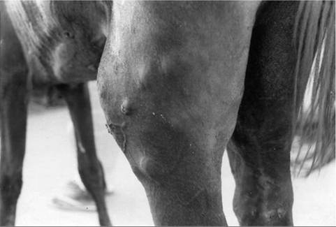
FIG. 31.38 Typical cutaneous form of glanders (known as farcy) showing skin nodules.
Nasal and pulmonary signs are usually seen in the acute form. The symptoms include a high fever, decreased appetite, coughing, progressive dyspnea, nasal discharge, and ulcers and nodules on the nasal mucosa. Bloody crusting may be seen on the nostrils, and there may be a purulent ocular discharge. The submaxillary lymph nodes are usually swollen and painful. Central nervous system signs have been reported in experimentally infected horses, possibly as the result of secondary bacterial infection from a compromised blood-brain barrier.11 Animals with the acute form of glanders usually die in a few days to a few weeks.
The chronic form develops insidiously and results in progressive debilitation. The symptoms may include coughing, dyspnea, intermittent fever, enlargement of the lymph nodes, a chronic nasal discharge, and ulcers, nodules, and stellate scars on the nasal mucosa. The skin and lymphatics may also be involved. The chronic form is often fatal, but affected animals may live for years before succumbing to the disease.
In the latent form, there may be few symptoms other than a nasal discharge and occasional labored breathing. Lesions may be found only in the lungs.
The differential diagnoses for glanders include strangles caused by Streptococcus equi; epizootic lymphangitis, which is a chronic granulomatous and suppurative fungal infection caused by Histoplasma farciminosum; sporotrichosis, a chronic subcutaneous lymphatic mycosis caused by a gram-positive fungus Sporotrichum schenckii; ulcerative lymphangitis, a bacterial disease caused by Corynebacterium pseudotuberculosis; and melioidosis (Whitmore’s disease), which is caused by B.
pseudomallei. Melioidosis is particularly difficult to differentiate because B. pseudomallei and B. mallei cross-react in many serologic and hypersensitivity tests.1,2■ Diagnosis A diagnosis of glanders based on clinical signs can be made when B. mallei is isolated from samples or an immunologic reaction to the organism is detected serologically or on the basis of hypersensitivity test results. Isolation and identification of B. mallei in cultures of samples from lesions or various exudates, including respiratory secretions, is regarded as a gold standard for the diagnosis of glanders. B. mallei can be stained with methylene blue, Wright’s stain, or Gram stain, but the staining can be weak or irregular, and bipolar staining of the gram-negative organism may be observed. They can be pleomorphic in older cultures, but in clinical samples and young cultures they typically form straight or slightly curved rods that are 0.3 to 0.8 μm in width and 2 to 5 μm in length. Some authors report that, in tissues, the organism is identified best with Giemsa stain.12
B. mallei can be grown on routine culture media, including nutrient, blood, and MacConkey agars; growth is slow, but colonies are usually visible within 48 hours. Optimal growth occurs at 37° C (98.6° F).
Automated bacterial identification systems do not always correctly identify this organism. All isolations must be performed in biosafety level 3 laboratories. Inoculation into laboratory animals, particularly guinea pigs (the Strauss reaction) or hamsters, was used for identification of B. mallei in the past, but this technique is now rarely practiced.
A cell-mediated hypersensitivity reaction test (mallein intradermal skin test) was used extensively to screen horses for glanders. This test is still used to certify horses as negative for glanders during importation to some countries. It may also be used in the diagnosis of clinical cases. The mallein test is based on hypersensitivity reaction to mallein, a commercially available, purified protein derivative of B.
mallei. The test is performed most often by intradermal palpebral injection. Reactors are identified by marked eyelid swelling that develops within 24 to 48 hours, accompanied by fever and occasionally by purulent ocular discharge. No reaction or minimal swelling of the eyelid develops in unsensitized animals. False-positive reactions can occur in animals that are sensitized to other closely related Burkholderia spp., including B. pseudomallei, and cross-reactions with S. equi have been described.Serologic tests are sometimes used to screen horses for importation to glanders-free countries or to identify clinically affected animals. Measurable titers of serum antibody against B. mallei develop approximately 1 week after infection. Complement fixation was the preferred serologic test in past eradication programs because of its ability to detect subclinically or chronically infected animals. This test was most useful when the prevalence of disease was high. Complement fixation is still used by some countries to assess imported equids for glanders. Enzyme-linked immunosorbent assay tests are also used to screen animals for international trade. Other serologic tests include indirect hemagglutination, counterimmunoelectrophoresis, immunoblotting, and indirect fluorescent testing, but these tests are not approved by the World Organisation for Animal Health (OIE) for use in international trade.1,2 Rapid DNA tests, such as PCR assays, are now also used.
■ Treatment Treatment of glanders is often not advised because infected animals may remain subclinically infected and shed B. mallei.1,2 The risk of zoonotic transmission also discourages treatment. Systemic treatment with antibiotics, including penicillin and streptomycin, is usually ineffective in controlling the disease. Some evidence has accumulated that enrofloxacin, erythromycin, ampicillin, sulfonamide, gentamicin, and tetracycline might be effective against B. mallei. This evidence has emerged from in vitro antibiotic sensitivity testing and from treatment studies in experimentally infected guinea pigs. Experimentally, in lab animals effective treatments also include doxycycline, ceftazidime, gentamicin, and trimethoprim sulfas.1
■ Prevention and Control Cases of glanders in animals must be reported to the OIE.1 The occurrence of this disease in any country can result in restrictions on international trade in horses and other affected animals. The mallein test and complement fixation test are approved assays recommended by the OIE for international trade purposes.1
If glanders is detected, control measures include strict quarantine of all infected and exposed animals, diagnostic testing of animals with clinical signs indicative of glanders, and assessment of apparently normal equids with the elimination of those that react positively in screening tests. Ill animals and those with positive results of mallein tests are euthanized. Horses exposed to B. mallei that yield negative results in mallein tests are isolated and retested 2 to 3 weeks later. Carcasses and contaminated bedding and feed should be burned or buried. Equipment or other items in contact with infected animals should be disinfected.
People who handle tissues from infected animals, bacterial cultures, or contaminated fomites should implement strict precautions to prevent infection. Protective clothing, including masks, gloves, face shields, and gowns, should be worn. In clinical and research laboratories, B. mallei requires biosafety containment level 3 accommodation. Precautions are indicated for activities that are likely to produce aerosols, droplets, or concentrations of bacteria. Currently there is no commercial vaccine against glanders for use in humans or any other animal species.
Exercise-Induced Pulmonary Hemorrhage

■ TABLE 31-12
Causes of Epistaxis in Horses. (Reproduced With Modifications by Permission.91)
| Disease | Epidemiology | Clinical Signs and Diagnosis | Treatment and Control |
| Hemorrhage Into Trachea or Bronchi, Sometimes With Epistaxis | |||
| Exercise-induced | Horses after strenuous exercise | Epistaxis is a rare but very specific | Efficacy of various drugs |
| pulmonary hemorrhage | Most common in | sign of EIP; only occurs after | used for treatment and |
| (EIPH) | Thoroughbred and Standardbred racehorses | exercise Endoscopic examination of the airways is diagnostic | control is debated Furosemide is used extensively before racing |
| Trauma | Sporadic; associated with trauma to head, neck, or chest | Physical examination reveals site and nature of the trauma; can require endoscopic examination of upper airways | Symptomatic treatment |
| Pneumonia | Recent transport or respiratory disease; can occur as outbreaks though usually individual animals | Fever, tachypnea, abnormal lung sounds, leukocytosis, radiography demonstrates lung lesions; cytological and microbiological examination of tracheal aspirate | Antimicrobials, NSAIDs, oxygen Control by vaccination and prevention of respiratory disease |
| Lung abscess | Sporadic; hemorrhage can occur after exercise | Sometimes no premonitory signs fever, depression, anorexia, cough hemogram demonstrates leukocytosis Hyperfibrinogenemia ultrasonography or radiography demonstrates lesion Tracheal aspirates | Antibiotics |
| Intrabronchial foreign body | Sporadic | Cough, hemoptysis, fever; endoscopy or radiography reveals foreign body | Removal of foreign body— often not readily achieved |
| Pulmonary neoplasia Sporadic; often older horse, but not always hemangiosarcoma Epistaxis (in Addition to the Above Diseases) | Cough, hemoptysis demonstrate mass on ultrasonographic or radiographic examination | None | |
| Guttural pouch mycosis | Sporadic; acute onset epistaxis | Severe, life-threatening epistaxis; tachycardia, anemia, hemorrhagic shock | Surgical ligation or occlusion of arteries in the guttural pouch |
| Ethmoidal hematoma | Sporadic | Epistaxis not associated with exercise Usually unilateral | Surgery or injection of mass with formaldehyde |
| Thrombocytopenia | Sporadic | Epistaxis, mild, intermittent Petechiation and ecchymotic hemorrhages Thrombocytopenia | Glucocorticoids |
| Coagulopathy | Sporadic, congenital or associated with ingestion of rodenticides | Epistaxis, increased bleeding from minor wounds, hemarthrosis, | Identify and correct underlying cause Vitamin K administration |
| Neoplasia | Sporadic | Neoplasia of upper airways | None |
| Trauma | Sporadic | Injury to head or pharynx | Symptomatic |
| Sinusitis | Sporadic | Endoscopic or radiographic examination of sinus | Drainage, antimicrobials |
presenting complaints for horses with EIPH. Although poor performance might be attributable to any of a large number of causes, epistaxis associated with exercise is almost always secondary to EIPH.
Epistaxis due to EIPH occurs during or shortly after exercise and is usually first noticed at the end of a race, particularly when the horse is returned to the paddock or winner's circle and is allowed to lower its head. It is usually bilateral and resolves within hours of the end of the race. Epistaxis might occur on more than one occasion, especially when horses are raced or exercised at high speed soon after an initial episode. It is important to recognize that epistaxis is an uncommon manifestation of EIPH, occurring in as few as 4% of horses with EIPH.16
EXERCISE-INDUCED PULMONARY HEMORRHAGE AND PERFORMANCE. Failure of racehorses to perform to the expected standard (poor performance) is often, accurately or not, attributed to EIPH.17 Many horses with poor performance have cytologic evidence of EIPH on microscopic examination of tracheobronchial aspirates or BAL fluid or have blood evident on endoscopic examination of the tracheobronchial tree performed 30 to 90 minutes after strenuous exercise or racing. However, it is important to recognize that EIPH is very common in racehorses, and it should be considered the cause of poor performance only after other causes have been eliminated. Severe EIPH undoubtedly results in poor performance and, on rare occasions, death of Thoroughbred racehorses.10,17,18 Thoroughbred horses with EIPH have impaired performance compared to unaffected horses.17,19,20 The severity of EIPH is negatively correlated to race performance, with more severely affected horses having a lower likelihood of finishing in the first three places; they are less likely to be elite money earners and finish further behind the winner than do unaffected horses.17,19,20
Results of studies in Standardbred racehorses indicate either a lack of effect of EIPH on performance or an association between EIPH and superior performance. There was no relationship between presence of EIPH and finishing position in 29 Standardbred racehorses with intermittent EIPH, each examined on at least two occasions, or in 92 Standardbred racehorses examined on one occasion. However, of 965 Standardbred racehorses examined after racing, those finishing first or second were 1.4 times more likely (95% confidence interval 0.9 to 2.2) to have evidence of EIPH on tracheobron- choscopic examination than were horses that finished in seventh or eighth position.
There is no clear association between EIPH and performance (time) in barrel racing Quarter Horses.21
■ Diagnostic Approach
PHYSICAL EXAMINATION. Apart from epistaxis in a small proportion of affected horse, there are few abnormalities detectable on routine physical examination of horses with EIPH. Rectal temperature and heart and breathing rates might be elevated as a consequence of exercise in horses examined soon after exercise, but values of these variables in horses with EIPH at rest are not noticeably different from horses with no evidence of EIPH. Affected horses might swallow more frequently during recovery from exercise than do unaffected horses, probably as a result of blood in the larynx and pharynx. Coughing is common in horses recovering from strenuous exercise and after recovery from exercise; horses with EIPH are no more likely to cough than are unaffected horses.10 Other clinical signs related to respiratory abnormalities are uncommon in horses with EIPH. Respiratory distress is rare in horses with EIPH and when present indicates severe hemorrhage or other serious lung disease such as pneumonia, pneumothorax, or rupture of a pulmonary abscess. Lung sounds are abnormal in a small number of EIPH-affected horses and, when present, are characterized by increased intensity of normal breath sounds during rebreathing examination. Crackles may be auscultated over the trachea in horses with EIPH but are also heard in unaffected horses.
TRACHEOBRONCHOSCOPY. Observation of blood in the trachea or large bronchi of horses 30 to 120 minutes after racing or strenuous exercise provides a definitive diagnosis of EIPH. The amount of blood in the large airways varies from a few small specks on the airway walls to a stream of blood occupying the ventral one third of the trachea. Blood might also be present in the larynx and nasopharynx. If there is a strong suspicion of EIPH and blood is not present on a single examination conducted soon after exercise, the examination should be repeated in 60 to 90 minutes. Some horses with EIPH do not have blood present in the rostral airways immediately after exercise but do have blood when examined 1 to 2 hours later. Blood is detectable by tracheobronchoscopic examination for 1 to 3 days in most horses, with some horses having blood detectable for up to 7 days.
Bronchoscopic examination can be used to estimate the severity of EIPH through the use of a grading system. The interobserver repeatability of tracheobronchoscopic assessment of severity of EIPH using a 0 to 4 grading scale is excellent, and this scoring system has been widely adopted (Table 31.13).16,17,20-23
It is assumed that a higher score represents more severe hemorrhage, but while the repeatability of this scoring system has been established, the relationship between the amount of
■ TABLE 31.13
Validated System for Grading the Severity of Exercise-Induced Pulmonary Hemorrhage Detected on Tracheobronchoscopic Examination of Thoroughbred Racehorses92
Grade 0 No blood detected in the pharynx, larynx,
trachea, or main stem bronchi
Grade 1 Presence of one or more flecks of blood or
≤2 short (2 short streams occupying less than one third of the tracheal circumference
Grade 3 Multiple, distinct streams of blood covering
more than one third of the tracheal circumference. No blood pooling at the thoracic inlet
Grade 4 Multiple, coalescing streams of blood
covering >90% of the tracheal surface, with pooling of blood at the thoracic inlet
blood in the large airways and the actual amount of hemorrhage has not been established.
RADIOGRAPHY. Thoracic radiography is of limited use in detecting horses with EIPH. Radiographs can demonstrate the presence of densities in the caudodorsal lung fields of some horses, but many affected horses have minimal to undetectable radiographic abnormalities.10 Examination of thoracic radiographs of horses with EIPH can be useful in ruling out the presence of another disease process, such as a pulmonary abscess, contributing to the horse's pulmonary hemorrhage or poor athletic performance. The utility of thoracic ultrasound for detection of EIPH has not been extensively reported. One report of detecting EIPH in 81% of 157 horses in which EIPH was diagnosed by relatively liberal assessment of BAL fluid reported sensitivity of presence of comet-tail lesions for diagnosis of EIPH of 86% and specificity of 25%,24 indicating that ultrasound examination of the thorax has limited utility in confirming the presence of EIPH.
■ Clinical Pathology
EXAMINATION OF AIRWAY SECRETIONS OR LAVAGE FLUID. The presence of red blood cells or macrophages containing either effete red blood cells or the breakdown products of hemoglobin (hemosiderophages) in tracheal or bronchoalveolar lavage fluid provides evidence of EIPH.25 Detection of red blood cells or hemosiderophages in tracheal aspirates or BAL fluid is believed to be both sensitive and specific in the diagnosis of EIPH, although in the absence of a gold standard for diagnosis, this claim is yet to be validated. Examination of airway fluids from a variety of breeds and uses of horses, including barrel racing horses,21 indicates the presence of EIPH in a greater proportion of horses than does tracheobroncho- scopic examination after strenuous exercise or racing. The greater sensitivity of examination of airway fluid is probably attributable to the ability of this examination to detect the presence of small amounts of blood or its residual products and the longevity of these products in the airways. Although endoscopic examination can detect blood in occasional horses up to 7 days after an episode of EIPH, cellular evidence of pulmonary hemorrhage persists for weeks after a single episode. Red blood cells and macrophages containing red blood cells are present in BAL fluid or tracheal aspirates for at least 1 week after strenuous exercise or instillation of autologous blood into airways, and hemosiderophages are present for at least 21 days and possibly longer.26
Recent studies have reported on the use of red blood cell numbers in BAL fluid as a quantitative indicator of EIPH.21,25,27-29 The diagnostic cutoff has not been determined, although 1000 cells∕μL was used in one study,21 and there is only a weak correlation between EIPH score determined by tracheobron- choscopic examination and red blood cell count in BAL fluid (r2 = 0.15).21 However, this indicator of EIPH severity has not been validated nor demonstrated to be more reliable or repeatable than tracheobronchoscopic examination and visual scoring. Furthermore, considerable concern exists over the suitability of red blood cell counts in BAL fluid for assessment of severity of EIPH given that an unknown area, although presumably small, of the lung is examined by lavage and there is a risk that this area of lung might not be representative of the lung as a whole, similar to the situation of examination of BAL fluid of horses with pneumonia.7 BAL of sections of both lungs, achieved using an endoscope, might obviate some of these concerns given that BAL results of each lung in a horse differ.30
Tracheal aspirates can be obtained any time after exercise by aspiration either during tracheobronchoscopic examination or through a percutaneous intratracheal needle. Aspirates obtained through an endoscope may not be sterile, depending on the collection technique. BAL fluid can be obtained through either an endoscope wedged in the distal airway or a cuffed tube inserted blindly into a distal airway. Collection of fluid through an endoscope has the advantage of permitting examination of the distal airways and selection of the area of lung to be lavaged. However, it does require the use of an endoscope that is longer (2 m) than those readily available in most equine practices. Use of a commercial BAL catheter does not require use of an endoscope, and this procedure can be readily performed in field situations.
■ Necropsy EIPH is a rare cause of death of racehorses, but among racehorses that die during racing for reasons other than musculoskeletal injuries, EIPH is common.18 Necropsy examination of horses is usually incidental to examination for another cause of death. Grossly, horses examined within hours of strenuous exercise, such as horses examined because of catastrophic musculoskeletal injuries incurred during racing, can have severe petechiation in the caudodorsal lung fields. Horses with chronic disease have blue/gray or blue/brown discoloration of the visceral pleural surfaces of the caudodorsal lung fields that is often sharply demarcated, especially on the diaphragmatic surface. The discoloration affects both lungs equally with 30% to 50% of the lung fields being discolored in severe cases. Affected areas do not collapse to the same extent as unaffected areas and in the deflated lung have a spleen-like consistency. On cut surface, the discolored areas of lung are predominantly contiguous with the dorsal pleural surface and extend ventrally into the lung parenchyma. Areas of affected lung can be separated by normal lung. There is proliferation of bronchial vessels, predominantly arteries and arterioles, in affected areas. Histologically, affected areas exhibit bronchiolitis, hemosiderophages in the alveolar lumen and interstitial spaces, and fibrosis of interlobular septa and pleura and around vessels and bronchioles.
■ Pothogenesis The cause of EIPH is rupture of alveolar capillary membranes with subsequent extravasation of blood into interstitial and alveolar spaces. The source of blood in such instances is the pulmonary circulation. Bleeding from bronchial circulation during exercise has been suggested, based on histologic evidence of bronchial angiogenesis in horses that have experienced previous episodes of EIPH, but contribution of the bronchial circulation to EIPH has not been demonstrated. Regardless of the contribution of bronchial circulation to blood in the airways, the likely proximate lesion is in capillaries associated with the pulmonary circulation, with the causative lesion likely in the pulmonary veins. In other words, pulmonary capillaries rupture, allowing blood to escape into the alveoli (the proximate lesion) due to lesions in the pulmonary veins that increase pulmonary capillary pressure. There is increasing evidence that the primary lesion is arteriovenous remodeling of pulmonary veins and that this remodeling differs in severity
3137 among lung regions and with the intensity of training.31-3 Remodeling of pulmonary veins results in loss of distensibility and partial occlusion to blood flow, with subsequent presumed increases in pulmonary alveolar capillary pressure.31,38 Hemorrhage into the interstitial space and alveoli, with subsequent rostral movement of blood in the airways, results in blood in the trachea and bronchi.
Rupture of alveolar capillaries occurs secondary to an exercise-induced increase in transmural pressure (pressure difference between the inside of the capillary and the alveolar lumen), which, as noted above, can occur as a result of venous remodeling and loss of distensibility of pulmonary veins.35 If the transmural stress exceeds the tensile strength of the capillary wall, the capillary ruptures. The proximate cause of alveolar capillary rupture is the high transmural pressure generated by positive intracapillary pressures, which are largely attributable to capillary blood pressure, and the lower intraalveolar pressure generated by the negative pleural pressures associated with inspiration.
During exercise, the absolute magnitudes of both pulmonary capillary pressure and alveolar pressure increase, with a consequent increase in transmural pressure. Strenuous exercise is associated with marked increases in pulmonary artery pressure in horses. Values for mean pulmonary arterial pressure at rest of 20 to 25 mm Hg increase to more than 90 mm Hg during intense exercise because of the large cardiac output achieved by exercising horses.39 The increases in pulmonary artery pressure, combined with an increase in left atrial pressure during exercise, probably result in an increase in pulmonary capillary pressure. Combined with the increase in pulmonary capillary pressure is a marked decrease (more negative) in pleural, and therefore alveolar, pressure during exercise. The pleural pressure of normal horses during inspiration decreases from approximately -0.7 kPa (-5.3 mm Hg) at rest to as low as -8.5 kPa (64 mm Hg) during strenuous exercise. Together, the increase in pulmonary capillary pressure and decrease (more negative) in intrapleural (alveolar) pressure contribute to a marked increase in stress in the alveolar wall. Although the alveolar wall and pulmonary capillaries of horses are stronger than those of other species, rupture may occur because the wall stress in the alveolus exceeds the mechanical strength of the capillary.40-42
Other theories of the pathogenesis of EIPH include small airway disease, upper airway obstruction, hemostatic abnormalities, changes in blood viscosity and erythrocyte shape, intra- thoracic sheer forces associated with gait, and bit-induced asphyxia.43,44 It is likely that the pathogenesis of EIPH involves several processes, including pulmonary hypertension, lower alveolar pressure, and changes in lung structure, that summate to induce stress failure of pulmonary capillaries.
Obstruction of either the upper or the lower airway has been proposed as a cause of EIPH. Inspiratory airway obstruction results in more negative intrapleural, and therefore alveolar, pressures. This effect is exacerbated by exercise, with the result that alveolar transmural pressure is greater in horses with airway obstruction. The higher transmural pressure in such horses can increase the severity of EIPH, although this has not been demonstrated. Moreover, although inspiratory airway obstruction might predispose to EIPH, the prevalence of this condition is much less than that of EIPH, indicating that it is not the sole factor inducing EIPH in most horses.
Horses with moderate to severe EIPH have histologic evidence of inflammation of the small airways, and there is a clear association between the presence of EIPH and inflammatory changes in bronchoalveolar or tracheal aspirate fluid. However, instillation of autologous blood into the airways does not induce a marked inflammatory response in normal horses, and it is therefore unclear whether inflammation alone induces or predisposes to EIPH.45,46 Theoretically, small airway inflammation and bronchoconstriction have the potential to produce intrathoracic airway obstruction and therefore a more negative alveolar pressure. Given that small airway disease is common in horses, there is the potential for an important effect of factors, such as viral infections, air pollution, and allergic airway disease, to contribute to the initiation or propagation of EIPH.
The characteristic location of lesions of EIPH in the caudodorsal lung fields has led to the proposal that hemorrhage is a result of tissue damage occurring when waves of stress, generated by forelimb foot strike, are focused and amplified into the narrowing cross-sectional area of the caudal lung lobes.47 According to the theory, the locomotor impact of the forelimbs results in transmission of forces through the scapula to the body wall, from where they pass into the lungs and caudally and dorsally. As the wave of pressure passes into the narrower caudodorsal regions of the lungs, it generates progressively greater shearing forces that disrupt tissue and cause EIPH. However, studies of intrapleural pressures have not demonstrated the presence of a systemic pressure wave passing through the lung and do not provide support for this hypothesis. Furthermore, track surface is not associated with incidence of EIPH in Thoroughbred racehorses.22,48
Horses with EIPH have been suspected of having defects in either hemostasis or fibrinolysis. However, although exercise induces substantial changes in blood coagulation and fibrino- lysis,49 these is no evidence that horses with EIPH have defective coagulation or increased fibrinolysis.50
Regardless of the cause, rupture of pulmonary capillaries and subsequent hemorrhage into airways and interstitium cause inflammation of both airways and interstitium, with subsequent development of fibrosis and alteration of tissue compliance. Heterogeneity of compliance within the lungs, and particularly at the junction of normal and diseased tissue, results in the development of abnormal shear stress with subsequent tissue damage. These changes are exacerbated by inflammation and obstruction of small airways, with resulting uneven inflation of the lungs. The structural abnormalities, combined with pulmonary hypertension and the large intrathoracic forces associated with respiration during strenuous exercise, cause repetitive damage at the boundary of normal and diseased tissue with further hemorrhage and inflammation. The process, once started, is lifelong and continues for as long as the horse continues to perform strenuous exercise.35
■ Epidemiology EIPH is primarily a disease of horses, although it also occurs in racing camels and Greyhounds.51,52 EIPH occurs in horses worldwide, and there does not appear to be any geographic distribution. It is a disorder of horses that run at high speed, such as Thoroughbred, Standardbred, or Quarter Horse racehorses; Quarter Horses used for barrel racing; and polo ponies.2,5,21,53-55 The disorder is uncommon in endurance horses and is rare in draft breeds.11,56 There is increasing recognition of its importance in sport horses, including 3-day event and show jumping, but there are no reports of the disease in horses used for dressage, although it is thought to occur.57,58
EIPH is considered to be associated with death during racing or in the hours after completion of racing in Thoroughbred and Standardbred horses,59,60 although there is minimal evidence of the relative risk of death during racing of horses with EIPH.10 Estimates of the incidence of such “sudden” deaths associated with EIPH in two large studies were 38% (60 of 157) and 35% (50 of 143) of deaths not attributable to musculoskeletal injury in Thoroughbreds, Standardbreds, and Quarter Horses or in solely Thoroughbreds, respectively.59,60 However, there is uncertainty among some pathologists in attributing the cause of death to EIPH, and the role of EIPH in causing death of horses during strenuous exercise is unclear. Recognizing the subjectivity of both the diagnosis and the attribution of cause of death, it is suggested that EIPH associated with death during racing should be classified as “exercise-associated fatal pulmonary hemorrhage.”59
The prevalence of EIPH varies with the method used to detect it and the frequency with which horses are examined, as discussed later in this section. Epistaxis associated with exercise is almost always attributable to pulmonary hemorrhage and occurs only in a small proportion of racehorses. Epistaxis occurs in only 3% of horses that have blood detected in the trachea by endoscopic examination performed within 2 hours of racing. The prevalence of epistaxis in racehorses varies between 0.1% and 9.0%, with the frequency depending on the breed, age, and sex of horses selected for study; the type of racing; and the timing and frequency of observation of horses after racing. Epistaxis is more common in older horses. There are conflicting reports of a sex predisposition, although epistaxis might be more common in female Thoroughbreds.61 Epistaxis is more common after races of less than 1600 m than in longer races, although not all sources agree on this point. However, horses in steeplechase races, which are typically longer than 2000 m, are at greater risk of epistaxis than are horses in flat races.47,61-63 Incidence of epistaxis in steeplechase horses in the United Kingdom is 5.3 per 1000 starts and 3.6 per 1000 starts in hurdle racing.1 Risk factors for horses in jumps races (steeplechase) include previous epistaxis (odds ratio [OR] 6.1 [4.4 to 8.3]), racing in a claiming race (OR 5.9 [1.4 to 25]), more than 9 starts in the previous 4 to 6 months (OR 10 [2 to 47]), and racing on firmer ground.1 In summary, epistaxis is relatively uncommon, and most horses with EIPH do not have epistaxis.
There are a variety of other methods of detecting EIPH, including endoscopic examination of the airways and microscopic examination of tracheal aspirates or BAL fluid, and the incidence, or prevalence depending on the study design, is influenced by the method used to detect EIPH and the definition of the disease.10
Almost all Thoroughbred racehorses in active training, and a large proportion of other types of horses undergoing regular high-speed exercise, have hemosiderophages in BAL fluid, indicating that all have some degree of EIPH.27,64-66 The prevalence of EIPH decreases when diagnosis is based on endoscopic examination of horses after exercise or racing.
EIPH is very common in Thoroughbred racehorses, with estimates of prevalence, based on a single endoscopic examination of the trachea and bronchi, of 43% to 75%.16,17 The prevalence increases with the frequency of examination, with more than 80% of horses having evidence of EIPH on at least one occasion after examination after each of three consecutive races.67 There can be considerable variability in severity of EIPH within an individual horse on repeated examination over a racing season, with this variability being greatest in horses that have EIPH of the greatest severity.16 Horses with evidence of EIPH on one examination are likely to have EIPH detected on subsequent endoscopic examinations, most commonly at low grade (Grade 1 or Grade 2).16 The prevalence of EIPH in Standardbred racehorses is assumed to be lower, with 26% to 34% of horses reported to have blood in the trachea after racing. However, these studies were based on a single examination, and one study only reported as positive those horses with blood covering more than one half the tracheobronchial tree. When examined after each of three races, 87% of Standardbred racehorses have evidence of EIPH on at least one occasion, suggesting that EIPH is as common in Standardbred racehorses as it is in Thoroughbred racehorses.68
EIPH occurs in approximately 62% of racing Quarter Horses and in 54% of Quarter Horses used for barrel racing when the diagnosis is based on tracheobronchoscopy and 66% when the diagnosis is based on a red blood cell count exceeding 1000 cells∕μL.21,55 Approximately 11% of polo ponies are affected with EIPH.5,6 The disease does not appear to occur with any important frequency in draft horses but is not well documented.56
Age is considered a risk factor for EIPH, with the prevalence of the disorder being higher in older horses, but the risk factor is the amount of racing a horse has completed, not its age.69,70 There is no consistent association of sex with prevalence of EIPH. Among Thoroughbred racehorses there is an unclear relationship between the speed of racing and the risk of EIPH.22,69 Lesions of EIPH are not detected in young Thoroughbred racehorses that have trained at speeds of less than 7 m/s.71
The risk of EIPH increases with racing at lower ambient temperatures22,69 and with the wearing of bar shoes during racing.22 There is no association between risk of EIPH and track hardness.22,69
■ Treatment Prevention of EIPH is contentious, as it can involve the administration of medications on the day of racing.8 The efficacy of various interventions and medications has recently been evaluated in two systematic reviews that concluded that there was moderately strong to strong evidence that administration of furosemide before racing reduces the frequency and severity of EIPH in Thoroughbred racehorses.10,12 There is either weak evidence or no evidence of efficacy of other interventions. There is a recommendation for use of furosemide, but because of the regulatory issues related to its use, this is only a weak recommendation.10 Were there not regulatory considerations, the recommendation for use of furosemide would likely be strong, based on evidence of efficacy. Clearly, the decision to permit administration of furosemide to horses on the day of racing is based not only on its clinical efficacy but also on considerations of the welfare of the horse, integrity of wagering, policies on medication use in racehorses, and opinions of owners, bettors, trainers, and the public.9
A comprehensive evaluation of the efficacy of medications putatively used to treat or prevent EIPH is available, including citations to research.10 Common features of many studies of interventions to reduce the incidence or severity of EIPH are use of a small number of horses, use of horses with signalment that is not typical of those affected by EIPH, an exercise model other than that which causes EIPH (e.g., treadmill exercise instead of overground exercise or racing), and lack of adequate reporting of results.10,72 A key shortcoming of small studies is that they might fail to detect a treatment effect when such an effect does exist - the so called Type 2 error rate of underpowered studies.72
Furosemide apparently reduces pulmonary capillary pressure in exercising horses and likely does so by reducing blood and plasma volume as a result of profound diuresis.73-76 There appears to be a direct effect of furosemide on pulmonary vasculature in some species,77 but prevention of diuresis by ligation of ureters abolishes the hemodynamic effect of furosemide in anesthetized horses, suggesting that this is not an important mechanism of its hemodynamic effect in horses.78
Furosemide administration as prophylaxis against EIPH is permitted in a number of racing jurisdictions worldwide, most notably in Canada, the United States, Mexico, and most of the South American countries.73 Within the United States and Canada, almost all Thoroughbred, Standardbred, and Quarter Horse racing jurisdictions permit administration of furosemide before racing. The vast majority (>90%) of Thoroughbred horses racing in the United States receive furosemide before racing, at an estimated annual cost of between $6 million and $20 million (USD, 1995 value). Although accurate numbers are not available, it appears that a smaller proportion of Standardbred and Quarter Horse racehorses receive furosemide before racing.
Therapy of EIPH is usually a combination of attempts to reduce the severity of subsequent hemorrhage and efforts to minimize the effect of recent hemorrhage. Treatment of EIPH is problematic for a number of reasons. First, the pathogenesis of EIPH has not been determined, although the available evidence supports a role for stress failure of pulmonary capillaries secondary to exercise-induced pulmonary hypertension. Second, with the exception of furosemide, there is a lack of information using large numbers of horses under field conditions that demonstrates an effect of any medication or management practice on EIPH. There are numerous studies of small numbers of horses (EIPH is either unknown or, in very small studies, not evident.10 Corticosteroids, including dexamethasone, fluticasone, and beclomethasone administered by inhalation, parenterally, or enterally, reduce airway inflammation and obstruction but have no demonstrated efficacy in preventing EIPH. Cromolyn sodium (sodium cromoglycate) has no efficacy in preventing EIPH.85
Water vapor treatment (inhalation of water-saturated air) has been proposed as a treatment for EIPH because of its putative effect on small airway disease. However, water vapor treatment has no effect on EIPH.86
The use of bedding of low allergenic potential (shredded paper) to prevent EIPH has no apparent effect on prevalence of the condition.87 While it is suggested that preventing or minimizing small airway disease might reduce the severity of EIPH, studies to demonstrate such an effect have not been reported. However, optimizing the air quality in barns and stables and preventing infectious respiratory disease appear to be sensible precautions.
INTERSTITIAL INFLAMMATION AND BRONCHIAL ANGIOGENESIS. Hemorrhage into interstitial tissues induces inflammation, with subsequent development of fibrosis and bronchial artery angiogenesis. The role of these changes in perpetuating EIPH in horses is unclear but is probably of some importance. Treatments to reduce inflammation and promote healing with minimal fibrosis have been proposed. Rest is an obvious recommendation, and many racing jurisdictions have rules regarding enforced rest for horses with epistaxis. While the recommendation for rest is intuitive, there is no information that rest reduces the severity or incidence of EIPH in horses with prior evidence of this disorder.
Similarly, corticosteroids are often administered, either by inhalation, enterally, or parenterally, in an attempt to reduce pulmonary inflammation and minimize fibrosis. Again, the efficacy of this intervention in preventing or minimizing severity of EIPh has not been documented.
COAGULOPATHY AND FIBRINOLYSIS. Exercise induces substantial changes in blood coagulation and fibrinolysis. However, there is no evidence that horses with EIPH have defective coagulation or increased fibrinolysis. Regardless, aminocaproic acid, a potent inhibitor of fibrin degradation, has been administered to horses to prevent EIPH. The efficacy of aminocaproic acid in preventing EIPH has not been demonstrated.10 Similarly, estrogens are given to horses with the expectation of improving hemostasis, although the effect of estrogens on coagulation in any species is unclear. There is no evidence that estrogens prevent EIPH in horses.
Vitamin K is administered to horses with EIPH, presumably in the expectation that it will decrease coagulation times. However, because EIPH is not associated with prolonged bleeding times, it is unlikely that this intervention will affect the prevalence or severity of EIPH.
PLATELET FUNCTION. Aspirin inhibits platelet aggregation in horses and increases bleeding time. Seemingly paradoxically, aspirin is sometimes administered to horses with EIPH because of concerns that increased platelet aggregation contributes to EIPH. There is no evidence that aspirin either exacerbates or prevents EIPH. There is evidence of considerable variability between horses in the effect of aspirin on platelet function.88
CAPILLARY INTEGRITY. Capillary fragility increases the risk of hemorrhage in many species. Various bioflavonoids have been suggested to increase capillary integrity and prevent bleeding. However, hesperidin and citrus bioflavonoids have no efficacy in prevention of EIPH in horses.89 Similarly, vitamin C is administered to horses with EIPH without scientific evidence of any beneficial effect.
SUMMARY OF TREATMENT OPTIONS. Selection of therapy for horses with EIPH is problematic.10 Given that most horses have some degree of pulmonary hemorrhage during most bouts of intense exercise, the decision must be made as to not only the type of treatment and its timing but also which horses to treat. Moreover, the apparently progressive nature of the disease with continued work highlights the importance of early and effective prophylaxis and emphasizes the need for studies of factors such as air quality, respiratory infections, individual differences in susceptibility, and frequency and duration of high-intensity exercise in inciting the 35
disorder.35
The currently favored treatment for EIPH is administration of furosemide before intense exercise.10 Its use is permitted in racehorses in a number of countries but is contentious in many. A frequent practice is to administer furosemide before high-speed training, and not on the day of racing, in jurisdictions that do not permit race-day administration of medications. There is increasing interest in the effect of furosemide administered 24 hours before racing on the incidence or severity of EIPH.90 Its efficacy in this situation remains to be determined, although a recent study found that furosemide administered 24 hours before simulated racing did not reduce the incidence or severity of EIPH compared to a saline placebo treatment.29 However, the study did not have sufficient statistical power to detect important differences in EIPH grade or red blood cell count in BAL fluid; there is a high risk of type 2 error (failure to detect a difference when one actually exists), as is the case with many studies of treatments for EIPh that include small numbers of horses (in this case, 15).10 The association between furosemide administration and superior performance in Standardbred and Thoroughbred racehorses should be considered when recommending use of this drug.
■ Prevention and Control There are no documented preventive strategies. Rest is an obvious recommendation for horses with EIPH, but the hemorrhage is likely to recur when the horse is next strenuously exercised. The duration of rest and the optimal exercise program to return horses to racing after EIPH is unknown, although some jurisdictions require exercise no more intense than trotting for 2 months. Firm recommendations cannot be made on duration of rest because of a lack of objective information.
Although a role for lower airway disease (either infectious or allergic) in the genesis of EIPH has not been demonstrated, control of infectious diseases and minimization of noninfectious lower airway inflammation appear prudent.
Concern about the role of impact waves in the genesis of EIPH has led to discussion of “low-stress” training protocols, but these have not been adequately evaluated.
■ Prognosis Horses that have experienced one episode of epistaxis are more likely to have a second episode. For this reason most racing jurisdictions do not permit horses with epistaxis to race for a period of weeks to months after the initial instance, with more prolonged enforced rest after a subsequent episode of epistaxis and retirement from racing after a third bout. The recurrence rate after one episode of epistaxis in Thoroughbred horses is 20% to 30%.13,62 This high rate of recurrence suggests that the inciting pulmonary lesions have not healed.
Long-term examination of performance of horses with EIPH indicates that horses with Grade 4 EIPH have shorter racing careers, but it is not clear if this is a function of the biology of the disease or a management decision by owners and trainers.70 There is no association between measures of long-term performance measured over 10 years and Grades 1, 2, or 3 of EIPH in Thoroughbred racehorses.70 Racehorses in Hong Kong that are EIPH positive on one or more occasions do not have fewer starts than horses for which EIPH was not detected (18 versus 15, P = 0.17) on endoscopic examination.16