Infection Control Issues Related to Specific Pathogens
Although all contagious diseases have the potential to be important hazards for the health of individual patients, there is more empirical evidence on the importance of some diseases.
Further, while safeguarding the health of veterinary personnel must be a priority for all veterinary practices, some zoonotic diseases have been shown to be of greater concern. Information follows on control of diseases of particular importance as hazards for HAIs and zoonotic disease.Salmonella
S. enterica is the agent most commonly reported in association with outbreaks of HAIs and closure of large animal hospitals. In two different surveys of teaching hospitals at AVMA- accredited institutions, 20 of 38 (65%) and 6 of 20 (30%) participating institutions reported identifying epidemics of Salmonella-associated HAIs during the previous 5 years.16,17 Many of these institutions had to restrict admissions or even close completely to facilitate management of the outbreaks. S. enterica is recognized as a pathogen around the world, and HAIs have been identified in many countries. However, the average risk among large animal patients varies tremendously among species, production practices, and even geographically within a country. In North America, the general risk of shedding is very low among animals raised in the northernmost regions and can be very high in warmer latitudes. All species are susceptible to infection, but large, intensively managed populations generally often have the highest prevalence of shedding, and the likelihood of shedding in patients while hospitalized is greatly influenced by the prevalence of infection in animals at home premises. Cattle and swine from large populations often have a higher likelihood of shedding compared with horses, camelids, and small ruminants. Given these factors that affected variability in infection rates, it is prudent for large animal hospitals to objectively assess the risk of shedding among their specific patient populations in order to appropriately adjust the rigor of surveillance and control efforts.
A notable number of patients shed Salmonella without evidence of associated illness, but infected patients are significantly more likely to shed if they show signs of GI illness or are systemically ill. Numbers of organisms shed per gram of feces are generally much greater in clinically affected animals. Animals known or suspected to be infected with Salmonella generally should be managed in isolation with strict barrier precautions and hand-hygiene protocols. Shedding tends to be more commonly detected in the summer and fall and may also be more common in warmer climates. Whereas outbreaks of HAIs are usually detected because of clusters of clinical disease, subclinical infections can be more common than clinical infections during outbreaks. Zoonotic infections in veterinary personnel have been commonly detected in association with outbreaks of HAIs. There is apparent variability among strains in virulence, infectivity, and ability to persist in the environment, and this strain variability may be a very important determining factor regarding outbreaks of HAIs. Environmental contamination near infected patients is the rule rather than the exception,64 and active surveillance has shown that contamination can become disseminated to quite distant areas of the hospital from only a single infected patient. In one study, 12% of 452 environmental samples collected over 10 weeks in a nonepidemic period using electrostatic household wipes were positive for SalmonellaJ6 Experience has shown that environmental contamination is even greater during outbreaks. In nonepidemic situations, there tends to be great variability in phenotypic markers (serogroup, serotype, and antimicrobial susceptibility) among isolates recovered from patients over time, which often makes it possible to differentiate isolates in epidemiologic investigations. In other situations, especially when certain strains are circulating actively in the region of a hospital, genetic analysis may be necessary in order to differentiate strains of Salmonella for purposes of epidemiologic investigations.
Methods used in diagnostic laboratories for culture and molecular diagnostics are highly variable, which can significantly affect the reliability of assays. As such, veterinarians should seek out laboratories that are known to have optimized laboratory methods for use in diagnostic situations. PCR assays for Salmonella are available commercially at a number of laboratories. However, because of the importance of phenotypic and genotypic comparisons of isolates for epidemiologic investigations conducted as part of prevention and control efforts, PCR assays are not recommended for sole use in surveillance programs without parallel analysis using culture. Active surveillance programs are commonly used in large hospitals as a management tool to detect clinical and subclinical infections in large animal patients. In addition, environmental surveillance is sometimes used as an adjunct to detect environmental contamination. All common disinfectants are effective against Salmonella organisms under optimal conditions. However, the common association of Salmonella with fecal material, other organic matter, and dirt requires careful adherence to good cleaning (physical disruption of surface matter using detergents) and disinfection procedures in order to minimize the likelihood of environmental persistence. Mitigation in response to outbreaks of HAIs requires thorough decontamination of all environmental surfaces, which may be possible only after closure to new admissions, although disinfectant misting may be a useful alternative in some situations. 9,80Clostridium difficile
Although less commonly implicated in hospital outbreaks of diarrheic disease than S. enterica, C. difficile can be an important pathogen in horses and is often associated with outbreaks of neonatal diarrhea on breeding farms. C. difficile should be considered a differential diagnosis in horses with diarrhea and duodenitis-proximal jejunitis. Standard infection control methods used for salmonellosis should be adequate to control transmission of C.
difficile, with the exception of disinfection. Because C. difficile is a spore-forming bacterium, sporicidal disinfectants (e.g., accelerated hydrogen peroxide, bleach) should be used after thorough cleaning. An additional albeit unsubstantiated concern is the potential for zoonotic transmission of C. difficile from horses or cattle to humans. The strains of C. difficile isolated from animals are often indistinguishable from those that cause disease in humans. Therefore animals infected with C. difficile should be considered potential sources of zoonotic infections in humans.Cryptosporidium parvum
Cryptosporidiosis may be the most common zoonotic disease affecting large animal veterinary personnel. Although it usually causes self-limiting diarrhea in healthy individuals, cryptosporidiosis can be severe (including life-threatening) in certain immunocompromised populations.
Animals of all ages in a variety of host species have been shown to shed C. parvum oocysts, but the shedding prevalence is much greater in young animals, and the primary infection control hazard involves shedding by diarrheic neonates, especially calves. This is because of the extraordinary numbers of oocysts shed by affected young animals, the small infectious dose, and the hardiness of organisms. Affected calves can shed more than 107 oocysts per gram of feces during peak shedding, whereas humans and other animals can be clinically infected with fewer than 100 oocysts, although there does appear to be some difference among individuals as well as among strains. While there has been less study, Cryptosporidium also infects foals, and foal-human transmission has been documented in a veterinary hospital.81 To further complicate control, oocysts are profoundly resistant to all disinfectants that can be regularly used in hospitals.
Because of the high prevalence of Cryptosporidium in calves, all calves should be considered a source of exposure, with additional concern about diarrheic calves because of the likely greater level of shedding.
While less is known about foals, some degree of risk should be considered. It is very prudent to house diar- rheic neonates in isolation and handle them with strict barrier precautions and hand-hygiene protocols. Personnel cleaning these housing areas should avoid using high-pressure water to minimize the risk of aerosol and droplet exposure. This is complicated by the inherent resistance of coccidian parasites to all disinfectants, which necessitates reliance on vigorous scrubbing with soap and rinsing with copious amounts of water in decontamination efforts. The likelihood of inadvertent oral exposures while cleaning can be reduced by using face shields or disposable masks along with gloves and fastidious attention to hand hygiene. Strict adherence to basic practices to prevent inadvertent oral exposure (e.g., good hand hygiene; preventing contamination of items such as clothing, pens, and cell phones) is critical.Equine Rotavirus
Although outbreaks of rotavirus are fairly common on breeding farms, outbreaks in veterinary clinics are rare. Equine rotavirus is of most concern in facilities with a large neonatal caseload. Rotavirus may be shed in the feces of affected foals, clinically normal foals, and adult horses. Thus prevention of exposure is difficult. However, it is likely that clinically affected foals are the most common source of infection, through direct contact or common vehicle exposure. As a result, isolation of affected animals and the use of barrier precautions are the most important infection control measures. With the exception of disinfectants, protocols directed at control of S. enterica should be adequate for rotavirus control. Little specific information is available regarding the relative effectiveness of different disinfectants on equine rotavirus. However, as a nonenveloped virus, equine rotavirus should be expected to be resistant to environmental degradation and many disinfectants commonly used in veterinary medicine. While phenolics have been recommended in the past because of their better activity in the presence of organic debris, use of oxidizing agents such as accelerated hydrogen peroxide is probably better because of similar to superior effectiveness and a much lower potential for toxicity.
Bovine Viral Diarrhea Virus and
Border Disease Virus
The closely related members of the genus Pestivirus are not commonly considered disease hazards caused by HAIs, but this may be more because of a lack of detection than a lack of occurrence. The main infection control hazard is related to exposure of susceptible pregnant cattle, sheep, goats, or camelids to persistently infected animals, which continuously shed large amounts of virus. The long period between infection of pregnant females and the birth of affected offspring complicates the ability to make relevant observations about the origin and frequency of HAIs. As such, it may be important to encourage or require vaccination of valuable pregnant cattle or alpacas prior to admission. In addition, animals known or suspected to be persistently infected should be managed in isolation with barrier precautions to minimize transmission. This includes neonates showing signs of congenital infection. Increased infection control precautions should also be used with animals from herds with a recent history of disease related to these viruses. Direct contact with persistently infected animals is an efficient method of transmission, but limited research shows that calves can be infected through contact with contaminated stalls and through droplet or aerosol exposure over a distance of at least 1.5 m.82 Appropriate use of cleaning and disinfection methods should readily decontaminate the environment, and all common disinfectants are effective against these viruses under optimal conditions.
Strangles (Streptococcus equi subsp. equi)
An important aspect of S. equi control is identification and management of subclinically infected animals.78 Syndromic guidelines for isolation upon admission (i.e., isolation when patients are admitted from farms with a recent history of clinical S. equi infections, or isolation of horses with fever of unknown origin or unexplained nasal discharge) can help in managing potentially infectious horses so that they can be isolated pending the results of screening. However, the ubiquitous nature of S. equi and the possibility that essentially any hospitalized horse could be a subclinical carrier mean that there is an ever-present likelihood of S. equi introduction into the hospital environment, and clinicians should be alert to history and clinical signs that may be indicative of previous infection in hospitalized horses. Screening and isolation of horses from farms with endemic S. equi is a reasonable control strategy. Nasal rinsing or guttural pouch lavage performed for diagnostic testing should be conducted only in facilities where drainage and aerosolized fluids can be contained, and thorough environmental cleaning and disinfection should be conducted before use with other patients. Standard infection control measures, including preventing direct contact of hospitalized animals, optimizing hand hygiene, using appropriate cleaning, and disinfection, should be useful for reducing the risk of S. equi transmission should a colonized horse be admitted. Prompt examination and isolation of horses developing fever of unknown origin during hospitalization can indicate which horses should be removed from the general population before the onset of bacterial shedding. Vaccination during an outbreak with vaccines that are currently available is not recommended because of a lack of proven efficacy and concerns regarding development of purpura hemorrhagica. There is also no evidence of a need to require vaccination of elective cases prior to hospital admission. S. equi is susceptible to all common disinfectants when used properly.
Equine Influenza
Influenza is one of the most contagious diseases affecting horses. Immunity is transient, and horses can be repeatedly affected during their lifetime. There is no carrier state, and maintenance in a population depends on transmission from one acutely infected horse to another. In horse populations aggregated at racetracks, shows, or other venues, attack rates can reach 15% to 30%.83 The incubation period from exposure to onset of clinical signs is typically about 2 to 4 days, which commonly coincides with the onset of virus shedding, although viral shedding can sometimes start before the onset of clinical signs. Horses are often febrile and obtunded at the onset of disease. Paroxysmal coughing is a classical sign of influenza infection that develops in some horses as disease progresses. Because of its contagious nature, identification of multiple acutely febrile horses can be an early indication that influenza virus is spreading in the population. Rapid identification and confirmation of animals shedding virus allows initiation of efforts to minimize contagious spread. Antigen detection assays are commercially available and very useful for rapid confirmatory testing (e.g., Directigen EZ Flu A+B [BD Diagnostic Systems, Franklin Lakes, NJ]). However, this assay has limited sensitivity; virus shedding was detected in only approximately 30% of clinically affected horses during outbreaks using this assay, so tests should be performed on multiple clinically affected horses and interpreted in the aggregate for the population.84,85 Negative test results for individual samples should be interpreted with caution because of the consequences associated with not using appropriate control measures during an outbreak of a highly contagious disease. Reverse transcription PCR (RT-PCR) tests are available and are likely more sensitive than antigen detection assays,85 but there is limited published validation of the highly variable RT-PCR assays used in diagnostic laboratories. Influenza virus can be transmitted through aerosol, droplet, and contact transmission and can easily be transmitted over several feet in respiratory aerosols generated by coughing horses. As such, horses confirmed to be infected should be managed in complete isolation with full barrier precautions, paying strict attention to hand-hygiene protocols. Influenza virus can survive on surfaces at most for a few days in a cool, moist environment. As an enveloped virus, influenza is susceptible to damage from extreme environmental conditions and is readily inactivated by all common disinfectants if they are properly applied. Whereas clinically affected animals are the most likely to shed large amounts of virus, in nonaffected horses sampled during large outbreaks, seroconversion and virus shedding can be found in about 30% and 5% of the exposed populations, respectively.83,84 As such, it is prudent to increase infection control precautions for all exposed but apparently unaffected horses in order to minimize risks of transmission. Early vaccination of all horses with intranasal vaccine may be of value in abbreviating the course of an epidemic. If new admissions are allowed when there is an elevated risk of influenza virus infection, horses should be required to be recently vaccinated with vaccines having proven efficacy prior to admission (preferably a minimum of 2 weeks prior to admission). Regardless of vaccination history, it is unwise to admit very young or immunocompromised horses when there is an increased risk of infection with influenza virus. Influenza virus is susceptible to all common disinfectants when used properly. Zoonotic risks of currently circulating H3N8 equine influenza viruses seem to be very low, but there is always some potential for infection.
Equine Herpesviruses
Equine herpesvirus types 1 to 5 are ubiquitous in horse populations and are highly contagious. EHV-1 and EHV-4 are the most widely recognized as important causes of outbreaks of disease in horses, and HAIs have been noted for both agents. EHV-4 infections are associated with contagious respiratory disease that principally affects horses under 3 years of age. EHV-1 is of greatest concern regarding HAIs. Infections are associated with respiratory disease, neurologic disease, peracute pulmonary vasculitis, abortion, and neonatal mortality in congenitally infected foals. The potential for contagious spread of herpesvirus myeloencephalitis was dramatically highlighted in 2011 when infections among horses at a horse show in Ogden, Utah, resulted in subsequent spread to documented clinical infections in 180 horses (29% with neurologic disease) on 63 premises located in 14 states and Canadian provinces, and subsequent euthanasia or death of 14 horses.18,19 Documented spread of infections within hospitals as well as at the home premises of previously hospitalized horses further highlights the risk for HAIs with EHV-1.8-10 Immunity is relatively short lived, and infections likely occur throughout the lives of horses. Many if not most of these infections are undetected or occur with only mild clinical signs. The most common clinical sequela of infection is mild respiratory disease during the first 2 years of life. More infrequently, EHV-1 infections can result in more severe complications such as abortions in pregnant mares or paralysis. All EHV-1 and EHV-4 infections originate in the respiratory tract, but epidemiologic evidence suggests that contact and droplet transmission among horses in relative proximity is much more common than aerosol transmission over greater distances. Fever is commonly the initial clinical sign exhibited by infected horses, and identification of multiple acutely febrile patients can be an early indication of nosocomial spread of equine herpesvirus in the population. Most horses can be shown to be latently infected with EHV-1 and EHV-4 by the time they reach adulthood, and subsequently any hospitalized horse might serve as a source of infection for other patients by recrudescing virus in response to stresses of disease, hospitalization, and transport. However, clinical experience suggests that EHV-associated HAIs most commonly originate from clinically affected horses. As such, rapid identification of animals suspected of being infected, isolation of affected horses with complete segregation of materials used in their care, and use of barrier precautions with appropriate hand-hygiene protocols are generally effective for minimizing risks for HAIs. All horses exhibiting ascending weakness, paresis, or paralysis should be suspected of EHV-1 infection and managed in isolation until the animal can be proven to have stopped shedding or another diagnosis is confirmed. Herpesvirus infection should also be promptly ruled out in horses with upper respiratory disease or fever of unknown origin. Use of PCR testing as an adjunct to thorough clinical investigation is often extremely useful for this purpose. Herpesviruses are enveloped viruses, and routine cleaning and disinfection procedures should be adequate for decontamination of surfaces, assuming that appropriate protocols are followed. PCR testing of nasal secretions is the most useful assay for rapid confirmatory testing. Serial testing of horses is useful for establishing that shedding has stopped because field data and study involving experimental infection have shown that shedding can be intermittent and negative results for individual tests must be interpreted with caution.10 Although many laboratories offering PCR testing now provide information about the ORF30 genotype (D versus N, or sometimes labeled as “neurotropic” and “non-neurotropic” genotypes), both genotypes can cause neurologic disease, both genotypes of virus are contagious, and both genotypes can cause epidemics of disease. As such, this information should not affect the level of caution taken with infected or exposed horses.86 Without adjunctive testing, infected horses should be quarantined for a minimum of 28 days after cessation of disease.9,10 Using serial testing as a confirmatory adjunct, this quarantine period might be shortened to 14 days after cessation of disease.10,86 Field data from one outbreak suggest that about 60% of horses can be shedding virus at the onset of clinical disease irrespective of the severity of clinical signs, and shedding can persist for 10 to 14 days after disease onset.10
EHV-3 is less commonly considered a problem regarding HAIs, but it has been noted to spread through contact with contaminated materials during reproductive procedures as well as between horses during coitus. As such, it is a notable hazard for practices that specialize in reproductive services. EHV-3 causes coital exanthema, a pustular disease affecting the vulva and vagina of mares and the penis and prepuce of stallions. Appropriate cleaning and disinfection of materials used in reproductive examinations, avoiding the use of examination sleeves and gloves with multiple horses, and rigorous adherence to sound infection control precautions for breeding populations should control the spread of EHV-3. The prevalence of latent EHV-3 infections is not well documented.
EHV-5 has been associated with equine multinodular pulmonary fibrosis (EMPF); however, the epidemiology of this disease has been poorly described. The risk posed by horses with EMPF is unclear, and healthy horses may also shed this virus, so optimal hospital preventive approaches are unclear.
Enveloped viruses, including all herpesviruses, are susceptible to all common disinfectants when used properly.
Methicillin-Resistant Staphylococcus aureus
MRSA is emerging as an important veterinary and zoonotic pathogen. In large animals, MRSA infections have been most commonly identified in horses, veal calves, and pigs. Transmission of MRSA between these species and attending personnel can occur, and MRSA colonization and infection appear to be an emerging occupational risk associated with large animal veterinary practice, particularly in northern Europe where livestock-associated clones (e.g., sequence type 398) are more common and nonzoonotic MRSA infections are rare. Transmission of MRSA is thought to mainly occur through direct or indirect contact between infected or colonized people or horses and hospitalized horses. Aerosol transmission from MRSA-contaminated dust is a concern in intensive indoor housing situations, particularly swine barns. The potential for droplet and airborne transmission in less intensive situations is unknown and may be limited. The largest problem related to HAIs with MRSA in large animals has been the infection and colonization of equine patients. Infections are principally noted at surgical sites and wounds, whereas colonization predominantly occurs in the nasal passages, although GI colonization has also been noted. If not identified by active surveillance, colonized horses can be silent yet prolific reservoirs for infection of humans and horses. Prevention of MRSA transmission requires careful attention to practices that prevent contact and common vehicle transmission, including use of good hand-hygiene practices, restriction of horse contacts and isolation of infected or colonized animals, and use of specific measures aimed at identifying carriers. All colonized or infected horses should be treated as infectious, housed in isolation, and handled with strict attention to contact barrier precautions. Screening by culture of nasal swabs collected at the time of admission can help control MRSA in areas where it is endemic in the community or the horse population; however, the need for routine screening varies depending on the facility, the MRSA prevalence in the patient population, and other factors. Although colonized personnel can be a source of infection of patients, staff screening is fraught with complex issues (e.g., privacy, occupational health) and is difficult to justify in the absence of an ongoing outbreak that is uncontrolled with improvement in routine infection control practices. Various studies have demonstrated high rates of MRSA carriage among equine veterinary personnel, so it is reasonable to assume that all equine facilities regularly have one or more colonized staff. For this reason, good attention to standard hygiene and infection control practices is critical. S. aureus is susceptible to most disinfectants if used properly.
Coxiella burnetii
C. burnetii is the causative agent associated with Q fever in people and coxiellosis in animals. Although most infections in humans are subclinical, personnel who contact livestock have a greater risk of infection and disease that can be severe and even fatal in a small portion of cases. It is also highly infectious, has a low infective dose, and can be transmitted through contact as well as by droplet and airborne routes. C. burnetii can be found in a variety of animal species, but zoonotic transmission is most commonly associated with periparturient ruminants (especially small ruminants) and aborted fetuses. Serum antibody tests, antigen detection assays, and PCR can be used to identify infected animals, but the sensitivity of these assays has been questioned. Because of the potential significance of clinical infections in humans, all attending personnel should be appropriately aware of the zoonotic potential and risks associated with Q fever.28 The risk of serious clinical consequences should be noted by pregnant women, people with valvular heart disease, and those who are immunosuppressed. Medical histories for individuals and flocks should be considered to determine if there is an indication that C. burnetii may be a greater than average risk. Infection control measures that can be used to minimize hazards to people with a high risk of clinical infection include isolating periparturient small ruminants, paying strict attention to hand-hygiene protocols, using respiratory protection and barrier nursing precautions when handling potentially infected animals, and even excluding periparturient small ruminants from care. Particular care should be taken during parturition and when handling aborted fetuses and newborn small ruminants. For animals known to be infected, care should be used when cleaning and disinfecting stalls or other housing environments, and contaminated bedding and other materials should be handled cautiously. C. burnetii is susceptible to most disinfectants if used properly.
Antimicrobial Resistance and Antimicrobial Stewardship
Although antimicrobials are undoubtedly required for proper management of a significant percentage of hospitalized large animals, any use of antimicrobials creates some degree of selection pressure for antimicrobial resistance. This can be relevant on multiple levels, including resistant infections in individual patients, outbreaks of infections caused by resistant pathogens, and increasing the pool of resistant organisms and resistance genes in the horse population.87,88 Of additional concern is the potential transmission of resistant pathogens to people or the less well understood potential for transmission of resistance genes from animal-associated pathogens or commensals to human pathogens.
Antimicrobial use is a required part of management of many veterinary patients. The need for access to a range of antimicrobials is critical for maintenance of the health and welfare of animals, economic production of food, food safety, and food security. However, the potential consequences of antimicrobial resistance on human and animal populations cannot be ignored in this era of antimicrobial resistance. There has been (and will continue to be) increasing scrutiny of antimicrobial use in animals, particularly livestock. All of these factors highlight the need for “antimicrobial stewardship,” which is a coordinated approach to optimizing the use of antimicrobials and maximizing patient care while also minimizing the risk of resistance, toxicity, or other adverse events. It involves a multifaceted approach to determine when to prescribe antimicrobials; what drug, dose, and duration is appropriate, how antimicrobials are administered, and whether other approaches (e.g., surgery, wound care, management of underlying disease) can be used to complement or replace antimicrobials.
Veterinarians have practiced some form of antimicrobial stewardship for decades when making decisions about how and when to use antimicrobials, as well as through efforts to reduce the need for antimicrobials (e.g., preventive medicine). However, a more formal and comprehensive approach to antimicrobial stewardship is required.
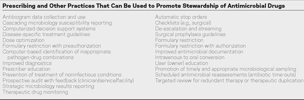
Although veterinarians have practiced some form of stewardship, the overall field of antimicrobial stewardship is in its infancy in veterinary medicine. Fortunately, most aspects of antimicrobial stewardship are not complex, costly, or time consuming. There is no standard approach to antimicrobial stewardship, in part because of a lack of data but also because of the marked differences in needs, threats, and practical aspects between facilities. Some facilities may require few components while others (e.g., larger, tertiary care, greater prevalence of antimicrobial resistant pathogens) may require a more comprehensive program. Examples of potential stewardship practices are outlined in Box 46.4.
■ BOX 46.4
Management of Donor Animals
It is increasingly common to use animals owned by hospitals or veterinary personnel as donors for blood and ingesta (e.g., rumen fluid, equine feces) transfer to patients. Animals used for this purpose should be known to have a very low risk for contagious diseases (e.g., by virtue of their age, clinical status, facility origin), should be housed away from hospitalized patients, should be clinically healthy at the time of specimen recovery, and should be screened periodically for diseases that could be transmitted by use of transfer products. Diseases of concern that might be transferred by blood products from ruminants include bovine leukemia virus, bovine viral diarrhea and border disease viruses, and anaplasmosis; similarly, horses should be screened for equine infectious anemia infection. Bovine ingesta donors should be screened for Johne's disease (Mycobacterium avium subsp. paratuberculosis) and S. enterica, and equine fecal donors should be screened for Salmonella and potentially for other pathogens, such as C. difficile, equine coronavirus, and fecal parasites.