Lameness and Stiffness
RandaH B. Eggleston • John Maas • Carter E. Judy
Lameness is the term used to describe a condition in which an animal is incapable of normal locomotion. Generally, lameness is characterized by an inability to maintain a normal gait, manifested by asymmetry in movement, apparent incoordination or weakness, and inefficient or ineffective motion of the limbs.
Lameness can usually be assessed only when the animal is moving under its own power, although lameness severe enough to cause an inability to bear weight can be assumed at a standstill. The onset of lameness can be acute (e.g., fracture), chronic (e.g., degenerative joint disease), or acute on chronic (e.g., catastrophic fracture secondary to stress fractures).Mechanisms of Lameness and Stiffness
The ultimate effects of any cause of lameness are restricted movement of the limbs or body, reduced performance, and abnormal gait. Causes of lameness are generally associated with conditions of the musculoskeletal system or nervous system. Most causes of lameness have both a musculoskeletal component (e.g., atrophy of the supraspinatus and infraspinatus muscles) and a neurologic component (e.g., suprascapular neurapraxia). Some causes of lameness have only a musculoskeletal component (e.g., upward fixation of the patella) and are not principally associated with either afferent nerve signs (i.e., pain) or efferent nerve signs (i.e., motor dysfunction). Similarly, other causes of lameness are solely related to a motor nerve deficit (e.g., radial neurapraxia).
Unlike the usual definition of lameness, stiffness refers to a generalized restriction in freedom of movement in a limb, the neck, or back. Stiffness is manifested by a limited range of motion by a joint, reduced length of stride, or decreased flexibility during bending or turning. For example, cellulitis and soft tissue swelling in the area of the tarsocrural joint can cause restricted freedom of movement of the hind limb and an apparent lameness, yet there may be no specific musculoskeletal or neurologic cause.
Stiffness may have either congenital or acquired causes, and the clinical signs may be mild and transient or severe and persistent. Stiffness may or may not be associated with pain.Approach to Diagnosis of Lameness and Stiffness in Horses
The lameness examination is the most commonly performed assessment of the musculoskeletal system in the horse. The examination should be well planned, consistent, and thorough. Knowledge of all diseases capable of causing lameness is not required, as long as the examiner maintains an open mind and objectivity during the examination (Box 13.1). The goals of the lameness examination are to determine which limbs are affected, differentiate between supporting limb and swinging limb lameness, and establish the musculoskeletal and/or neurologic components producing the lameness.
1. History. The lameness examination begins with the client interview. A summary of the important historical features of the lameness should include answers to basic questions about the following:
• Onset (e.g., When was the last time the horse was seen sound? Was the lameness acute in onset, or did it have a slow, insidious onset?)
• Characteristics of the lameness (e.g., Is the lameness seen more in hand, at the lunge, or under saddle?)
• Associated or inciting factors (e.g., injury) that may have contributed to or caused the lameness.
• Changes in the characteristics, intensity, and duration of the lameness.
• Responsiveness to treatment (e.g., Has the horse received any type of treatment, and, if so, what was the response?)
• Time since the last hoof trimming and shoeing, and whether or not the horse's shoeing was changed.
• In addition, the signalment and activity that the horse undertakes (e.g., jumping vs. racing) should be ascertained and may be a guide in determining potential causes of the lameness (e.g., stress fractures are more common in racing Thoroughbreds, and osteochondrosis is more commonly diagnosed in young animals).
2. Observe from a distance—stationary phase. Observing the horse from a distance while it is stationary permits an assessment of the horse's conformation, position, and posture. The horse should be viewed from the front, from behind, and from both sides. Special note should be made of any abnormality in the following:
■ BOX 13.1
Causes of Lameness and Stiffness in Horses
Common Causes
Infections of the foot Bruised or punctured sole Hoof wall defects
Fractures
Septic (infectious) arthritis
Laminitis
Secondary (degenerative) joint disease Navicular disease
Osteomyelitis
Fibrotic or ossifying myopathy Rhabdomyopathy (tying up) Sprain
Strain
Tenosynovitis Contracted tendons (flexural deformity)
Ankylosis or arthrogryposis Osteochondrosis or bone cyst Cruciate or meniscal rupture
Luxation or subluxation (dislocations) Upward fixation of the patella (locking patella) Sesamoiditis
Muscle injury, soreness, bruise, trauma, compartment syndrome Subcutaneous abscess, cellulitis
Angular limb deformities Disruption of the suspensory apparatus (broken down) Postanesthetic equine myasthenia
Tendon rupture, damage, tendonitis (bowed tendon) Osteomalacia, osteodystrophy (rickets)
Bucked shins
Epiphysitis (physeal injuries) Purpura hemorrhagica
Less Common Causes
Shivers (shivering) Borreliosis (Lyme disease)
Equine monocytic ehrlichiosis (Potomac fever) Chronic selenium toxicity
Hemangioma, hemangiosarcoma, angiosarcoma Skeletal neoplasia
Rabies Spondylitis, Cliskospondylitis Spinal or vertebral neoplasia
Vertical column malformation
White muscle disease (nutritional myodegeneration) Gunshot injury
Corynebacterium pseudotuberculosis Hypothyroidism (goiter)
Actinobacillosis Hyperparathyroidism Ulcerative lymphangitis
Myotonia congenita
Vesicular stomatitis
Fistulous withers (Brucella abortus or other organisms) Sporadic equine lymphangitis
Acute necrotizing equine vasculitis (with or without thrombocytopenia) Peripheral arteriovenous fistula
Hypertrophic osteopathy or osteodystrophy
Uncommon Causes
Nocardiosis
Cutaneous blastomycosis Pemphigus foliaceus
Tuberculosis
Multisystemic postexhaustion syndrome
Generalized steatitis
Cutaneous vasculitis
Sterile nodular panniculitis Multiple clotting defects in ill foals Salmonellosis
Factor VIII deficiency (hemophilia A) Idiopathic equine aplastic anemia Idiopathic equine thrombocytopenia
Hemimelia (radial, tibial, ulnar hypoplasia, agenesis) Lupus erythematosus (rheumatoid arthritis) Phycomycosis
Bone fragility disorder
Poisons, Toxins, Deficiencies, and Excesses
Moldy sweet clover poisoning Strychnine toxicity
Tetrachlorodibenzodioxin (dioxin) toxicity Warfarin (dicumarol) toxicity
Vitamin K-induced renal toxicity Calcinosis resulting from plant poisoning
Zinc toxicity Phosphorus toxicity Phosphorus deficiency
Vitamin D toxicity
Locoweed-associated limb deformities or stringhalt-like gait Chronic fluoride toxicity
Conformation.
A number of conformational abnormalities have been associated with lameness. For example, upright pastern conformation predisposes to pastern disease and foot lameness; offset or bench knees predispose to carpal disease; and straight through the hocks or post-legged conformation predisposes to upward fixation of the patella, suspensory desmitis, and fetlock disease. Poor conformation can affect the young horse when it is put into training or can cause a slow insidious onset of lameness. Recognizing these conformational abnormalities at the time of examination can be helpful in diagnosing potential causes of lameness. When a horse is evaluated for purchase, recognition of poor conformation should be noted and discussed as a source of future lameness problems.Position of the head (e.g., tilted, turned).
Distribution and equality of muscle mass along the neck and trunk.
Topographic symmetry of the front limbs, from the dorsal region of each scapula to the hoof. From the rear, the height and mass of the hip musculature and the symmetry between the hindlimbs should be assessed. From each side, abnormalities in stance (e.g., camped out in front) or load bearing (e.g., dropped elbow) and the position of the head and neck (e.g., hyperflexed poll) should be compared.
3. Physical examination and palpation. Palpation enables a closer inspection of the horse and identification of abnormalities that may or may not otherwise be noticed. A thorough examination of the musculoskeletal system allows for identification of palpable abnormalities and also offers the opportunity for the practitioner to refine the identification of normal structures; subtle abnormalities cannot be appreciated unless the examiner is skilled at recognizing normal anatomy. There are also many instances in which normal structures palpate abnormally but are not necessarily associated with lameness (e.g., flexor tendon sheath [windpuffs, windgalls] and palmar or plantar metacarpophalangeal joint [MCPJ] pouch effusion [wind puffs]).
The examination should be conducted consistently and thoroughly starting with the cervical neck and concluding at the tail. Abnormal findings should be described to identify their location on the limb, their size, and their orientation relative to normal anatomic landmarks. Palpation of the upper limb is often limited to the overlying muscle mass, with identification of any atrophy, hypertrophy, pain, or fibrosis. Articular structures and surrounding ligamentous structures can be difficult to palpate because of the overlying muscle. Deep palpation of the thoracolumbar and gluteal musculature can provide clues to potential hindlimb lameness and tack or rider issues. The pelvis, iliac arteries, and sublumbar musculature can be evaluated by rectal palpation while the horse is standing quietly; movement or crepitation can be assessed while swaying the horse from side to side.Particular attention should be directed toward palpation of the limbs. The majority of lameness will originate from the carpus distally in the front limb. Common sources of lameness in the hindlimb can be identified from the stifle distally. All palpable structures should be evaluated, including skeletal structures, synovial structures, and soft tissue structures (tendons and ligaments). This portion of the examination can be performed using different methods, by either palpating each tissue in one pass of the limb or making multiple passes of the limb, palpating each tissue structure separately. Regardless of the preferred technique, a consistent and complete examination should be performed. Once the limbs have been palpated in the weight-bearing position, the examiner should palpate them in the non-weight-bearing position. This allows for separation of the soft tissue structures and facilitates deep palpation of the suspensory apparatus. Comparing limbs is often useful for distinguishing an abnormality from an unusual or unique conformation.
The relative size, shape, and condition of the feet (e.g., contracted heel, scuffed toe); length of heel; and pattern of shoe wear (e.g., thinner branch on the outside of the shoe than on the inside) give clinically significant but often overlooked clues to the site and cause of lameness.
Evaluation of the feet with hoof testers is mandatory; most lameness arises from problems in the forefeet.Certain signs indicating trauma (e.g., wounds, swelling, hair loss, pain) may lead to more important findings such as underlying evidence of a fracture (e.g., bony crepitus, warm or cold areas, bony protuberance). Observe from a distance—the mobile phase. Observations made from a distance while the horse is moving can be evaluated critically once clues provided by the history, as well as observations made of any postural deformities, direct the practitioner’s attention to a specific area of the horse's body. This part of the examination is conducted while the horse is moving in at least two gaits, the walk and the trot. Sometimes it is also helpful diagnostically to observe the horse move at other gaits (e.g., canter) or while under saddle. It may also be beneficial to observe the horse on different surfaces to amplify different lameness issues. If possible, the horse should be evaluated under conditions similar to those under which it performs. At a walk, the horse should be observed moving toward and away from the examiner. The breakover point of the foot at the toe, the arc of the foot flight, the distance covered by the foot in the swing phase, and the placement of the foot should be evaluated for each limb and should be compared between pairs of limbs. Although many abnormalities can often be observed only during a trot, some conditions may cause a subtle alteration in gait that can be observed only at a walk (e.g., fibrotic myopathy, stringhalt).
If a fracture is suspected or if there is the possibility of exacerbating preexisting trauma, this part of the examination should either be abbreviated or not performed at all to preclude further damage or trauma. In such cases, immediate
■ BOX 13.2
Causes of Spontaneous Fractures in Horses and Ruminants
Pathologic fractures Subclinical stress fractures Tumors
Infection
Inflammation
Osteoporosis
Copper deficiency Molybdenum excess Phosphorus deficiency Protein deficiency Osteomalacia Osteodystrophy (rickets) Rapid growth
Lactation
Advanced pregnancy Bone fragility disorder
radiographic or other definitive diagnostic tests should be performed (Box 13.2).
Recognizing the asymmetric movement of the head and neck for frontlimb lameness and the asymmetric movement of the pelvis for hindlimb lameness is a common method of lameness identification. Hindlimb lameness issues often present the greatest diagnostic challenge.
Sound horses at a trot show a perfect sinusoidal pattern for all midline body locations, including the head, withers, and tubera sacrale. The height of these structures falls from the beginning of the diagonal stance phase, reaching the lowest position at midstance, and then rising to the highest level at or shortly after the end of the stance phase (suspension). Correlating head and neck movement with the correct frontlimb lameness is relatively easy. It is well recognized that the head is elevated during the stance phase of the lame limb, with an increase in downward motion during the stance phase of the sound limb—“down on sound.” Lameness can also be recognized by changes in the distal limb, including changes in the motion of the MCPJ. During the stance phase, the hyperextension of the MCPJ is decreased with increasing lameness in the lame limb, whereas in the contralateral sound limb an increase is seen. With respect to stride length and foot flight, with forelimb lameness the caudal phase of both the lame and the sound limbs becomes shortened, whereas the cranial phase remains unchanged. In the hindlimbs the opposite is seen; the cranial phase is shortened and the caudal phase remains unchanged. This may be explained by the significantly decreased suspension phase following the lame diagonal. In the forelimbs the arc of the lame front foot is unchanged, but there is an increase in the arc of the sound front foot. In the hindlimbs, the arc of the foot flight in the lame hindlimb is lower than the sound limb in most cases. The change in maximal hoof height during the swing phase appears to be the result of changes in trunk height and is no indication for reduced flexion in the upper joints or an effort to reduce the pain when the hoof lands. Medial (winging) or lateral (paddling) deviation of the distal limb during the flight phase can result in interference and trauma to other limbs and potential lameness. Conformational abnormalities, most commonly toeing in or toeing out, give rise to an alteration in the point of breakover and a change in the flight of the distal limb. Poor foot balance caused by either poor conformation or poor trimming can result in similar flight patterns.
Plaiting describes adduction of the lame limb directly in C C 1 1 1 ∙ 1 ∙ 1 τ 1 C 1'1
front of or lateral to the opposite limb. In the front limbs,
plaiting is commonly the result of faulty conformation, but in the hindlimbs it is more commonly associated with lameness. This pattern of travel is often associated with upper limb lameness but can also be seen with distal hock or proximal metacarpal disease.
A dampening effect also appears to occur as an adaptation to lameness. This effect is more pronounced in the hindlimb than the frontlimb. Flexion of the shoulder and hock joints actually increases during weight bearing in the lame limb. This is probably an increase in the function of the shock-absorbing mechanism. The increased flexion cannot be related to increased loadings but has to be attributed to a gentler braking of the flexion by the extensor muscles. In such a way the loading of the lame limb with the body weight occurs more gradually, reducing the peak forces in the hoof.
The tubera coxae are typically the landmark of choice in evaluating hindlimb lameness. Because the tubera coxae are more laterally located, the pattern is different from that seen in the head. Also, because the hindlimbs lack closely located segments, such as the neck and head, an enhancement of the vertical movements must be found in a rotation of the back around a longitudinal axis. Such a rotation is indicated by different vertical displacements of one tuber coxae during both stance phases. The vertical movement of the tuber coxae exhibits a characteristic pattern of a double-waved, slightly asymmetric line during one stride. The lowest point of the hip is reached in the middle of the stance phase of the contralateral limb. The highest point of the hip is reached shortly after the stance phase of the contralateral limb.
Kinematic studies have more clearly defined the notion of “hip hike” and “hip drop” and have recorded regular patterns of pelvic movement in lame horses. Consistent findings in the overall pelvic movement in the lame horse include less downward movement during the midstance phase and less upward movement at the end of and after the stance phase of the lame limb. This can give the appearance of an overall pelvic elevation during the stance phase of the lame limb as compared with pelvic height during stance of the sound limb; a similar exaggerated pattern is seen in the tubera coxae. The tubera coxae also exhibit less downward movement during the midstance phase and less upward movement at the end of the stance phase in the lame limb. More notably, there is more downward movement during midstance of the sound limb (midflight of the lame limb) and more upward movement at the end of stance of the sound limb (impact of the lame limb), giving rise to the notion of a “hip hike.” These changes result in an increase in the overall vertical movement of the tuber coxae on the lame side as compared with the sound side. Clinically, many find it easier to identify the exaggerated excursion of a tuber coxae to identify the side of the lameness.
Lateral movement or drifting of the hind end can also be seen in horses with unilateral hind limb lameness. Horses tend to drift or move away from the side of the lameness. Subtle lameness with an absence of asymmetric pelvic movement may present with a consistent drifting to one side or the other.
Thorough and useful systems for grading the severity of lameness are available. Most systems are designed to enable the practitioner to compare how lameness changes with time, assess the characteristic of lameness among horses, and accurately record information and communicate information to other veterinarians. Simple and consistent schemes that are easy to remember and modify can be developed (Table 13.1).
Once the initial standing and mobile examinations are completed, the affected limb is identified, and the lameness is graded, identification of the specific region of the limb is the next goal. Manipulative tests or stressing of articulations and associated soft tissue structures can provide additional information as to the location of the source of lameness. Flexion and extension tests are designed to stress selective regions of the
■ TABLE 13.1
Five-Grade Lameness Scheme
Grade Description
1 An inconsistently observable lameness visible
under special circumstances (e.g., in a circle, flexion tests, hard surface)
2 A consistently observable lameness visible only
under special circumstances (e.g., in a circle, flexion test, hard surface)
3 A consistently observable lameness at a trot in a
straight line
4 A consistently observable lameness at a walk
5 A non-weight-bearing lameness; horse is unable
to use the leg
Modified from the Am Assoc Equine Pract Newsl, March 12, 1983.
limb and observe the effects of the manipulation on the lameness. These tests are also commonly performed on the sound horse to reveal potential areas of concern, particularly during prepurchase examinations. Flexion and extension manipulations also enable an assessment of range of motion. Interpretation of these tests should be approached with caution. They are seldom specific for one particular joint. For example, the fetlock flexion test not only stresses the fetlock joint but also places stress on the proximal and distal interphalangeal joints; the hock flexion test also flexes and stresses the stifle joint because of the presence of the stay apparatus. If a flexion test results in a positive response, the horse should be walked out of the response and observed before additional manipulations. Occasionally exacerbation of the lameness will persist for an extended period of time, which changes the baseline lameness and clouds the interpretation of additional manipulations.
It is common for horses to be presented with multiple lameness issues. Secondary lameness or compensatory lameness is the result of increased stress or overloading of the other limbs in response to the primary lameness. This most commonly occurs in the contralateral limb but can also occur between frontlimbs and hindlimbs. The secondary lameness can also be the result of shifts in body mass that produce an apparent or phantom lameness. Phantom lameness is less severe than the primary lameness. The following guidelines can be used to aid in the differentiation between a real or compensatory and an apparent or phantom lameness.
• Address the most severe lameness first.
• Horses with primary hindlimb lameness and apparent or phantom contralateral frontlimb lameness: Each lameness should be considered as real.
• Horses with a primary forelimb lameness and apparent or phantom ipsilateral hindlimb lameness: Each lameness should be considered as real.
• Primary forelimb lameness may produce asymmetric pelvic movement causing the perception of a contralateral hindlimb lameness. Example: left foreleg lameness (head elevation) causing apparent or phantom right hindleg lameness (hip drop).
• Horses with a primary forelimb lameness and apparent contralateral hindlimb lameness: Isolate out the front limb lameness first.
• Primary hindlimb lameness (>3 to 5/5) can mimic ipsilateral forelimb lameness. Example: A horse shows a cranial load shift during the stance phase of the lame limb that causes the head and neck to shift forward and nod down, giving the perception of ipsilateral forelimb lameness—“down on sound.”
• Horses with a primary hindlimb lameness and apparent ipsilateral forelimb lameness: Isolate the hindlimb lameness first.
■ TABLE 13.2
Structures Desensitized by Commonly Performed Nerve Blocks
| Nerve Block | Nerve(s) Affected | Structures Desensitized |
| Palmar (plantar) digital Abaxial sesamoid Low palmar (volar) High palmar (volar) High 2-point | Palmar (plantar) digital Palmar (plantar) Palmar, palmar metacarpalb Palmar, palmar metacarpalb Lateral palmar, medial palmar | Heel bulbs; frog; bars; navicular bone and bursa; palmar regions of the third phalanx, distal interphalangeal joint, sole, and soft tissues Coronary band, interphalangeal joints, lamellar and solar corium Skin of medial and lateral pastern, metacarpophalangeal joint, proximal sesamoids, flexor tendons, tendon sheath Skin and deep structures of palmar cannon region (flexor tendons, suspensory ligament except origin, interosseous ligaments of splint bones) Origin of suspensory ligament |
aIncludes all structures listed up to and including the particular block; first structure listed in each block: is also the area that can be tested with point pressure to evaluate the effectiveness of the block.
bFor hindlimbs, additional anesthetic (i.e., ring block) is necessary at the level of the particular perineural block to achieve the desired effect.
Assumptions as to the cause of a horse's lameness based solely on the physical examination and visual inspection should be avoided unless obvious signs are present, such as severe swelling or crepitus. After the physical and visual examination, evaluation of the horse with perineural analgesia is mandatory for accurate isolation and diagnosis. The clinician must possess a thorough knowledge of anatomy and the structures desensitized by blockade of the appropriate peripheral nerves or synovial structures (Table 13.2). When performing perineural analgesia, it is important to remember to block from distal to proximal. Perineural anesthesia of proximal structures first may inadvertently anesthetize more distal pathology, resulting in misinterpretation of the region of pain affected. An improvement in gait indicates a favorable response to a nerve or joint anesthesia; complete elimination of gait asymmetry is unusual and generally should not be expected after intraarticular or peripheral nerve analgesia. If necessary, improvement in gait can be confirmed by repeating the successful block the next day. By that time residual effects from multiple blocks performed previously should be absent.
Common local anesthetics used in horses include solutions of lidocaine, mepivacaine, and bupivacaine. These solutions all share a common mechanism of action, specifically the ability to block or inhibit nociceptive nerve conduction by preventing the increase in membrane permeability to sodium ions. Lidocaine and mepivacaine are considered to be fast acting and have a duration of action of 12 to 3 hours and 2 to 3 hours, respectively. Bupivacaine, on the other hand, is intermediate in onset and has a much longer duration of action of 3 to 6 hours. Mepivacaine is reportedly less irritating to tissues than lidocaine.
Intrasynovial analgesia can be used to more specifically isolate lameness to a joint, tendon sheath, or bursa. It can be used in combination with perineural analgesia or alone depending on the suspected source of the lameness. Proper patient restraint and strict aseptic technique, including aseptic preparation of the skin, wearing sterile gloves, and use of a new bottle of anesthetic, are imperative to avoid iatrogenic synovial sepsis. Lameness may be erroneously associated with a joint if intraarticular analgesia of several joints is performed within a short period of time; ample time (30 to 60 minutes) must be allowed between joint blocks to allow for adequate articular desensitization. Some intraarticular and intrasynovial anesthetic techniques may mimic the results of perineural anesthesia. An example is that of the coffin joint, where intraarticular anesthesia of the joint has the same regions of anesthesia as that of the palmar digital nerve block. It is important not to overinterpret the results in such cases.
When performing intrasynovial analgesia, it is not necessary to follow the distal-to-proximal rule. If intraarticular analgesia of a proximal joint results in no improvement in the lameness, immediate follow-up with distal limb perineural blocks is still possible. Exceptions to this rule exist with intrasynovial analgesia to the foot. When performing intrasynovial analgesia of the distal interphalangeal joint (DIPJ) or navicular bursa, it is important to take into consideration the volume of anesthetic used and the timing at which the lameness is reevaluated. The recommended volume of anesthetic for the DIPJ is 4 to 5 mL, and for the navicular bursa 3 to 4 mL. Once injections into these structures have been performed, the horse should be evaluated at 5-minute intervals to help with the interpretation of the response to the block because they may result in inadvertent anesthesia similar to the palmar digital nerve block.
Significant improvement in experimentally induced lameness to the navicular bursa can be seen at 5 minutes after intraarticular anesthesia of the DIPJ with 5 mL of 2% mepivacaine hydrochloride. Amelioration of bursal lameness is most likely caused by diffusion of the anesthetic into the bursa via an indirect or functional communication, or by diffusion of anesthetic into the periarticular tissues. The proximal palmar pouch of the DIPJ lies in close proximity to the palmar digital (PD) neurovascular bundles as they course along the medial aspects of the collateral cartilages, making it possible for anesthetic diffusion to block nerve conduction at that level.
Experimentally induced solar toe pain can also be ameliorated by intraarticular blockade of the DIPJ with 10 mL of 2% mepivacaine hydrochloride. The structures innervated by the deep branch of the PD nerves include the DIPJ, navicular bursa, distal navicular ligament, laminar corium, and corium of the sole. The DIPJ capsule contacts the PD neurovascular bundle, and a local anesthetic injected into the DIPJ likely desensitizes the PD nerves below the level of the coronary band, as well as the structures innervated by them.
Variable responses are also seen with blockade of the DIPJ when different volumes of anesthetic are used. Blocking the DIPJ with 6 mL of 2% mepivacaine results in significant improvement in lameness originating from the dorsal margin of the sole; however, lameness originating from the palmar sole shows no improvement. Using 10 mL of mepivacaine reduces lameness originating from the dorsal margin of the sole, as well as the palmar heel regions of the sole, but only after 30 minutes. The difference in response to analgesia of the DIPJ in attenuating pain at the dorsal margin of the sole versus the angles of the sole may be because these regions are innervated by different branches of the PD nerve. This may help distinguish between pain arising from the DIPJ or the navicular apparatus and palmar solar pain.
In contrast to the responses seen with blocking the DIPJ in the presence of navicular bursa disease, blocking the navicular bursa with 3.5 mL of mepivacaine hydrochloride in the presence of experimentally induced DIPJ lameness results in a significant improvement in lameness but only after 30 minutes. Experimentally induced lameness from the dorsal sole is improved by blockade of the navicular bursa; lameness originating from the palmar sole does not show significant improvement.
Knowledge of the previously described responses to intra- synovial analgesia of the DIPJ and the navicular bursa is helpful in localizing and interpreting lameness commonly seen in the horse. Similar responses can be encountered with intraarticular analgesia of the carpus and distal tarsal joints. Instillation of anesthetic into the middle carpal joint and the tarsometatarsal joints can result in desensitization of the proximal suspensory ligament, a common site for soft tissue injury and lameness in the horse.
Recently, objective methods of lameness evaluation have become prevalent in equine veterinary practice. The development of a body-mounted inertial sensor system using accelerometers and gyroscopes to measure acceleration and angular velocity of the head and torso during locomotion has been shown to facilitate lameness evaluation. Results of a recent study indicated that lameness evaluation of horses obtained by use of a body-mounted inertial sensor system was significantly associated with results obtained via subjective lameness evaluation by expert clinicians. The objective measurements provided by this system may be beneficial for evaluation of horses with mild, intermittent lameness; difficult, multi-limb involvement; and for documentation of response to regional or intraarticular anesthetic blocks.
Once the lameness has been described and localized, a radiographic or ultrasonographic examination can be performed as the next step to confirm a clinical diagnosis. Radiography should be performed using proper technique, an ideal film/ screen combination, and multiple views to construct a thorough study (Table 13.3). Comparing radiographs of affected and unaffected limbs can help confirm or refute a suspected abnormality, evaluate the severity of the disease, and identify possible bilateral limb involvement.
Although standard radiographic techniques are well documented and described, ultrasound examination has become a
■ TABLE 13.3
Recommended Radiographic Views of Extremities
| Radiographic Series | Minimum Radiographic Views |
| Distal extremity | 45 degrees DP, 65 degrees DP (2), |
| (navicular) | LM, flexor tangentiala |
| Pastern | 45 degrees DP, LO, MO, LM |
| Fetlock | 45 degrees DP, LO, MO, LM, flexed LM |
| Metacarpal or | DP, LO, MO, LM |
| metatarsal | |
| Carpus | DP, LO, MO, LM, flexed LM, flexed skylines (distal radius, proximal and distal rows of carpal bones) |
| Tarsus | 0 degrees DP, 10 degrees DP, LO, MO, LM |
| Radius-ulna or | Cr-Cd, LO, MO, LM |
| tibia-fibula | |
| Elbow | Cd-Cr, LO, LM |
| Shoulder | ML |
| Stifle | Cd-Cr, LM, flexed LM, Cd 30° L-CMO, patellar skyline |
aView to highlight the flexor cortical margin of the navicular bone (50 degrees proximal palmarodistal oblique).
Cd-Cr, Caudocranial; Cd 30o L-CMO, caudal 30-degree lateral-craniomedial oblique; Cr-Cd, craniocaudal; DP, dorsopalmar (dorsoplantar); LM, lateromedial; LO, lateral oblique; ML, mediolateral; MO, medial oblique.
mainstay in musculoskeletal imaging. Indications for ultrasonographic evaluation of a lameness include diagnosis of soft tissue injuries, including muscular, vascular, tendon, tendon sheath, ligament, joint capsule, or bursal defects; evaluation of articular surfaces (articular cartilage thickness, osteochondritis dissecans lesions); assessment of fluid accumulation (synovial effusions, seromas, or infection); evaluation of bony surfaces; monitoring of the progression of healing; and monitoring of the effects of training on soft tissue injuries such as tendonitis or desmitis.
When radiographic or ultrasonographic techniques are nondiagnostic, other methods such as thermography, nuclear scintigraphy, treadmill evaluation, computerized videographic gait analysis, force plate evaluation, body-mounted inertial sensors examination, computed axial tomography (CAT), or magnetic resonance imaging (MRI) may be useful.
Approach to Diagnosis of Stiffness, Lameness, and Abnormal Gait and Posture in Ruminants
Sarel R. Van Amstel • Jan K. Shearer
Clinical signs can be caused by neurologic deficits, pain, and muscular or skeletal disorders. Diagnosis requires a complete history, including review of information on lameness in dairy records; careful observation during standing and walking, including locomotion scoring; and examination and palpation of the foot and upper limb.
History
Historical details should be obtained in terms of duration, severity, onset of clinical signs, and previous treatment. Past history may help establish if the condition is acquired or congenital. This is particularly important in order to distinguish among conditions with similar signs such as spastic paresis, upward patella fixation, and spastic syndrome. History should also include information regarding management and housing such as free stall size and bedding; evidence of overcrowding; types of walking surfaces; number of times a day milking; and accumulation of mud, manure, gravel, and stones on tracks in cases of pastured cattle. Finally, historical information regarding nutrition including the use of supplements, water source, and potential toxin exposure should be included.
Observation
Detection of lameness may present a challenge because cows are adept at disguising discomfort. Therefore careful observation both during standing and walking is important. Changes in weight bearing, posture, and conformation may be indicative of both the presence and type of lameness. Nearly 90% of lameness involves the foot; thus particular attention should be paid to alterations in weight bearing between claws. Observation is best conducted on a flat, hard surface because subtle lameness may not show when the animal is walking on a softer earthen surface. Stiffness and rigidity may be major components of altered gait and posture. Common causes in which stiffness may be an important component are shown in Box 13.3.
Palpation
For upper leg conditions, hands-on examination includes palpation during standing and walking, as well as manipulation of the leg with the animal in lateral recumbency. For examination of the hip, the examiner attempts to detect crepitation in the joint by placing both hands over the greater trochanter while an assistant abducts, adducts, and rotates the affected limb. For the stifle, examination in the standing animal involves palpation of the joint for fluid effusion and stability. Stability of the joint can be assessed in the following ways: (1) Standing
■ BOX 13.3
Alterations in Gait in Ruminants in Which Stiffness Can Be a Major Component
Chronic hypophosphatemia
Chronic fluorosis
Tetanus
Lupine alkaloid intoxication
Copper deficiency or molybdenosis
Acute laminitis
Nutritional myodegeneration (white muscle disease) Locoweed toxicity
Sweet clover poisoning
Rickets, osteomalacia, osteodystrophy
Bovine virus diarrhea (coronitis and cerebellar atrophy) (B) Sarcocystis
Vesicular stomatitis (B)
Overgrown claws
Bluetongue (coronitis) (O)
Degenerative joint disease
Muscle trauma
Ionophore toxicity
Hemlock poisoning
Generalized calcinosis
Fescue intoxication (fescue foot)
Frostbite
Contracted tendons
Selenium toxicosis
Clostridial myositis
Claviceps spp. mycotoxicosis (ergotism; Bahia grass and Dallis grass staggers)
Mycoplasma bovis polyarthritis and tenosynovitis (B)
Mycoplasma mycoides subsp. mycoides polyarthritis (C) Caprine arthritis encephalitis virus (CAE) (C)
Muscle abscess
Ruptured anterior cruciate ligament or torn collateral ligament of stifle
Luxations and subluxations
Upward fixation of the patella
Epiphysitis
behind the animal, the examiner reaches around the leg with both hands locking the fingers over the tibial crest while leaning into the back of the thigh. The leg is stabilized against that of the examiner while the tibia is pulled caudally (Video 13.1). (2) Alternatively, the examiner stands in front of the affected leg and attempts to demonstrate laxity by pushing on the tibial crest. It is important to stabilize the foot during this procedure. Distention of the joint capsule can cause swelling between the patellar ligaments despite the presence of the fat pad (see Video 13.1). One should also note that there are extensions of the joint capsule between the quadriceps femoris muscle and the femur and distally around the tendons of the peroneus tertius and the long digital extensor muscle.
When evaluating the tarsus or hock, one should always assess the degree of flexion possible in this joint. Overflexion of the hock indicates gastrocnemius rupture, whereas overextension may be indicative of rupture of the peroneus tertius.
Swelling of joints and legs should be palpated for heat, pain, and consistency. Peritarsal bursitis causes a soft, nonpainful swelling on the lateral side of the hock, whereas a hard, painful swelling may be associated with septic arthritis or degenerative joint changes. In cases of septic arthritis, a discharging tract may also be present.
In some cases it may be difficult to distinguish upper from lower leg problems. This can be done by placing a tourniquet at or slightly above the level of the fetlock joint followed by an intravenous injection of 20 mL of 2% lidocaine into a vein
below the tourniquet (Video 13.2). Remove the tourniquet after a few minutes or so, and immediately allow the animal to walk. In cases of an upper leg problem, the lameness will persist. Pain and lameness associated with upper leg problems tend to become worse after a period of flexion.
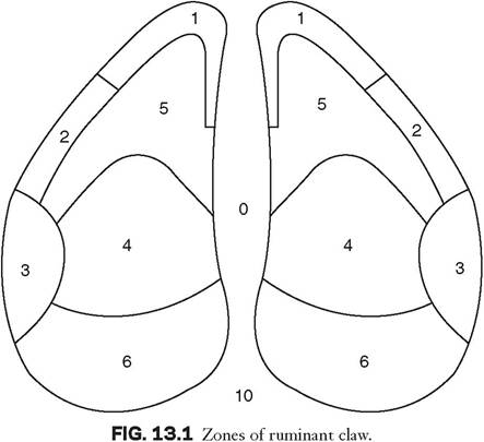
Foot and claw conditions are diagnosed by visual appearance after appropriate restraint. The claw including the interdigital space is thoroughly cleaned. Claw horn and foot lesions should be identified, taking into consideration the location and the claw zone affected (Fig. 13.1). The presence of pain should be investigated if no lesions are visible. This can be done with a hoof tester. Important areas in which to apply pressure include the sole ulcer site in the axial region of zone 4 and the toe ulcer site in zone 5. Sole hemorrhages occurring in zone 4 should also be tested for pain.
The skin of the interdigital space and interdigital cleft (zones 0 and 10, respectively) should also be carefully examined for evidence of fissures or lesions indicative of digital dermatitis.