Liver
Hepatic cytology can be classified into normal, hyperplastic, neoplastic, inflammatory, degenerative / metabolic, and cholestatic abnormalities; extramedullary hematopoiesis; and mixed results.4,6,7
1.7.3.1 Normal liver cells
Normal liver cells show only slight anisocytosis.
The cytoplasm often contains fine granules. The nuclear: cytoplasmic ratio ranges from 1:4 to 1:5. A nucleolus is clearly identifiable within the nucleus. Some normal liver cells may contain two nuclei. Bile duct epithelium and mesothelial cells can also be found in some liver FNA specimens. Leucocytes found on FNA cytology may originate from blood contamination and do not necessarily indicate infiltration of the liver with inflammatory cells. In unclear cases, the leucocyte:erythrocyte ratio should be compared between peripheral blood and the FNA cytology specimen. It should also be noted that FNA cytology can be contaminated with neutrophilic granulocytes originating from a purulent peritonitis, and leading to a false interpretation of liver cytology.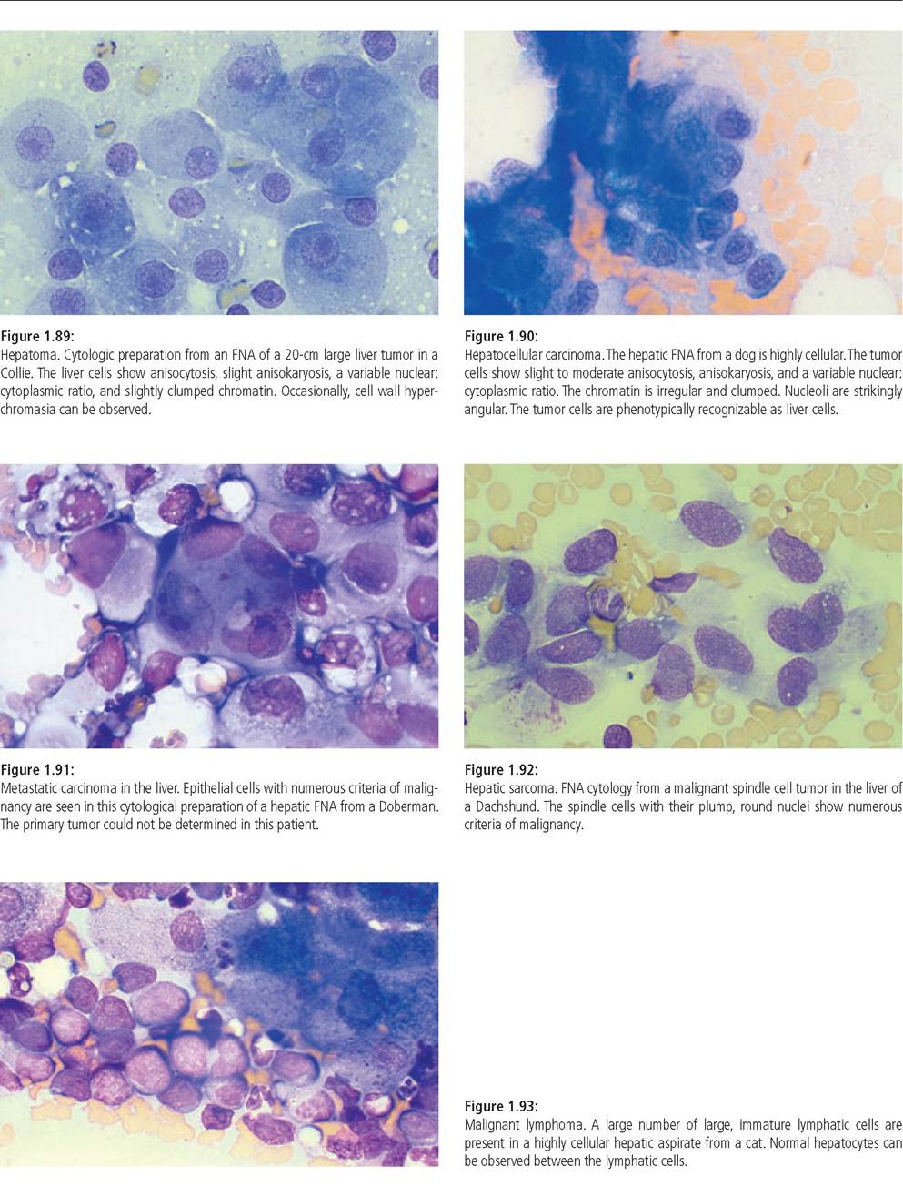
1.7.3.2 Hyperplasia
Hyperplastic areas contain only few pleomorphic cells. It can be difficult to distinguish hyperplastic from normal liver cells or from cells from a well-differentiated hepatocellular carcinoma.
1.7.3.3 Inflammation
The different types of inflammation are classified based on the predominant leucocyte population. An increased number of neutrophilic granulocytes may be caused by suppurative hepatitis. Bacterial infection is a common cause and bacteria can be found on microscopic examination in some cases. Bacterial toxins will often cause degeneration of the neutrophil granulocytes.
Also, in many cases bacteria can no longer be identified after treatment with antibiotics has been initiated. In these cases, fewer degenerated neutrophils are often observed.Lymphoplasmacytic inflammation is characterized by the presence of small lymphocytes and plasma cells. Subtle indications for hepatitis are the presence of intra- or extracellular bile pigment, vacuolar degeneration, and hyperplasia or proliferation of the bile duct epithelium. Other indications for hepatitis include signs of fibrosis (presence of a pink, proteinrich substance that is infrequently infiltrated with fibrocytes), and signs of hepatocellular regeneration.
Eosinophilic granulocytes can be nonspecific and can be observed during inflammation with various other predominant cell types or they can represent primary eosinophilic inflammation as would occur during eosinophilic enteritis, systemic mast cell tumors, or liver fluke infestation in cats (Amphimerus pseudofelineus) in some states of the USA.
1.7.3.4 Neoplasia
Malignant tumors can be classified either as primary (hepatocellular carcinoma or bile duct carcinoma), or secondary (malignant lymphoma, metastasis of sarcoma or carcinoma, mast cell tumor, malignant histiocytosis, and myeloproliferative disease). The most common hepatic tumors in the dog are hepatocellular carcinomas, malignant lymphomas, sarcomas, and undifferentiated carcinomas. The most common hepatic tumors found in cats are bile duct carcinomas and malignant lymphomas (Figures 1.89-1.93).4,6,7
1.7.3.5 Other abnormalities of the liver
Extracellular hematopoiesis is characterized by the presence of precursor cells and various maturation stages of hematopoietic cells (Figure 1.94). Metabolic and degenerative abnormalities include hepatic lipidosis and steroid-induced hepatopathy. Hepatic lipidosis occurs mainly in cats and is characterized by round cytoplasmic vacuoles (Figure 1.95). Steroid-induced hepatopathy, which predominantly occurs in dogs, is characterized by a moth-eaten appearance, which starts at the cell edges and is due to glycogen incorporation (Figure 1.96).
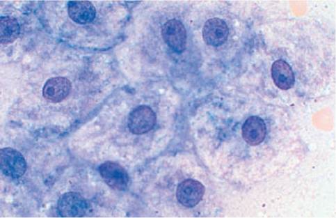
Figure 1.96:
Steroid-induced hepatopathy.
Hepatocytes from this dog have a moth-eaten appearance, which is more severe at the cell edges but extends towards the nuclei. These abnormalities occur irregularly. In some areas of the smear, they occur frequently while in other areas they are absent. These changes in the cytoplasm are typically seen with steroid-induced glycogen incorporation, but can also be caused by ischemia or hepatic toxins. A specific characterization of glycogen storage could be achieved using PAS stain.
Figure 1.97:
Bile pigment cholestasis. Intra- and extracellular bile pigment can be seen as a black-green pigment in this cat with a cholestatic disorder.
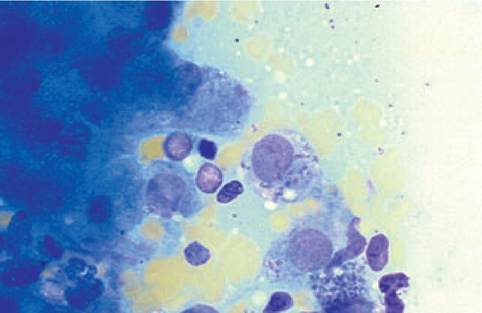
Figure 1.98:
Leishmaniasis. Macrophages in the liver of a dog, also known as Kupffer cells, have phagocytized parasites. Leishmania have a bright cytoplasm, an oval-shaped nucleus, and a small dark kinetoplast.
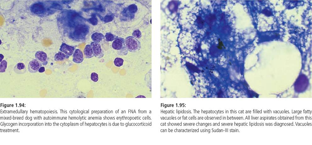
Necrosis is characterized by vacuolization of the cytoplasm and nuclear degeneration and fragmentation. Different types of pigments may also be seen, either dark green bile pigment (cholestasis, hepatitides), gold-brown hemosiderin, or lightgreen copper (mainly in Bedlington Terriers). Lipofuscin granules, a normal finding in hepatocytes of older cats, should not be confused with intra-cytoplasmic bile pigment (Figure 1.97). Differentiation of various cytoplasmic abnormalities may be achieved using a variety of different stains (Table 1.14).4,8 Due to the fact that hepatic cirrhosis is characterized by structural changes (lobular structure, fibrosis, and areas of reconstruction and regeneration), this condition can not be diagnosed based on liver cytology.9,10 Occasionally microorganisms can be observed on FNA cytology (Figure 1.98).
The diagnostic accuracy of hepatic cytology depends considerably on the experience of the observer.
Table 1.15 lists different disorders that are easy to diagnose even for the less experienced observer. Considerable experience is necessary to diagnose and differentiate the various types of inflammation or a well-differentiated hepatocellular carcinoma.1,2,4Table 1.14: Special stains used for differentiation of hepatic pigments4,8
| Pigment | Stain |
| Copper | Rubeanic acid |
| Fat | Sudan-III stain |
| Glycogen | PAS stain |
| Hemosiderin | Prussian blue stain |
| Lipofuscin | Luxol blue stain |
Table 1.15: Disorders that can easily be diagnosed based on FNA cytology1-2-4-6-7
| Diagnosis Steroid-induced hepatopathy | Cytologic features Moth-eaten appearance of hepatocytes | Possible problems |
| Hepatic lipidosis | Vacuolar cytoplasmic abnormalities | |
| Lymphoma of liver and pancreas; also in patients with circumscribed lesions of the intestinal wall | Frequently approximately 50% lymphoblasts; occasionally as few as 5% lymphoblasts | It can be difficult to distinguish lymphoma from lymphocytic cholangiohepatitis |
| Mast cell tumor | Mast cells | Presence of well differentiated mast cells may lead to formation of cell groups |
| Metastatic tumors | Epithelial or mesenchymal cells with criteria of malignancy surrounded by normal hepatocytes | Can be confused with mesothelial cells and bile duct epithelial cells |
| Suppurative hepatitis | Mostly degenerated neutrophils, in some cases with intra- and extracellular bacteria |
1.7.3.2 Bile
Bile can be obtained by FNA und subsequently evaluated under the microscope. Neutrophilic granulocytes or bacteria present in bile are evidence for an inflammatory or infectious condition, respectively.
1.7.4