Pancreas
Cytology of the pancreas, obtained either as FNA under sonographic guidance or intraoperatively using impression smears or scrapings, may yield a rapid diagnosis while the patient is still under anesthesia.
The risk for complications is minimal with these procedures. However, FNA under sonographic guidance requires experience.An indication for pancreatic FNA cytology is the evaluation of unexplained pancreatic enlargements observed during ultrasonographic examination. Neoplastic and cystic abnormalities need to be distinguished from an inflammatory process. Inflammation is characterized by the presence of numerous neutrophils, which frequently are degenerated, macrophages, and necrotic material. Inflammation also leads to slight dysplasia of pancreatic cells (Figure 1.99). However, it should be noted that pancreatitis is frequently localized, and a negative result of a single FNA does not rule out the presence of pancreatitis.
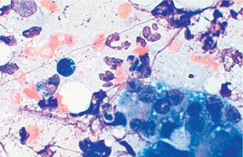
Figure 1.99:
Pancreatitis. This cytological preparation of an FNA from a pancreas of a Dachshund shows large numbers of mostly degenerated neutrophilic granulocytes, intermixed with some macrophages and lymphocytes. The pancreatic acinar cells exhibit mild dysplastic changes (slight anisocytosis, anisokaryosis, and vacuolization). Observed fatty vacuoles are due to steatitis and lipolysis. In some cases, lipophages can be seen (not on this slide).
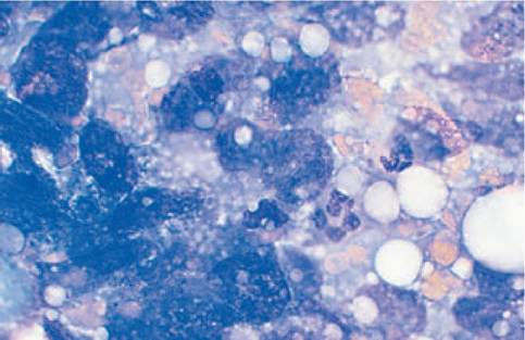
Figure 1.100:
Pancreatic adenocarcinoma. The FNA cytology of a pancreatic mass shows numerous epithelial cells with various criteria of malignancy (severe anisocytosis, anisokaryosis, macrocaryosis, variable nuclear:cytoplasmic ratio, and atypical nuclei and nucleoli). Only few inflammatory cells can be observed in the background.
Malignant neoplasia is characterized by the presence of so- called criteria of malignancy: anisokaryosis, pleomorphic cells, and excessive cellularity. A poorly differentiated pancreatic carcinoma is easy to diagnose (Figure 1.100). However, malignant tumors lead to extensive secondary inflammation, which makes the diagnostic differentiation of pancreatitis and pancreatic carcinoma by cytology extremely difficult, especially in cases where the pancreatic cells exhibit only a slight dysplasia with concurrent inflammation. In addition to primary tumors, metastatic tumors can also be diagnosed in the pancreas based on FNA cytology. Cysts und pseudocysts are characterized by the presence of encapsulated fluid that contains only few cells.5
1.7.5