Liver Flukes in Ruminants
John B. Malone
Etiology The common liver fluke, F. hepatica, causes a disease of production in ruminants that mimics the production effects and clinical appearance of the GI nematode-parasite complex.
The disease often has its maximum economic effect in late fall and winter when animals are most likely to be under seasonal nutritional stress. F. hepatica is unique among the common helminths of ruminants in that it has an asexual multiplication phase of the life cycle in snail intermediate hosts that is highly sensitive to environmental conditions. F. hepatica is well known for its exponential propagation of infective stages under favorable conditions, sometimes leading to explosive seasonal outbreaks of severe parasitism, especially in sheep. In addition, liver parenchyma migration forms leave necrotic tracts in their wake that are a primary predisposing factor in some areas for acute fatalities caused by C. novyi types B (black■ TABLE 33.5
Hepatotoxic Plants
| Common Name (Botanical Name) | Approximate Ld50 (%Bw)a | Geographic Distribution | Liver Lesion |
| For pyrrolizidine alkaloid- containing plants, see Table 33.4. | |||
| Lantana (Lantana camara) | 1 | Northern North America | Necrosis, canalicular collapse |
| Littleleaf horsebrush | 0.5 | Dry desert | Necrosis |
| (Tetradymia glabrata) | |||
| Sacahuista or bear grass (Nolina | 1.1 | Southwestern U.S. | Fatty and centrilobular necrosis |
| texana) | |||
| Lechuguilla (Agave lechuguilla) | 4-15 | Southern U.S., Mexico | Necrosis, photosensitivity |
| Bindii or puncture vine (Tribulus | Warmer regions | Biliary fibrosis, necrosis (big head) | |
| terrestris) | |||
| Whitebrush (Aloysia gratissima) | Southern U.S., Mexico | Fatty degeneration | |
| Panic grasses (Panicum spp.) | Texas-California, South America | Biliary fibrosis, necrosis | |
| Kleingrass (Panicum coloratum) | Southern U.S. | Necrosis, obstructed bile ducts | |
| Scutch grass or Bermuda grass | North America | ? | |
| (Cynodon dactylon) | |||
| Cocklebur (Xanthium orientale) | 0.75-3 | North America | Hemorrhagic centrilobular |
| Lupine (Lupinus spp.) | Varies | Western North America | necrosis Necrosis |
| Mycotoxin from Phoronopsis inferior | |||
| Cottonseed (Gossypium spp.) | Varies; gossypol content | Southern U.S. | Necrosis |
| Gossypol pigment Poisonous mushrooms (Amanita | Few mushrooms | North America | Cardiac lesions Necrosis, central nervous system |
| phalloides, Galerina venenata) Blue-green algae (Microcystis | ? 0.001 | Ponds, worldwide | signs Necrosis, dissociation |
| aeruginosa, Nodularia spumigena) | |||
| Alsike clover (Trifolium hybridum) | Varies | Cultivated | Portal fibrosis, biliary hyperplasia |
| Mexican fireweed (Kochia scoparia) | Southwest U.S. | ? | |
| Coffee senna (Cassia occidentalis) | 0.05 (horse); 0.5-2 | North and South America, | Pericentrolobular vacuolar |
| Cycad palm (Cycas/Zamia spp.) | (cattle) | Africa, Asia and Australia South America, Australia | degeneration and necrosis ? |
| Yellow-wood or rosewood | Australia, Africa | Centrilobular necrosis | |
| (Terminalia oblongata) | |||
| Sneezeweed (Helenium spp.) | 0.25 | Northern U.S., Canada | |
| Moldy alfalfa (Medicago sativa) | ? | Wet areas | Biliary hyperplasia, necrosis |
| Bitterweed (Hymenoxys spp.) | 1 | Southwestern U.S. | |
aLethal dose (as percentage of body weight).
?, Unknown; Bw, body weight; Ld50, median lethal dose; U.S., United States.
disease) and haemolyticum (bacillary hemoglobinuria), discussed earlier in the chapter.1
The geographic distribution of F. hepatica in the United States is limited mainly to areas in the south-central states and Florida and to the Pacific Northwest, where neutral well- buffered soils are found and local hydrologic conditions provide suitable habitats for lymnaeid snail intermediate hosts. The important vector species in temperate climates, recently renamed based on molecular evidence as Galba spp. or Lymnaea spp.,2 are semiaquatic “mud” snails found in disturbed mud banks and hoofprints in shallow depressions in fields, drainage channels, and temporary water bodies in alluvial river basins, bottomlands, and coastal regions of the southeastern United States. Springs, small streams, seeps, and sloughs serve as habitats in areas of greater terrain relief in the western states. Most infections occur by grazing in and around water in fluctuating habitats that stay wet for more than half the year.
The life cycle development of both the snail host and the parasite in the snail occurs only when temperatures are above 10° C (50° F) and snail habitats are wet. This leads to a distinct seasonal transmission in the mild, wet winter and spring of the south-central states and Florida and a late spring to fall transmission in the cooler climates of the western states (Figs. 33.3 and 33.4). It takes a minimum of 42 days at 25° C (77° F) or the accumulation of approximately 600 growingdegree days to complete intramolluscan development, with release of cercariae that encyst as infective metacercariae on pasture vegetation (600 growing-degree days = 25° C minus the base temperature of 10° C = 15 growing-degree days ? 42 days; an equivalent value for the life cycle is 15° C or 5 growing-degree days ? 120 days). Using soil water budget analysis to indicate the length of time habitats are wet and the accumulated sum of growing-degree days over 600, a climate forecast index can be calculated for use in describing the pattern of seasonal transmission at a given site and to provide an indicator of the risk of economic losses in a given climate year.
Two weeks of sustained summer heat and drought ends the transmission season by killing pasture metacercariae and forcing snail populations to estivate in soil or perish. In the cooler climates of the western United States, the season ends with drying of springs, seeps, sloughs, and other habitats or with the onset of sustained cold winter weather. Variation in the annual climate alone may lead to a hundredfold variation in fluke burdens in different climate years.3The amount of snail habitat present on individual farms related to pasture wetness conditions, such as premises with
■ TABLE 33.6
Hepatotoxic Chemicals
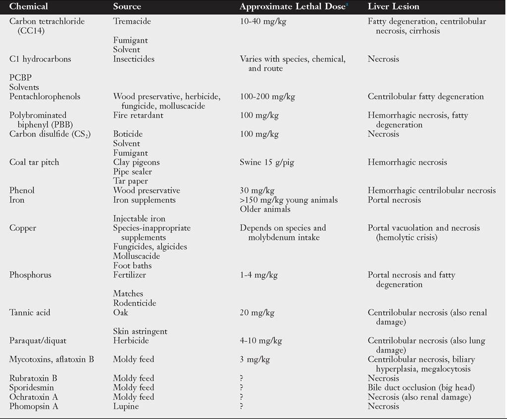
aLethal dose varies with individual, species, route of entry, rate or chronicity, enzyme induction, and many other factors. ?, Unknown.
low-lying heavy clay soils with a high water table, is also an important consideration in evaluating the need for fluke control. This factor has also been shown to lead to a hundredfold variation in the risk of fluke losses in the same year on different premises in otherwise ecologically similar areas.4 The snail habitat area on most enzootic livestock operations typically is only 1% to 5% of the total land area; operations with more than 5% habitat may be associated with a high risk of liver fluke losses.
Clinical Signs Subclinical production effects of F. hepatica include reduced rate of gain and feed efficiency in growing stock. Economic effects have been experimentally demonstrated in calves on a marginal nutritional plane in the first 5 to 6 months after infection, resulting in an 8% loss in the rate of gain with a mean of 40 flukes and a 28% loss with 140 flukes. In cattle it is estimated that herd economic losses are “negligible” with a burden of less than 10 flukes per animal, “possible” at 10 to 40 flukes, and “probable” with more than 40 flukes. Clinical disease is seen with a burden of more than 200 flukes.
In cow-calf operations, flukes often act in concert with periods of nutritional stress and concurrent heavy GI nematode infections, causing further reduction of body condition, poor milking ability, and slower return to estrus. This translates to reduced reproductive efficiency, prolonged calving interval, and lightweight calves at weaning.5In cattle, flukes may lead to chronic disease or, in rare cases, subacute disease. When clinical signs occur, the typical population “overdispersion” (individual variation in parasite burdens) is manifested first in the 10% to 20% minority of the herd that harbor most of the parasite numbers. Signs may consist of weight loss, emaciation, depression, anorexia, rough hair coat, anemia, hypoproteinemia, submandibular edema, and in rare cases, mild icterus. The anemia associated with F hepatica infections is more than can be accounted for by fluke blood feeding. Evidence indicates that depression, anemia, and biliary
| ■ TABLE 33.7 | |
| Drugs Used in Large Animals That Could Damage the Liver | |
| Drug | Lesion |
| Carbon tetrachloride | Centrilobular necrosis or fatty degeneration |
| Hexachloroethane | Centrilobular or massive necrosis |
| Carbon disulfide | Necrosis |
| Alcohol | Necrosis/cirrhosis |
| Tetracycline | Fatty degeneration (also renal damage) |
| Clindamycin | Cholestasis, icterus |
| Erythromycin | Cholestasis, icterus |
| Isoniazid | Active hepatitis → cirrhosis/ massive necrosis |
| Rifampin | Cholestasis, icterus |
| Rifampin/doxycycline | Hemolytic icterus, elevated |
| combinations | liver enzymes |
| Nitrofurantoin | Active hepatitis → cirrhosis |
| Anabolic steroids | Cholestasis, icterus |
| Phenothiazine | Cholestasis, icterus tranquilizers |
| Halothane | Active hepatitis → cirrhosis/ massive necrosis |
| Fluothane | Active hepatitis/massive necrosis |
| Isoflurane | Active hepatitis/massive necrosis |
| Some diuretics | Cholestasis, icterus |
| Diazepam | Cholestasis, icterus |
| Phenobarbital | Necrosis/cholestasis icterus |
| Aspirin | Active hepatitis → cirrhosis |
| Oil of pennyroyal | Centrilobular necrosis |
| Tannic acid | Centrilobular necrosis, fatty degeneration (renal damage) |
| Dantrolene | Active hepatitis → cirrhosis |
| Copper disodium edetate | Massive centrilobular necrosis |
| Iron (injectable) | Necrosis, portal, cirrhosis, hemosiderosis |
| Glucocorticoids | Hepatocellular vacuolization |
| Ionophores (salinomycin) | Hepatic lipidosis, necrosis |
aMay vary with dose and time.
hyperplasia result from high levels of proline, a product of fluke metabolism.6 Cattle are able to mount a protective immune response, with partial acquired resistance to F.
hepatica beginning at 5 to 6 months after initial exposure.7 This and the short life span of flukes in cattle typically lead to a linear reduction of fluke numbers, with few surviving by the end of 1 year. Sheep and goats are more susceptible, and fatal acute disease with ascites, abdominal hemorrhage, pallor, and icterus can occur in association with massive entry to the bile ducts at 6 to 10 weeks after infection by 1000 to 5000 or more migrating immature forms from the liver parenchyma. Subacute disease has been associated with a burden of more than 800 flukes acquired over time. Chronic clinical disease in sheep with submandibular edema, ascites, and emaciation has been associated with fluke burdens above 200.8Liver flukes in horses are sometimes reported as an incidental finding during postmortem examination. In one study, prevalence was 9.5% in a population of horses from Ireland.9 Horses with weight loss and diarrhea that respond to flukicide treatment provide anecdotal evidence that flukes may result in detectable clinical disease, although the association is unproven.
Necropsy findings attributable to migrating young flukes are caused by tortuous tunnels of coagulative necrosis that organize and fibrose, ultimately leading to a diffusely fibrotic liver parenchyma, especially in the ventral lobe, which in severe cases undergoes marked atrophy. Fibrous tags may result from fibrinohemorrhagic deposits left by large numbers of flukes at liver penetration 3 to 4 days after infection. Inflammatory events associated with penetration of the bile ducts at 6 to 8 weeks after infection are especially pathogenic. Once in the bile ducts, flukes grow rapidly from 1 mm to 2.5 cm or more and induce a proliferative cholangiohepatitis. The spines and suckers of flukes erode and denude the bile duct epithelium, leading to a fibrosed, thickened duct wall that is irregularly dilated and stenotic and begins to calcify in cattle (but not sheep) at about 20 weeks after infection.
The bile becomes darkly discolored and laden with regurgitated fluke ingesta, plasma proteins, and inflammatory cells. The extent of pathologic change is generally proportional to the fluke burdens of current or recurring previous infections.8■ Diagnosis Fecal sedimentation methods are the standard means of diagnosing liver flukes, although classical methods are cumbersome. A reusable commercial “nematode counting chamber” (1-mL volume of sediment fluid) is available (Chalex, Wallowa, Ore.) and can be examined on a standard microscope stage. It reduces sample processing time by half and is suitable for use by practitioners for quantitative examination of individual animals or 10- to 15-animal herd composite samples. For herd or lot evaluations in cattle, egg counts of less than 1 egg per 2 g (EP2g) and 25% prevalence 2 to 4 months after the transmission season ends have a “low probability” of economic losses; 1 to 3 EP2g indicates “possible” economic loss; and 10 or more EP2g indicates “high probability” of heavy infections and economic loss. It is important to differentiate F hepatica eggs from the eggs of Paramphistomum species, a nonpathogenic “rumen fluke” often found in the same herds in both the southern and the western enzootic areas. Flukecides used against F. hepatica are ineffective against rumen flukes, and egg counts thus persist after treatment. Paramphistomum eggs are gray (rather than amber), slightly smaller, and more pointed at the operculum end than F. hepatica. Paramphistomum egg counts can be used as a general indicator of the probable risk of F hepatica in the absence of control in the southern states because Paramphistomum flukes are known to be transmitted by the same snail vector in that region.5
Diagnostic enzyme-linked immunosorbent assays (ELISAs) have been developed to detect serum antibodies or coproantigen in the feces of infected animals or in bulk milk samples.10,11 These tests have not yet found wide use outside of research laboratories in the United States but are marketed in Europe.11 ELISAs for the major F. hepatica excretory-secretory protein, CL1, have high specificity, but low sensitivity in horses, compared to ELISAs for the liver fluke 2.9-kDa antigen, which have high sensitivity and specificity in horse serum.9 Blood and serum clinicopathologic results reflect the anemia, hypoproteinemia, and mild eosinophilia caused by F. hepatica. Pathologic changes in the liver bile ducts and parenchyma are reflected by an elevation in serum liver enzymes (e.g., GGT, GDH).
■ Treatment and Prevention Prevention is the key to control in foundation herds and flocks because even low numbers of eggs shed can multiply asexually in snails and lead to significant infection rates during the following transmission season. A single routine fall or late fall-winter treatment after the end of the transmission season with a highly effective drug is recommended to remove adult flukes before winter stress and to prevent egg shedding and snail infection in the next season (see Figs. 33.3 and 33.4). Most of the pathogenic and
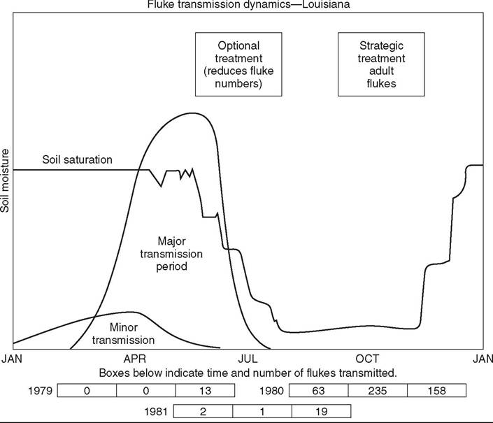
FIG. 33.3 Pattern of Fasciola hepatica transmission typical of the southern United States (based on 10 years of experimental data from Alexandria, Louisiana) and strategic treatment recommendations with adulticidal drugs. (From Malone JB, Loyacano AR, Hugh-Jones ME, et al.: A three-year study on seasonal transmission and control of Fasciola hepatica in cattle in Louisiana. Prev Vet Med 3:131, 1984.)

FIG. 33.4 Pattern of Fasciola hepatica transmission typical of the U.S. Pacific Northwest in natural rainfall and irrigated zones (based on experimental data from Langolis, Oregon) and strategic treatment recommendations with adulticidal drugs. (From Rickard LG, Z.i'mmeτma'n GL, Hoberg EP, et al.: Influence of ivermectin and clorsulon treatment on productivity of cow-calf herd on the Southern Oregon coast. Vet Parasitol 41:45, 1992.)
economic effects of flukes in cattle are reported to occur within the first 5 to 6 months after the major exposure period and may be related to metabolic products associated with the rapid growth and heavy egg production phases of the life cycle. This, coupled with linear loss of heavy fluke burdens in cattle and the onset of effective immunity at 20 weeks after exposure, suggests the value of early flukecide treatment within 2 to 3 months after the transmission season ends. In some herds an optional curative treatment in spring may be needed in very high-risk years or on high-risk premises, or as a second treatment to remove flukes acquired during late, extended transmission on irrigation pastures or wet coastal areas in the west.
Flukecidal drugs available in the United States are effective against mature flukes in bile ducts (albendazole, 10 mg/kg; closulon, 2 mg/kg). Closulon at 7 mg/kg has added efficacy against juvenile flukes over 6 weeks old in the bile ducts. The optimum time to treat should be based on the estimated susceptibility of mature fluke populations (>12 weeks old after end of transmission season) but early enough to remove flukes while they are most pathogenic (90% adult and 50% to 90% juvenile bile duct flukes), rafoxanide (>50% late migratory flukes, >90% juvenile and adult bile duct flukes), and diamphenathide (>90% migratory flukes, 50% to 80% bile duct flukes).
The economic benefit of routine flukecide treatment of feedlot calves from enzootic areas in the United States has not been consistently demonstrated industry-wide because of the high variability of fluke burdens in animals from enzootic areas or because lots are often of mixed origin. Fluke-related losses would be expected to be mainly absorbed by stocker operations, where lightweight calves are typically placed on small grain pastures for extended periods before feedlot, a common practice in the south-central states. The economic return of treatment at feedlot should be evaluated on a case-by-case basis because significant losses may occur if the lot originates from the same premises and the history suggests a high risk of recent heavy infection rates, such as in favorable climate years or use of irrigated pastures in fluke areas. Fecal egg counts on 10 to 15 randomly selected animals may aid in herd evaluation. An example case of the explosiveness of the life cycle is the greatly reduced feedlot performance and 100% liver condemnations of a lot from a stocker operation in the Pacific Northwest. The animals originated from irrigated pastures where some habitats stayed wet year-round, and untreated calves from fluke areas were allowed to contaminate pastures with eggs, which translated to infection later in the grazing season and to the next stocker group rotated onto the premises.
Other Flukes
Fascioloides magna, the large American liver fluke, may infect cattle and sheep that graze common areas with deer, the natural host. The life cycle is similar to that of F. hepatica, but with a wider variety of lymnaeid snail hosts and a broader geographic distribution in the Gulf States, the Great Lakes area, and the Northwest. Cattle are abnormal dead-end hosts that react with an intense encapsulation response, forming a closed cyst that does not allow escape of eggs and obviates diagnosis by fecal examination. The liver parenchyma and regional lymph nodes have a characteristic diffuse black pigmentation. The major economic effect of F. magna in cattle is condemnation of livers and other organs, such as lungs, in which aberrant migration sometimes occurs. In sheep and goats, however, F. magna does not encyst and migrates uninterrupted. One or two F. magna flukes kill sheep before the flukes have time to mature, and this parasite limits sheep production in some areas. Albendazole given at high dosage is moderately effective against F. magna.
In tropical regions, F. hepatica is replaced by Fasciola gigantica, a similar species that is somewhat larger, has a longer prepatent period (10 to 12 weeks), and is of somewhat greater pathogenicity. D. dendriticum occurs worldwide but in North America is an unimportant species, mainly limited to areas of central New York, with smaller foci in Pennsylvania, New England, Quebec, and British Columbia. Albendazole (20 mg/kg) and high doses of thiabendazole (150 to 300 mg/kg) are effective treatments.1