Hepatic Abscesses
T.G. Nagaraja
Hepatic abscesses occur sporadically in most animals but are most common in ruminants, particularly cattle fed high-grain diets. Hepatic abscesses occur at all ages and in all types of cattle but are most common and have the greatest economic impact in feedlot cattle.
The incidence in feedlot cattle ranges from 1% to 2% to a high of 60% to 80% but averages 10% to 20% in most feedlots.1 Because hepatic abscesses are generally a direct result of feeding practices, diet is an important factor influencing their incidence. Hepatic abscesses do occur in dairy cows, and in a survey of gross pathologic conditions in cull cows at slaughter, the incidence of liver abscesses was reported to be 32.1%.2 In the United States, it has become increasingly common to raise Holstein bull calves in feedlot production systems for beef production. Liver abscess data collected at slaughter have shown that the incidence of liver abscesses in Holstein feedlot steers is greater than in crossbred beef cattle.1,3 In addition, the proportion of more severe abscesses (one or more large or multiple small abscesses, often with adhesions to diaphragm and visceral organs) is greater in Holstein feedlot steers than in beef breeds (50% to 60% versus 30% to 40% of the total abscesses, respectively).■ Etiology Hepatic abscesses in cattle are generally polymicrobial infections, and in most cases the organisms are anaerobes.4,5 Fusobacterium necrophorum, a gram-negative, pleomorphically rod-shaped anaerobe, is the primary etiologic agent.3 The organism also is implicated as the primary pathogen in necrotic laryngitis (calf diphtheria), foot rot, and foot abscesses in cattle.6,7 F. necrophorum is a normal inhabitant of the rumen, and its role in ruminal fermentation is mainly to use lactic acid to produce volatile fatty acids and also digest ruminal epithelial and feed proteins.8 The organism is in higher concentration in grain-fed cattle than forage-fed cattle, possibly because of the increased availability of lactic acid from fermentation of starch in the grain.
Several virulence factors have been implicated in the pathogenesis of F. necrophorum infections.8,9 These include leukotoxin, endotoxic lipopolysaccharide (LPS), hemolysin, hemagglutinin, platelet aggregation factor, dermonecrotic toxin, adhesins (outer membrane proteins), and extracellular enzymes (e.g., proteases, deoxyribonucleases). Leukotoxin is believed to be the major virulence factor.9 The leukotoxin is cytotoxic to polymorphonuclear neutrophil leukocytes (PMNs), macrophages, hepatocytes, and ruminal epithelial cells. Because of leukotoxin, the organism is able to survive, proliferate, and establish to set up infection of the ruminal wall and liver. There are two subspecies of F necrophorum, subsp. necrophorum (formerly biotype A) and subsp. funduliforme (formerly biotype B). These two subspecies differ in cell morphology, colony characteristics, growth patterns in broth, and most importantly, in virulence factors. Subspecies necrophorum is more virulent (produces more Ieukotoxin) and thus more frequently encountered in liver abscesses than subsp. funduliforme, which tends to occur more often in mixed infections.7,8In most situations, Trueperella pyogenes (formerly Arcanobac- terium pyogenes), a gram-positive rod-shaped organism, is the second most frequent pathogen isolated from liver abscesses.10,11 The ruminal wall appears to be the niche for T. pyogenes, a facultative bacterium, because the wall provides an aerobic microenvironment in an otherwise anaerobic environment of the rumen.12 The occurrence of T pyogenes in liver abscesses is likely indicative of the ruminal damage that allows the entry of the organism into the portal circulation to reach the liver. In liver abscesses and other infections (foot rot in cattle, metritis in dairy cows), the organism acts in synergy with F. necrophorum.1 In recent years, Salmonella enterica has been found be associated with liver abscesses of cattle.
In a study of bacteriology of liver abscesses of crossbred beef cattle and Holstein steers collected at slaughter, the prevalence of S. enterica was reported to be 25.3%.13 A predominant serotype of Salmonella isolated from liver abscesses was Lubbock with the antigenic designation 6,7:g,m,s:e,n,z,15, a novel serotype closely related to Mbandaka. The Lubbock serotype has been isolated from lymph nodes of cattle at slaughter.14 The contribution of Salmonella to the cause of abscesses is not known, but there are reports of Salmonella causing liver abscesses in humans.15 A couple of features of Salmonella make its presence in liver abscesses not surprising. First, it is able to grow robustly in anaerobic condi- tions,16 and second, it is a facultatively intracellular pathogen, hence able to reach the liver through phagocytic cells.17A variety of other anaerobic and facultative bacteria, such as Bacteroides spp., Clostridium spp., Escherichia coli, Klebsiella pneumoniae, Mobiluncus spp., Mitsuokella spp., Pasteurella spp., Peptostreptococcus spp., Porphyromonas spp., Prevotella spp., Propionibacterium spp., Pseudomonas aeruginosa, Staphylococcus spp., Streptococcus spp., and many unidentified gram-positive and gram-negative bacteria are often isolated from liver abscesses of cattle.3,5,10
■ Pathophysiology Abscesses in the liver result from entry and establishment of F necrophorum either alone or with other bacteria. The routes by which bacteria can gain access to the liver include the portal vein, hepatic artery, umbilical vein (in newborns with omphalophlebitis), bile duct system, and direct extension. Entry through the hepatic artery (after an episode of septicemia) or the bile ducts (usually from obstruction, infection ascending from duodenum, or migration of flukes) is a rare occurrence. Direct extension of infection from adjacent tissues and organs, usually of traumatic origin (e.g., direct puncture of the liver by a foreign body lodged in the reticulum), is more likely to occur in dairy cows.
Traumatic reticuloperitonitis caused by metallic objects lodged in the reticulum and perforating through the reticular wall, rarely involving the ruminal wall, is often a predisposing factor for liver abscesses in dairy cows. However, in both dairy cows and feedlot cattle, the most common route of entry of bacteria into the liver is the portal vein.Liver abscesses are secondary to the primary foci of infection in the ruminal wall. Evidence to support this is the high correlation between ruminal wall lesions (rumenitis) and liver abscesses, thus the term rumenitis-liver abscess complex (Fig. 33.5). In a recent study of the relationship between ruminal wall lesions and liver abscesses in beef cattle at slaughter, Rezac and colleagues2 observed that 32% of cattle with mild or severe rumenitis had liver abscesses compared with 19% of cattle with healthy ruminal walls. However, not all studies have shown an association of liver abscesses with ruminal lesions.18 More direct evidence for the pathogenesis of liver abscesses was obtained by restriction fragment length polymorphism analysis of rRNA genes (ribotyping) of F. necrophorum and

FIG. 33.5 Pathogenesis of liver abscesses in cattle fed high-grain diets. (Modified from Nagaraja TJ, Chengappa MM: Liver abscesses in feedlot cattle: a review. J Anim Sci 76:287, 1998.)
T. pyogenes isolates from the rumen and liver abscesses of the same animal. The genetic similarity between the isolates from liver abscesses and ruminal walls supported the hypothesis that F. necrophorum and T. pyogenes isolates of liver abscesses originated from the rumen.12,19 It is well accepted that ruminal lesions resulting from acidosis are the predisposing factors for hepatic abscesses. Acid-induced rumenitis and damage of the protective surface usually are associated with a sudden change to high-energy diets and other dietary indiscretions, such as a change in feeding patterns, letting cattle become overly hungry, feeding unpalatable diets, and feeding very little roughage.
The ruminal damage often is aggravated by foreign objects in the feed, sharp feed particles, or hair.20 The ruminal wall that is damaged from acidity or penetration of foreign objects becomes susceptible to invasion and colonization by F. necrophorum. Once colonization has occurred, F. necrophorum can gain entry to the blood or cause ruminal wall abscesses and subsequently shed bacterial emboli to the portal circulation. Bacteria from the portal circulation are filtered by the liver, leading to infection and abscess formation.The virulence factors of F. necrophorum play a critical role in the penetration and colonization of the ruminal epithelium and entry and establishment of infection in the liver.8,9 The protease activity, dermonecrotic activity, and cytotoxic effect of leukotoxin on ruminal cells may aid in penetration and colonization of the ruminal wall. F. necrophorum, being an anaerobe, must overcome both high oxygen concentrations and phagocytic mechanisms to survive, proliferate, and initiate abscess formation. The leukotoxin may protect it from phagocytosis. Also, the release of cytolytic products such as lysosomal enzymes and oxygen metabolites, resulting from destruction of phagocytes, has a detrimental effect on the liver parenchyma. Synergism with facultative bacteria, intravascular coagulation induced by endotoxic LPS and platelet aggregation factor, formation of fibrin-encapsulated abscesses, and impairment of oxygen transport by damaged erythrocytes (action of hemolysin) all may contribute to the establishment of an anaerobic microenvironment conducive to the growth of anaerobic bacteria within the ruminal wall and liver.
■ Pathology Abscesses found in the liver at slaughter or necropsy often are well encapsulated, possessing thick, fibrotic walls (Fig. 33.6). The earliest lesion is a microabscess induced by an embolus of F. necrophorum in the sinusoid, and the lesion then progresses to coagulative necrosis by involving adjacent hepatocytes.
Subsequently, the lesion gradually changes into a pus-filled, encapsulated, true abscess. Histologically, a typical abscess is pyogranulomatous with a necrotic center, encapsulated, and often surrounded by an inflammatory zone.11 Hepatic abscesses are pus filled, have capsules that vary in thickness, and range in size from a minute pinpoint to more than 15 cm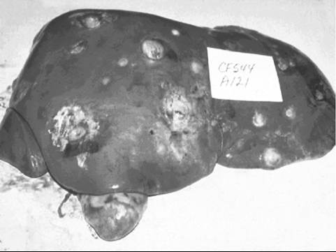
FIG. 33.6 Liver from a feedlot steer demonstrating a large liver abscess.
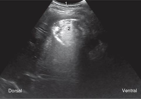
FIG. 33.7 Ultrasound image from the eleventh intercostal space of a cow. Abdominal wall (1), liver abscess (2).
(6 in) in diameter. The number of abscesses in the liver can vary from one to hundreds, and sizes range from less than 1 cm (0.4 in) to greater than 15 cm in diameter, often encircled by a hyperemic zone. The larger abscesses may result from small abscesses coalescing early in development. The distribution of abscesses in the liver shows no consistent pattern, with superficial and deep abscesses distributed almost evenly. Often small abscesses are scattered throughout the organ, whereas large abscesses are located close to the portal entry. Abscesses eventually become sterile and are replaced by fibrous scars, with eventual resorption.
■ Clinical Signs and Diagnosis Liver abscesses are detected only at the time of slaughter. Cattle, even those that carry hundreds of small abscesses or several large abscesses, seldom show any clinical signs. Occasionally the rupture of a superficial abscess or erosion and perforation of the caudal vena cava could lead to extensive spread and massive infection of other organs and eventual death. Generally, hematology and liver function tests have not proved to be good indicators of liver abscesses.21 In animals in which abscesses were induced by experimental inoculation of F. necrophorum, hepatic dysfunction was documented by elevated serum protein, bilirubin, and enzymes such as GGT and SDH concentrations.22 In a group of Holstein dairy cattle where the presence of liver abscesses was diagnosed via ultrasound or exploratory laparotomy, the
22
most common clinical sign was anorexia.22
Ultrasonography is a useful technique in the diagnosis of various hepatic diseases in cattle because of the location and tissue consistency of the liver (Fig. 33.7).23 The technique has been shown to be useful for monitoring the onset and progression of experimentally induced abscesses where the site of injection is known.22 The development of abscesses in feedlot calves has been recorded by ultrasonographic examination at regular intervals from weaning to slaughter.21 However, its application to the diagnosis in feedlot cattle is limited because the ultrasonographic scanning cannot visualize the whole liver, particularly the left side facing the internal organs, and parts of lobes are covered by other organs (e.g., lungs, kidneys).
Treatment and Prevention Both F necrophorum and T. pyogenes, but not S. enterica, are susceptible to penicillins and macrolides.5,24 Although successful treatment of cattle with liver abscesses has been described,25 antibiotic treatment is seldom practical in cattle because complete recovery is difficult to attain. Cattle with sequelae (e.g., caudal vena cava thrombosis syndrome) are generally culled for slaughter, for economic reasons. According to the U.S. Feed Additive Compendium, five antibiotics (bacitracin methylene disalicylate, chlortetracycline, oxytetracycline, tylosin, and virginiamycin) are approved for prevention of liver abscesses in feedlot cattle.26 Of these, only chlortetracycline, oxytetracycline, and tylosin are used for prevention. Tylosin, a macrolide, is the most effective antibiotic and the most commonly used feed additive (8 to 10 g/ton or 60 to 90 mg/head/day) in the feedlot. Studies show that tylosin feeding reduces abscess incidence by about 40% to 70%. Although there is no evidence of resistance development in F. necrophorum,5 the future of tylosin use as a feed additive in feedlot cattle is uncertain. According to the Center for Veterinary Medicine (CVM) of the U.S. Food and Drug Administration (FDA), the judicious use of medically important antimicrobial drugs in food-producing animals should be based on two principles: (1) limit medically important antimicrobial drugs to uses in animals that are considered necessary for assuring animal health, and (2) limit medically important antimicrobial drugs to uses in animals that include veterinary oversight or consultation. Because tylosin is used to prevent liver abscesses and not intended for growth promotion, it could be categorized as use to assure animal health. As of January 2017, the use of tylosin in feedlot cattle for the prevention of liver abscesses is under veterinary oversight.27 There is considerable interest in evaluating antibiotic alternatives, such as probiotics, essential oils, and vaccines, to control liver abscesses. In a feedlot study in cattle fed a finishing diet, inclusion of a product containing limonene, an essential oil, tended to reduce the incidence of liver abscesses compared to the control, but the difference was not significant.28
Because the pathogenicity and virulence factors of F. nec- rophorum have been studied widely, there has been considerable interest in and efforts to develop an effective vaccine.1 The use of vaccines has dual benefits: control of liver abscesses and alleviation of public health concerns associated with the continuous use of medically important antimicrobials in the feed. A bacterin vaccine (Fusogard [Elanco Animal Health, Greenfield, Ind.]), injected subcutaneously in 2-mL doses 21 days apart, is commercially available and has been shown to be effective in reducing the incidence and severity of liver abscesses in feedlot cattle. In a randomized and blinded field trial, vaccination reduced prevalence of liver abscesses in cattle with a low prevalence (10%) but was not effective in cattle with a high (30%) prevalence of liver abscesses.29 Two antigens of F nec- rophorum that have been targeted as potential candidates for vaccine development are leukotoxin, a critical virulence factor for bacterial survival and establishment of infection,30 and outer membrane proteins, which mediate adhesion of F. necrophorum to bovine cells.31 Both proteins have been recombinantly expressed and efficacy has been demonstrated in vitro and in a mouse model.31,32 However, the efficacy of the recombinant proteins in cattle has not been evaluated. Machado and col-
33
leagues33 have reported a vaccine formulation containing different combinations of proteins of E. coli, F necrophorum, and T. pyogenes to prevent puerperal metritis in dairy cows, leading to improved reproductive performance.
In addition to inclusion of antimicrobial compounds in the feed or vaccination, prudent bunk management to minimize fluctuations in intake and ruminal acidosis is well accepted as a key factor for effective control of liver abscesses. Management recommendations include gradual adaptation to high-grain diets, avoiding either underfeeding or overfeeding, increasing feeding frequency to spread out intake, increasing roughage content of the feed, inclusion of grain by-products like distiller’s grains, imposing quality control in mixing feeds, and providing adequate bunk space and fresh clean water.34
■ Sequelae Septic cardiac and pulmonary emboli are also associated with liver abscesses in feedlot and dairy cattle. Occasionally, sudden death has been reported in cattle secondary to rupture of liver abscesses, with septic embolization in the right side of the heart. Generally, the condition starts as phlebitis caused by the extension of liver abscesses involving caudal vena cava. The phlebitis leads to thrombus formation anywhere between the liver and right atrium but most often at the point of entry of caudal vena cava into the diaphragm. The clinical syndrome and the extent of lesions observed depend on the degree of thrombosis and type of organisms involved. The syndrome can range from death caused by rupture of caudal vena cava to various degrees of pulmonary embolism, pneumonia, infarction, endocarditis, hemoptysis, and epistaxis. Collectively, these lesions are categorized under caudal vena cava thrombosis (CVCT) syndrome.35,36
■ Economic Importance Liver abscesses are a major economic liability to the producer, the packer, and ultimately the consumer of beef. According to a National Beef Quality Audit report, 20.9% of livers were condemned at slaughter, and liver abscesses accounted for approximately two thirds of liver condemnations.37 The greatest economic impact of hepatic abscesses is not from the condemnation of liver but from the reduced animal performance and reduction in carcass yield. Cattle with abscessed livers have reduced feed intake, reduced weight gain, decreased feed efficiency, and decreased carcass dressing percentage. These effects are evident only in cattle with the most severe abscesses (one or more large abscesses or multiple small abscesses).38
Hepatic Abscesses in Horses
Hepatic abscesses occur sporadically in horses and are generally part of the intraabdominal abscess complex. In studies that have evaluated horses with intraabdominal abscesses, 12% (3 of 25)39 and 5% (3 of 61)40 of horses had abscesses in the liver. Hepatic abscesses in horses are usually associated with gastrointestinal diseases, including proximal enteritis, inflammatory bowel disease, or ileal diverticulitis, and may develop as a sequela to abdominal surgery or following intestinal perforation with foreign bodies.41,42 A variety of bacterial species, particularly anaerobes, have been cultured from the pus. The most frequently isolated bacteria include Streptococcus equi, E. coli, Bacteroides fragilis, Corynebacterium pseudotuberculosis, F.
3941
necrophorum, and Peptostreptococcus species.39 41
Clinically, horses with hepatic abscesses cannot be differentiated from those with other intraabdominal abscesses. Generally, horses have a history of fever, loss of appetite, signs of colic, depression, and weight loss. Clinicopathologic changes are typical of chronic bacterial infection and include leukocytosis, thrombocytosis, hypoalbuminemia, and decreased A/G ratio.39-41 The prognosis is generally poor because of failure to respond to antimicrobial treatment. Percutaneous or surgical drainage is an effective treatment in humans with hepatic abscesses and has been reported as a successful treatment in one horse.43
More on the topic Hepatic Abscesses:
- Hepatic Abscesses
- References
- REFERENCES
- Other Pneumonias
- References
- REFERENCES
- Liver and biliary tract
- Smith Bradford P., Van Metre David C., Pusterla Nicola (eds.). Large Animal Internal Medicine. Part 2. 6th edition. — Elsevier,2020. — 2279 p., 2020
- Gallbladder and Biliary Tract Disease
- Ultrasonography of the Ruminant Abdomen