Malignant Catarrhal Fever (Bovine Malignant Catarrh, Malignant Head Catarrh)
Robert J. CaUan
■ Definition and Etiology MCF is a severe lymphoproliferative disease caused by a group of herpesviruses collectively referred to as malignant catarrhal fever viruses.1'6 The most recent taxonomic nomenclature places these viruses in the order Herpesvirales, family Herpesviridae, subfamily Gammaherpesvirinae, genus Macavirus.1'3 Ten genetically and antigenically related viruses have been identified and constitute the group of MCF viruses.
Six of these viruses are associated with clinical disease, and the remaining four viruses have not yet been associated with disease (Table 32.13).5-9 Each virus has a reservoir host in which infection is typically asymptomatic. Clinical disease occurs in aberrant hosts and is characterized by acute onset of fever, inappetence, profuse ocular and nasal discharge, ocular signs such as corneal opacity and conjunctivitis, alimentary erosions, enteritis, and enlarged lymph nodes. The disease is typically progressive and often fatal.Clinical MCF is observed in cattle, deer, bison, buffalo, several other domestic and wild ruminants5,6 (>33 species to date), pigs,10-13 and alpaca.14 It has worldwide distribution and can be a problem in zoos and game parks. There are two primary etiologic forms of MCF that are designated by the host reservoir of the virus. Wildebeest-associated MCF is caused by alcelaphine gammaherpesvirus 1 (AlHV-1). The principal reservoir of AlHV-1 is the blue wildebeest (Connochaetes taurinus) and the black wildebeest (Connochaetes gnou).5,i5 Wildebeest- associated MCF occurs primarily in Africa or in zoologic parks where other ruminants have contact with wildebeest. Sheep- associated MCF is caused by ovine gammaherpesvirus 2 (OHV-2). Domestic and wild sheep and goats are asymptomatic reservoirs of the virus.
Sheep-associated MCF is the primary form of MCF that is observed outside of Africa and affects primarily domestic and wild ruminants. MCF caused by OHV-2 has also been identified in pigs.10-13 Goats are a reservoir for caprine gammaherpesvirus 2,16 which has been isolated from cases of MCF in white-tailed deer,17-20 sika deer,21-24 roe deer,25 moose,25 pudu,26 and water buffalo.27 Both AlHV-1 and OHV-2 can be experimentally transmitted to rabbits and hamsters, in which infection causes an acute, fatal lymphoproliferative disorder similar to natural MCF.28,29■ Clinical Signs and Differential Diagnosis The clinical signs and pathologic process of MCF have been reviewed.5,6 Cattle from 4 weeks of age to adulthood can be infected and develop clinical illness. After an incubation period of 2 to 30 weeks, the disease attacks vascular endothelium, causing a lymphocytic vasculitis. The incubation period can be prolonged; in at least one case, the incubation period was 200 days. High fever, alimentary erosions, diarrhea, dysentery, corneal opacity, anterior uveitis, conjunctivitis, mucopurulent nasal discharge, thickened cracking skin, encephalitis, lymphadenopathy, and hematuria may be present (Fig. 32.93). Oral and nasal erosions may be evident but are often too far back in the nasal or
■ TABLE 32.13
Ten Antigenically and Genetically Related Viruses in the Malignant Catarrhal Fever Virus Group and Their Respective Reservoir and Clinical Hosts5-8
| Current Name | Previous Name | Primary Reservoir Host | Clinically Susceptible Hosts |
| Ovine gammaherpesvirus 2 (OHV-2) | Ovine herpesvirus type 2 (OvHV-2) | Domestic and wild sheep | Cattle, bison, deer and other cervids, swine, moose, alpaca, and other domestic and wild ruminants |
| Alcelaphine | Alcelaphine herpesvirus | Wildebeest (Connochaetes | Cattle, deer, and other domestic and |
| gammaherpesvirus 1 (AHV-1) | type 1 (AlHV-1) | spp.) | wild ruminants |
| Alcelaphine | Alcelaphine herpesvirus | Jackson hartebeest (Alcelaphus | Barbary red deer (Cervus elaphus |
| gammaherpsevirus 2 (AHV-2) | type 2 (AlHV-2)64,65 | buselaphus lelwel), topi (Damaliscus lunatus) | barbarus), bison |
| Caprine gammaherpesvirus 2 (CHV-2) | Caprine herpesvirus type 2 (CpHV-2)9,16,21,23,25,27 | Goat | White-tailed deer, sika deer, roe deer, moose, pudu, water buffalo, pronghorn |
| Unassigned | MCFV of white-tailed deer (MCFV-WTD)19, Caprine herpesvirus 3 | Goat | White-tailed deer, red brocket deer |
| Unassigned | Ibex-MCFV66 | Ibex (Capra nubiana) | Bongo (Tragelaphus eurycerus), anoa, pronghorn antelope |
| Hippotragine gammaherpesvirus 1 (HHV-2) | HiHV-167,68 | Roan antelope (Hippotragus equinus), scimitar-horned oryx (Oryx dammah) | None identified |
| Unassigned | Oryx-MCFV4,9, or Gemsbok MCFB | Gemsbok (Oryx gazella) | None identified |
| Unassigned | Muskox-MCFV9 | Muskox (Ovibos moschatus) | None identified |
| Unassigned | Aoudad-MCFV4 | Aoudad, Barbary sheep (Ammotragus lervia) | None identified |
MCFV, Malignant catarrhal fever virus.
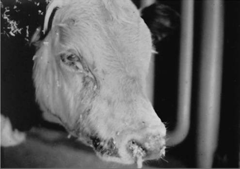
FIG. 32.93 A yearling steer with malignant catarrhal fever. Note the corneal opacity, lacrimation, and mucopurulent nasal discharge.
oral cavity to be observed externally. Ocular lesions are often present and consist of corneal opacity that originates in the periphery (centripetal) and progresses to the center. Corneal ulceration is generally not present, and there is no uptake of fluorescein dye. Affected animals exhibit generalized weakness and dyspnea. Ropy saliva may be dropped from the painful mouth, and scabs may develop on the muzzle. Swelling and hyperemia of the coronary bands may be observed, characteristic of coronitis. The hoof or horns may be shed, and lameness may be pronounced. Hematuria often is present and caused by a hemorrhagic cystitis. Nonsuppurative arthritis can also be observed. Neurologic signs include ataxia or altered mentation and are caused by a nonsuppurative encephalitis. Cutaneous lesions consist of multifocal coalescing alopecia with dermatitis and necrosis. When particular signs predominate, the condition may be labeled the alimentary form, the encephalitic form, the head and eye form, or the skin form. One strain of virus can cause all forms, and animals with most of these signs can be identified in a given outbreak. Clinical signs in any single animal may progress rapidly or over a course of weeks. Disease in Sitka deer (and perhaps other species) may consist primarily or dermatitis and alopecia.,,,30,
The course of the acute disease is usually 3 to 7 days; some animals survive longer. Disease is generally more severe and progresses more rapidly in bison than in cattle. The mortality rate is very high. Peracute deaths without any visible symptoms can also occur. A mild form, with transient fever and mild oral and nasal mucosal erosions followed by recovery, has been observed in experimentally infected cattle.
Some animals survive clinical disease and, in a few cases, may return to normal productivity. The case mortality rate is generally high and may exceed 90%. However, chronic and recovered cases have been described, and in one report, recovery occurred in 50% of cases.32MCF must be differentiated from BVD-related mucosal disease, rinderpest, bluetongue, vesicular stomatitis, and FMD. The last two diseases are not usually associated with diarrhea, and with vesicular stomatitis and FMD, morbidity rates tend to be high and mortality rates low. Clinical bluetongue is rare in cattle and not usually associated with dysentery. Arsenic toxicity and chlorinated naphthalene toxicity have some clinical similarities to the intestinal forms of MCF. Ocular signs, including corneal opacity, conjunctivitis, and discharges, must be differentiated from bovine keratoconjunctivitis (pinkeye), in which the lesion starts at the center of the cornea and corneal ulceration is present. In contrast, the corneal opacity of MCF starts at the limbus and is caused by edema secondary to the loss of the internal endothelial layer of the cornea.33,34
■ Clinical Pathology and Serology Some affected animals show leukopenia caused by lymphopenia and neutropenia if tested early in the course of disease, but this is an inconsistent finding in natural outbreaks.35 Joint fluid is cloudy and may contain increased protein and mononuclear cell numbers. Cerebrospinal fluid may have elevated protein concentrations and elevated WBC counts with mononuclear pleocytosis in animals displaying neurologic signs. Serologic tests include ELISA, indirect immunofluorescence, complement fixation, and virus neutralization.1,5 Virus isolation can be attempted for cases of MCF caused by AlHV-1. Serologic tests identify antibodies directed to a conserved MCF virus glycoprotein and do not discriminate between the different MCF viruses. Serologic tests are best used to screen the infection status of reservoir animals or to identify asymptomatic infection in clinically susceptible hosts.
Diagnostic PCR tests have been developed for AlHV-1 and OvHV-2.1,5 PCR tests are best used to distinguish between AlHV-1 and OvHV-2 in animals with clinical MCF. PCR tests for the identification of other MCF viruses, including a multiplex PCR assay, are available from specific research laboratories.1,5A presumptive diagnosis is based on a history of exposure, clinical signs, and evidence of infection supported by serologic or PCR results. A definitive diagnosis can be made only if characteristic histopathologic lesions, including lymphocytic vasculitis, are present, along with supportive diagnostic test results demonstrating infection.1,5
■ Pathophysiology Natural infection occurs through inhalation of aerosolized virus or through direct or indirect contact with ocular or nasal secretions from reservoir hosts. The exposure dose plays a role in the likelihood of developing clinical MCF and the duration of the incubation period.1,5,6,36-40 Experimental transmission studies in sheep and bison show initial replication oι the virus in respiratory epithelium.1,5,6,36,37,39,40 OvHV-2 DNA can be identified in the respiratory tract 9 to 12 days after infection in bison experimentally infected by intranasal inoculation.41 OvHV-2 infection of lymphocytes is observed 14 to 21 days after infection. The incubation period for clinical MCF is highly variable and ranges from 14 to more than 200 days. MCF virus infection causes infiltration of lymphocytes, particularly T cells, into tissues.2,5,6 This T cell proliferation is predominated by CD8+ gamma-delta T cells and, to a lesser extent, CD4+ T cells.5,42 There is also an increase in large granular lymphocyte cells that appear to have cytopathic activity. It is believed that the fundamental disease is caused by an autoimmune-like process involving dysregulation of cytotoxic T cells associated with expression of specific viral transcripts.2,5,42,43
■ Epidemiology Wildebeest-associated MCF remains an important disease in cattle and other ruminants that encounter wildebeest.5,6 AlHV-1, unlike OvHV-1, can be isolated in cell culture, and this has aided the ability to study the disease epidemiologic behavior and transmission.
Wildebeest calves are generally infected with AlHV-1 in utero or soon after birth and shed the virus in ocular and nasal secretions until the age of 4 to 6 months.5,15 Adult wildebeest become latently infected and transmit the virus to their offspring. Recrudescence and shedding of AlHV-1 in wildebeest can occur after treatment with corticosteroids. Cattle are infected with AlHV-1 when exposed to young, actively shedding wildebeest calves. The incubation period of wildebeest-associated MCF under natural exposure conditions in cattle can range from 2 weeks to 6 months and perhaps longer. In general, the incubation period of wildebeest-associated MCF is shorter than that of sheep-associated MCF. Some cattle that become infected with AlHV-1 do not develop clinical MCF. Diseased cattle do not shed free infectious virus and thus are not a risk to contact animals.5Sheep-associated MCF is the predominant form of MCF outside of Africa. The prevalence rates of OvHV-2 infection determined by PCR or competitive inhibition ELISA in healthy adult North American domestic sheep is reported to be 99% and 94%, respectively.44 Sheep serve as the primary reservoir of OvHV-2, although sheep do not show clinical signs of infection. Less than 5% of lambs are infected at birth.5,45 The majority of lambs remain uninfected up to 2.5 months of age. After 3.5 months of age, the lambs develop infections through close contact with adult sheep; the prevalence rate approaches 100% by the age of 5.5 months. OvHV-2 is shed and transmitted through nasal and ocular secretions.46,47 Seroconversion and detection of OvHV-2 DNA in nasal secretions lags behind the detection of viral DNA in blood by several months.5,45 Lambs separated from adults at 2 months of age remain reliably OvHV-2 free.5,48 These findings indicate that newborn lambs are not a major source of virus infection for cattle. Nasal viral shedding peaks at approximately 6 to 9 months of age, and it is these infected adolescent lambs that pose the greatest transmission risk to other animals.5,46
Infected adult sheep continue to shed low numbers of virus throughout life.5,46 Adult sheep may shed OvHV-1 in larger amounts during the periparturient period or while they feed in feedlots, which raises the risk for commingled cattle. OvHV-2 is readily transmitted between in-contact sheep; however, transmission by inoculation of whole blood or peripheral blood mononuclear cells is inconsistent.49 This suggests that infectious virus is produced and shed primarily in nasal or ocular secretions and that infection of blood cells may be largely nonproductive.
Positive results of competitive inhibition ELISA tests have been identified in bison (2% to 23%), domestic goats (74%), elk (9%), mule deer (2%), white-tailed deer (3%), pronghorn antelope (25%), bighorn sheep (37%), muskox (40%), and mouflon sheep (62%). Black-tailed deer, llamas, and mountain goats have been tested and were not found to be seropositive for MCF.50 However, it is not clear whether these species play any role as reservoir species.
Sheep-associated MCF can be acute or chronic, and infected animals may recover. Cattle and American bison (Bison bison) are the two primary species affected by sheep-associated MCF. The disease usually occurs sporadically, with only one or a few animals affected, but many large outbreaks have been reported.5,6,51-55 Transmission probably occurs through direct contact, indirect contact, and aerosol of nasal secretions from sheep. In one report, exposure of bison to sheep for less than a day at an auction resulted in a high mortality rate.55 There is evidence of transmission from sheep to cattle or bison extending over distances of 70 m up to 5.1 km.53,56,57 In one example of natural infection of bison, the incidence rates of disease and mortality were inversely related to the distance of exposure.53 The incubation period after experimental or natural exposure ranges from 14 to 220 days.6,38,41,51,54,55 Cattle and bison are considered dead-end hosts and do not spread the infection to other animals.2,5,55
Bison are more susceptible to developing clinical MCF than are cattle by both experimental inoculation studies and natural exposure.5,36,38,54 In bison, OvHV-2 infection and clinical MCF is observed with intranasal nebulization at doses as low as 103 DNA copies of OvHV-2.5,36,37 Cattle become infected with doses as low as 106 OvHV-2 copies but require at least 108 copies to become infected with clinical MCF.5,38 In contrast, the minimum infectious dose for sheep is between 102 and 103 DNA copies, but sheep require a dose of 3 ? 109 copies to develop nonfatal clinical signs of MCF.5,39,40 Several studies have shown that both cattle and bison can be infected with OvHV-2 and not develop clinical MCF.5,52,56,58 These studies show that infection, morbidity, and mortality are related to species susceptibility, inoculation dose, and probably other factors not yet determined. Both asymptomatic and clinically affected cattle and bison do not shed infectious virus and are considered dead-end hosts.5
■ Necropsy Findings In an infected animal, the nasal mucosa is hyperemic and may have hemorrhagic superficial erosions. The oral mucosa has necrotic papillae and large areas of necrosis and ulceration. Multiple focal ulcerations are seen in the esophagus. Parts of the forestomachs and intestines are thickened, edematous, and occasionally ulcerated and hemorrhagic. The lymph nodes, tonsils, and Peyer's patches are enlarged, moist, and friable. Splenic lymphoid follicles are prominent, and the liver is swollen. The adrenal glands are hemorrhagic. The mucosal surface of the bladder has focal areas of hemorrhage. The eyes are hyperemic and show severe corneal edema. The brain may have intersulcal cloudiness and meningeal petechiation consistent with encephalitis. Joints may have swollen and reddened synovia with an increased quantity of cloudy fluid.6,59
Histologically, there is a marked lymphoid accumulation around blood vessels, as well as necrosis of the tunica media and intima in multiple tissues. Generalized lymphoid hyperplasia and lymphoid infiltrates are present in subepithelial and intraepithelial locations, in association with epithelial necrosis and sloughing. Similar changes occur in all epithelial tissues.59 Chronic cases develop chronic obliterative arteriopathy.32
■ Treatment and Prognosis There is no specific treatment for MCF beyond supportive care. Fluid therapy and nursing care may allow some animals to survive and recover from disease. Antimicrobial treatment to help prevent secondary bacterial pneumonia or septicemia may provide some benefit. Treatment with NSAIDs may provide comfort but could increase the degree of alimentary erosions or decrease their healing. Gastrointestinal protectants such as bismuth subsalicylate (Kaopectate) may provide support to the gastrointestinal tract and help maintain appetite. Maintenance of appetite seems to be one of the key prognostic indicators in clinical cases of MCF. Although some animals that exhibit mild disease may survive, close to 100% of those with severe clinical signs die.
In one report, the severity and progression of ocular lesions associated with MCF in cattle were evaluated in relation to disease outcome.33 The degree of corneal edema at presentation did not show prognostic value in predicting disease outcome (death versus survival). However, improvement of corneal edema over the course of the disease was associated with a better outcome. In addition, animals in which anterior uveitis worsened were more likely to die or be euthanized.
■ Prevention and Control The primary method of control is to reduce the exposure of susceptible hosts to reservoir hosts.5,6 Transmission is related to the distance between reservoir hosts and susceptible hosts, so these groups of animals should be physically separated. This can be particularly difficult because, as mentioned, transmission from sheep to bison over distances as far as 5 km have been reported.53 Adult wildebeest and sheep pose a lesser risk of transmission than do neonatal wildebeest and adolescent sheep, which shed higher amounts of virus from nasal secretions than do the adults. These younger groups of reservoir animals should be kept at a greater distance from susceptible hosts. Feedlot sheep from 6 to 9 months of age pose a significant risk. Natural transmission of OvHV-2 between feedlot sheep and dairy cattle or bison has been reported and is probably related to the younger age of the feedlot sheep in comparison with adult breeding animals.53,54,56
Currently, no commercial vaccines are available for prevention of wildebeest-associated or sheep-associated MCF. Attenuated AlHV-1 vaccines have demonstrated some protection against virulent AlHV-1 challenge and natural infection in cattle.60,61 Respiratory tract mucosal immunity appears to be critical in preventing initial infection and development of clinical MCF.2,60,62,63 Further research identifying critical antigens and methods to induce strong, long-lasting mucosal immunity for both AlHV-1 and OvHV-2 are necessary for developing effective vaccines for bison and cattle.