Vesicular Stomatitis
Pamela J. HuHinger • DaneHe A. Bickett-Weddle
Definition and Etiology
Vesicular stomatitis virus (VSV) is a rhabdovirus in the genus Vesiculovirus that causes sporadic (cyclic) outbreaks in cattle, horses, donkeys, mules, and pigs.
Clinical signs include vesicles, erosions, and ulcers. Although deaths are rare, these lesions can result in pain, anorexia, and secondary bacterial mastitis. Some animals may lose their hooves after developing laminitis.1 Affected cattle younger than 1 year of age rarely show clinical signs. Affected sheep and goats rarely have clinical signs, and South American camelids are intermediate in susceptibility. Humans can become infected with VSV and develop a mild influenza-like disease.Vesicular stomatitis viruses are endemic from southern Mexico to northern South America, but they regularly spread north and south from these regions, causing outbreaks and epidemics. Although these viruses are no longer endemic in the U.S., they are introduced periodically into the southwestern states and can sometimes spread further north.1
Four distinct viruses are now recognized: VS Indiana (formerly called Indiana 1), VS New Jersey, Alagoas virus (formerly called Indiana 3), and Cocal virus (formerly called Indiana 2). Indiana 1 and New Jersey occur in the United States.1 Viral strains vary in virulence.
Historically, epizootic waves of vesicular stomatitis tended to occur at approximately 10-year intervals, usually in the summer or fall. However, outbreaks have become more frequent since the 2000s. An outbreak of vesicular stomatitis in the United States lasted from April 29, 2015, to March 4, 2016. A total of 823 premises were confirmed or suspected to have VSV infections (VS New Jersey serotype) in eight U.S. states: Arizona (36 premises in three counties), Colorado (441 premises in thirty-six counties), Nebraska (38 premises in ten counties), New Mexico (52 premises in thirteen counties), South Dakota (50 premises in seven counties), Texas (4 premises in four counties), Utah (56 premises in eight counties), and Wyoming (146 premises in ten counties).2
Although the VS New Jersey serotype has at least 14 distinct genotypes, only a few of these have been found in outbreaks in the United States.
The VS New Jersey serotype should be considered a collection of serologically related but genetically variant viruses.State and federal regulatory veterinarians should be contacted immediately when vesicular stomatitis is suspected so that quarantine and disease identification measures can quickly contain an outbreak.
Clinical Signs and Differential Diagnosis
The incubation period is usually 3 to 7 days but can be shorter or longer, depending on route of transmission and viral load. In the 1984 California outbreak, it was unusually long (9 days).1 Vesicles occur in the mouth, on the teats, and in interdigital areas. Vesicles are only occasionally visible because the epithelium rapidly becomes necrotic and turns into painful ulcers. Lesions on the gums and tongue may coalesce to form large
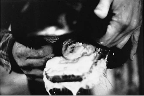
FIG. 32.94 Severe ulceration of the tongue of a dairy cow caused by vesicular stomatitis. A plaque of dying mucosa covered by fibrin is visible at the tip of the tongue, and excessive salivation is obvious. (Courtesy Dr. Mark Thurmond.)
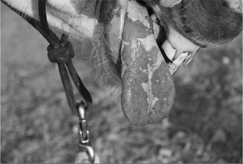
FIG. 32.95 Tongue of horse with vesicular stomatitis, demonstrating severe lingual erosions. (Courtesy Dr Skip Aaroe.)
eroded areas that cause dysphagia and reluctance to eat, frothing at the mouth, drooling, and weight loss (Figs. 32.94 and 32.95). Milk production falls quickly. In dairy cattle, teat lesions are common and cause mastitis. Small ulcers in the interdigital area and on the coronary band are occasionally present and cause lameness. Recovery varies from 2 to 21 days, depending on the severity of the lesions and management factors such as the type of feed and milking sanitation.3 Actual healing of the lesions may take 34 to 59 days.4
In cattle, morbidity rates are generally high (5 and 60%)3,4 and low mortality rates (1% to 5%).
Although many exposed cattle do not show obvious clinical signs of disease, many more are found to have oral lesions if closely examined, and most animals in the herd seroconvert.The major differential diagnostic consideration is FMD, which causes identical clinical signs, except that VSV can affect horses. Other diseases of cattle that result in oral lesions include BVD, BPS, bluetongue, and epizootic hemorrhagic disease. These usually appear, not as epidemics in herds, but rather in one or a few animals in the herd (although with BPS, morbidity rates may be high). Other causes of oral lesions such as bristle grass irritation and toxins should be ruled out.
Clinical Pathology and Laboratory Diagnosis
VSV can be found in vesicular fluid, swabs of ruptured vesicles, the epithelium of unruptured vesicles, and epithelial flaps from freshly ruptured vesicles.1 Many cell lines and other methods can be used to isolate VSV from clinical samples. The identity of cultured virus can be accomplished by RT-PCR, immunofluorescence, complement fixation tests, or antigen-capture ELISA. Antigen-capture ELISA can also be used for serotyping.1 Electron microscopy of tissue samples may be helpful in distinguishing VSV from some other viruses that cause vesicular lesions, such as the FMD virus. 1
Vaccination with inactivated virus results in a rapid rise in titer, followed by a gradual decline for a year.5 Serumneutralizing antibody titer rises rapidly after exposure and then gradually falls over the first year. Serum-neutralizing antibody titers that appear after natural exposure may persist for years. The presence of a serum-neutralizing antibody titer does not prevent reinfection or development of clinical signs. Hematologic and clinical chemistry findings generally reflect an acute to chronic inflammatory disease and are nonspecific.
Pathophysiology
After a short incubation period, fever occurs, and the virus invades the germinative layer of oral epithelial cells. Oral abrasions or trauma may increase susceptibility.
Contact of virus with teats or feet can result in lesions in these areas, especially if the teats are chapped or cracked or the feet are traumatized. The lesions progress rapidly from blanched macules to vesicles and soon rupture, leaving sloughed epithelium and ulcerated areas. Healing occurs quite rapidly if feed is soft and does not cause oral trauma.■ Epidemiology Older, high-producing dairy cows that have been in milk longer are more susceptible to clinical disease caused by VSV than are younger herdmates.5 Because the virus cannot penetrate intact mucosa, cattle fed coarse feeds or hard pellets that traumatize the oral mucosa are at higher risk. Cows with chapped or cracked teats and those on farms with poor milking hygiene are more likely to get teat lesions.5 Cow-to-cow contact is a major mode of transmission in outbreaks, and increased interpen movement of cattle, as well as sharing of feed and water troughs (unless cleaned frequently), increase the risk of exposure to VSV The virus can be transmitted by milking machines and human hands during outbreaks. In addition to being biological vectors, insects can also contribute to the mechanical spread of the disease.
The transmission of VSV is not completely understood, and transmission routes in individual outbreaks are unclear. Outbreaks can be associated with the movement of animals from another area, but disease epidemics unrelated to introduction of new animals do occur. The infection tends to be seasonal (occurring in the summer and fall in temperate areas and at the end of the rainy season in the tropics) and behaves like an arthropod-borne virus. Insect vectors are thought to introduce VSV into populations of domesticated animals. Sand flies (Lutzomyia sp.), black flies (family Simuliidae) and Culicoides midges can act as biological vectors. Sand flies seem to be important vectors in endemic areas, but they have a limited flight range and are not thought to spread these viruses over long distances.1 Black flies are believed to be particularly important vectors in parts of the western United States.
It is still uncertain where the virus originates before entering livestock populations. Once animals develop lesions, however, insects may become infected by feeding on these lesions or contaminated secretions. Transovarial transmission has been demonstrated in sand flies and black flies in the laboratory and may be possible in Culicoides midges. It might contribute to virus overwintering in cold climates.1 Both migratory western grasshoppers and the plants they eat can be infected with VSV,6 which provides a means of livestock exposure. It is hypothesized that VSV may be a plant virus that strays into animals, in which it causes sporadic disease.Other insects act as mechanical vectors. Antibodies have been found in a number of wild species of animals (deer, raccoons, bobcats, monkeys) that may serve as virus reservoirs. Active cases of vesicular stomatitis occur in Mexico between U.S. epidemics; it is possible that cattle from Mexico arriving in the United States may also be sources of VSV Sheep and goats in contact often seroconvert, although clinical signs in these species are rare.
The virus survives several weeks in cool soils but is inactivated by sunlight and is very resistant to pH changes.
Necropsy Findings
Deaths from vesicular stomatitis are rare and usually attributed to secondary bacterial diseases, including environmental mastitis and pneumonia. Affected cattle become gaunt and weak as a result of dysphagia and anorexia. Erosive and ulcerative lesions are usually confined to the mouth. The teats are frequently involved in affected lactating cows, and lesions on the coronary band and interdigital area occasionally appear.
Histologically, intracellular and extracellular edema, ballooning and degeneration of epithelial cells, and vesicle formation accompanied by neutrophilic infiltration occur. There are no inclusion bodies. The characteristic bullet-shaped structure of the VSV can sometimes be seen with electron microscope examination of fresh lesions or vesicular fluid.
Treatment and Prognosis
Mortality can be almost completely prevented if ill cattle are offered shade, fresh water, clean bedding, and soft feed. Offering soft feed hastens recovery and reduces the anorexic period. Judicious use of appropriate antibiotics to control secondary bacterial pneumonia may be warranted. Cattle with teat lesions are at high risk for developing mastitis and should be carefully milked last and monitored closely for mastitis. The prognosis for survival is very good, but agalactia and mastitis may necessitate the culling of animals.
Prevention and Control
The websites of World Organization for Animal Health (OIE)7 and the USDA2 provide the latest information on active vesicular stomatitis outbreaks. During an outbreak, regulatory officials implement a quarantine of the premises and require testing and isolation of sick animals. Leftover feed should be removed from feed bunks twice daily, and the bunks should be disinfected. Water troughs should be cleaned and disinfected daily. According to Spickler, “VSV is susceptible to numerous disinfectants including 1% sodium hypochlorite, 40-70% ethanol, 2-propanol, aldehydes (e.g., 0.5%-2% glutaraldehyde, formaldehyde), 1% cresylic acid, phenolic disinfectants and detergents. These viruses appear to be more susceptible to inactivation by acid (e.g., pH 2) than alkaline conditions. VSV are also susceptible to UV [ultraviot] light including sunlight, or heat (e.g., 4 minutes at 55°C, or one minute at 60°C).n1
Vaccination with killed8 or live virus vaccines is rarely practiced preventively because the disease occurs as such rare epidemics in small areas and vaccination interferes with serologic testing and monitoring. Although VSV vaccine is available in some South American countries, it is not currently available in the U.S.