methods of providing NUTRITION ENTERALLY
Feeding Into the Stomach
When feeding into the stomach (i.e., oral, nasogas- tric/nasoesophageal, esophagostomy, pharyngostomy, or gastrostomy), the quantity of diet fed is determined by the patient’s stomach capacity.
In normal dogs and cats, stomach capacity is approximately 80 ml of fluid per kilogram of body weight. However, anorexic patients can accommodate only 30 to 40 ml of fluid or liquid diet per kilogram of body weight when feeding begins. A gradual increase in volume over a 2- to 3-day period will generally allow the stomach to accommodate larger volumes of nutrients. A minimum of three feedings daily should be used; however, if vomiting and abdominal distention occur, the volume should be reduced and the number of feedings per day increased.When using 5 Fr or smaller feeding tubes (i.e., nasoesophageal or nasogastric tubes), only liquid diets may be administered. When using 8 Fr feeding tubes, convalescent diets with a homogeneous consistency may be used if diluted. When using larger-bore feeding tubes, blended canned diets may be used (Figure 12-35). The volume of food administered per feeding is determined by the caloric requirements and the consistency of the gruel.To calculate the amount of food to administer, the clinician divides the energy requirements of the patient by the caloric density of the food. Although the amount of water administered using canned products can be calculated by multiplying the volume of food to be administered by the moisture
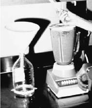
Figure 12-35 Blended canned diets can be administered through large-bore feeding tubes.
content of the diet, an alternative is to mix 1 part food with 1 part water.This usually exceeds the daily fluid requirements of the patient.
Although starter regimens have been recommended (e.g., feeding one third of calories on day 1, two thirds of calories on day 2, and full caloric intake on day 3), we recommend feeding full caloric intake on day 1. It is necessary to feed the total amount of diet and water divided over 6 to 8 feedings on day 1, however. On day 2, the volume of administered diet per feeding can be increased and the frequency of feedings can be decreased to four to six times. On day 3, the number of feedings can usually be decreased to 3.Feeding Into the Small Intestine
When feeding into the small intestine (i.e., gastro- duodenostomy or enterostomy), the clinician must carefully regulate the rate and volume of diet to avoid overdistention. Each patient is unique in the amount of fluid the small intestine will accommodate; therefore a guideline for feeding via enterostomy tube is presented. Enterostomy tube feeding is usually done by constant-rate infusion rather than bolus feeding. Because of this, the day is divided into two 12-hour cycles and the liquid diet is administered via constant-rate infusion over 8 to 10 hours, providing 2 to 4 hours to rest the GI tract. During the first 12-hour cycle, one quarter of the calculated diet is administered over 8 to 10 hours and then discontinued for 2 to 4 hours. If the patient tolerates the rate, one half of the daily calculated amount of diet is administered over the next 8 to 10 hours, again discontinuing for 2 to 4 hours. After that, one half of daily calculated diet requirements is administered per each 12-hour cycle, providing 2 to 4 hours of GI rest.
It is difficult to decrease the infusion time of diet to less than 8 hours or to bolus feeding, although both have been reported in enterostomy-fed patients.
Monitoring Tube-Fed Patients
Body weight and physical examination should be monitored daily while the patient is hospitalized. The tube site should be cleaned every day or every other day. Blood work should be performed as deemed necessary for optimum care of the patient (Box 12-4).
Transition From Tube Feeding to Oral Feeding
When the patient begins to eat voluntarily or is able to eat voluntarily, the clinician should consider making a transition from tube feeding to oral feeding. This should be done over several days. If the tube is not interfering with oral consumption of food, the clinician should leave it in for a few extra days to use if the patient is not consuming enough diet or if the patient stops eating again.
General Complications of Tube Feeding
Three types of complications can occur during the course of enteral nutritional support: mechanical, GI, and metabolic.
Mechanical Complications. Mechanical complications include inadvertent tube placement or displacement in the trachea (nasogastric/naso- esophageal, esophagostomy, pharyngostomy) or peritoneal cavity (gastrostomy, gastroduodenostomy, jejunostomy), gut perforation by the feeding tube
BOX 12-4
Protocol for Monitoring Tube-Fed Patients
1. Verify position of feeding tubes:
Continuous feeding: at least once a day Bolus feeding: before each meal
2. Monitor vital signs at least once a day initially, then as necessary
3. Weigh patient daily
4. Check urine glucose or blood glucose concentrations every 6 to 12 hours initially, then once a day
5. Monitor fecal frequency and consistency daily
6. Perform other laboratory tests as necessary (gastroduodenostomy, jejunostomy), regurgitating or vomiting the tube (nasogastric, esophagostomy, pharyngostomy), esophageal irritation (nasogas- tric/nasoesophageal, esophagostomy, pharyngos- tomy), infection at the tube exit site, occlusion with diet, or tube removal by the patient.
Inadvertent placement of feeding tubes in the trachea or peritoneal cavity can be prevented by careful attention during tube placement. A small amount of sterile aqueous contrast material should be injected through the feeding tube and an x-ray film taken if there is any question of tube location (Figure 12-36, A-C).
The possibility of gut perforation can be virtually eliminated by the use of small-bore Silastic or soft rubber gastroduodenostomy and jejunostomy feeding tubes.
Premature tube removal by the patient can be prevented by use of an adequate mechanical restraint device (e.g., bandaging and Elizabethan collar) and secure attachment of the tube to its exit site (i.e., Chinese finger-trap friction suture technique). Patient tolerance has been enhanced by the use of appropriate-size soft rubber feeding tubes. Cats do not tolerate large amounts of bandaging material. Alternatives to using bandage material include use of Ace bandages, use of a “sweater” made from stockinette, or use of “onesies” (sleeveless T-shirts with snaps used with neonatal humans). Most cats and many dogs do not require Elizabethan collars or excessive amounts of mechanical restraint devices.
Esophagitis secondary to nasogastric/naso- esophageal, esophagostomy, and pharyngostomy tube placement has been reported; however, use of Silastic, soft rubber, or soft polyvinyl feeding tubes prevents esophageal irritation. Also, midesophageal placement effectively eliminates reflux esophagitis.
Proper tube management can prevent infection at the tube exit. The area should be kept clean and covered with a bandage. Care should be taken when feeding the patient to keep diet formula from contaminating the exit site. Rhinitis secondary to nasogastric/nasoesophageal tube placement can be prevented by use of small-bore soft rubber feeding tubes.
Small- and large-diameter feeding tubes may become occluded with diet. Using a commercial liquid diet rather than a blenderized diet best prevents this problem in small-bore feeding tubes (i.e., 3.5 to 5 Fr).Taking care to flush diet out of the tube when feeding is complete and capping the tube to maintain a column of water will help prevent GI reflux and tube occlusion. If a tube becomes occluded, the use of a carbonated liquid (e.g., sparkling water, cola) can be infused into the tube.
It is felt that the effervescence of the liquid will help encourage removal of clogged material. If this is unsuccessful, tube replacement may be necessary.Gastrointestinal Complications. GI complications include vomiting, cramping, abdominal distention, and diarrhea. The most common causes are feeding too rapidly, feeding too large a volume, and feeding diets with high osmolality.Treatment is directed at decreasing the rate and volume fed or diluting the diet to a more acceptable osmolality. In addition, diet should be warmed to near body temperature before infusion because administration of cold diets may cause cramping and GI upset.
Metabolic Complications. Hyperglycemia secondary to rapid absorption of glucose is the most common metabolic complication; however, it occurs rarely in veterinary patients. Insulin may be used to control hyperglycemia. Regular insulin may be administered IV or as SQ boluses (0.12 U/lb q4-6h).Alternatively, NPH insulin may be administered as follows:
Dog: 0.25 to 0.45 IU∕lb NPH insulin SQ q12h Cat: 0.12 IU/lb NPH insulin SQ q12h
In most instances, complications can be prevented by proper tube placement technique, use of appropriate-diameter soft rubber feeding tubes, use of proper diets, carefully calculated feeding schedules, and proper tube management during and between feedings.