Oak (Acorn) Toxicosis in Ruminants and Horses
Bradford P. Smith
Definition and Etiology
Toxic signs can appear in ruminants,1,2 and occasionally in horses,3 that ingest large quantities of oak buds, oak leaves (green or dried), or acorns.
Most species of oak (Quercus spp.) cause similar signs when ingested, although there are marked differences in the amount of toxin among the 75 oak species.4 The metabolites of oak tannins and volatile phenols present in the buds, leaves, twigs, and acorns are responsible for causing toxicosis. The mouth, esophagus, gastrointestinal tract, and kidneys (renal tubular nephrosis) are the organs most affected. Because they are less selective in what they ingest, cattle seem to be the most frequently affected species. Signs begin shortly after ingestion of 50% or more of the diet as oak, and young cattle (weighing less than 300 kg) often appear to be more severely affected than adult cattle. Acorns tend to be relatively low in crude protein (2% to 7%) but high in starch, with variable levels of tannins.4Factors leading to toxicosis include the presence of large acorn crops when forage is scarce, wind or hail that causes large numbers of acorns to drop suddenly, or the sudden presentation of oak buds and young leaves to hungry cattle, as in spring windstorms or snowstorms that cover the grass and break branches. In the southwestern United States, range cattle regularly consume some oak species, which are apparently highly palatable and nutritious.5 The condition has been described wherever oak grows, including most of the United States, France, Great Britain, Germany, Sweden, Australia, China, and South Africa.
Acorn calf syndrome is completely different from the oak toxicosis described in this section. Acorn calves are congenitally malformed calves born to dams that ingest large numbers of acorns under poor forage conditions during the second trimester of pregnancy.
The cause appears to be a combination of poor nutrition and exposure to acorns. The calves have very short leg bones and may have abnormal hoof development and a short or long narrow head (Fig. 32.138). In badly affected herds, as many as 15% of calves are affected. The disease has been reproduced experimentally. Supplementation of the herd with adequate protein and energy eliminates the disease.Clinical Signs and Differential Diagnosis
The course of oak toxicosis usually is 1 to 12 days, but some cattle have a protracted, debilitating disease.1,2 In an outbreak in England, the first death from renal failure occurred 21 days after the animals had been removed from exposure and housed.6
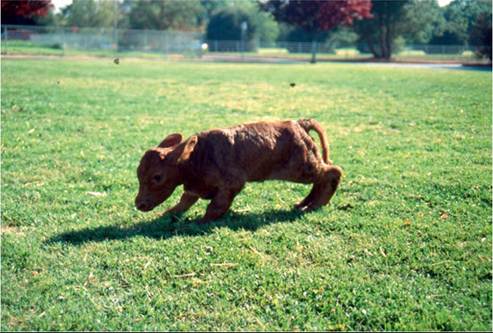
FIG. 32.138 Acorn calf, a congenital defect from dam which consumed many acorns and low quality forage during gestation. The calf has short legs, abnormal head shape and deformed hooves. This syndrome is different from acute oak/acorn toxicity.
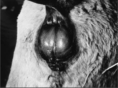
FIG. 32.139 Perirectal and vulvar edema in a 3-month-old calf with acute oak bud toxicity. Acute acorn or oak poisoning causes similar lesions.
In the peracute stages, cattle are recumbent, weak, anorexic, and listless.1 The rectal temperature is normal or below normal, and the heart and respiratory rates are elevated. Marked edema is present in the perineum and vulva (Fig. 32.139), and edema is obvious in the submandibular area, brisket, and ventral abdomen. Due to the edema, determination of hydration status is difficult, but anuria is present. Firm, dark, mucus-covered feces usually are produced. Evidence of hydrothorax, hydropericardium, and ascites may be noted on physical examination. Some affected cattle may simply be found dead.
A day or two after ingestion, affected animals appear anorexic and listless and have decreased ruminal motility.
Many affected calves have hemorrhagic diarrhea or dark diarrhea that tests positive for fecal occult blood. The feces may have a smell of phenol. Dehydration occurs rapidly, but vital signs may be remarkably normal until hypovolemia develops. As uremia progresses, scleral vessels become dark and engorged, and the breath may take on the smell of ammonia. The major differential diagnoses are other causes of renal failure and other toxins and clostridial diseases; viral diseases that cause ulceration of the alimentary tract should also be considered. Protracted cases most often result from renal failure and uremia, although some animals have chronic oral, esophageal, or gastrointestinal ulceration or perforation with abscessation.In horses, signs usually are peracute or acute; they include sudden death or colic, tenesmus, and hemorrhagic diarrhea.3 Acorn husks and shells may be noted in the feces. As with most colics, tachycardia, hyperpnea, and injected oral mucous membranes are seen. Abdominal borborygmi often is increased, and hemoglobinuria may occur. Determination of serum or urinary phenolic (hydrolyzed tannin) content, on the basis of a gallic acid standard, may be used in acute cases.3
Clinical Pathology
In cattle with peracute and acute signs, the serum urea nitrogen and creatinine levels are elevated, whereas other laboratory values may show considerable variation.2,3 Initially, hyponatremia, hyperkalemia, hypochloremia, hyperphosphatemia, marked hypocalcemia (5.1 to 6.8 mg/dL) and a mild metabolic acidosis with a very high anion gap (29 to 32) are found.1 Neutrophilia with mild hyperfibrinogenemia may be present. Although sorbitol dehydrogenase and GGT may be elevated, biopsy does not indicate that significant hepatic disease occurs with oak toxicosis. Affected animals may be anuric. If urine is being produced, often isosthenuria, proteinuria, and glucosuria occur. The urinary fractional excretion of sodium was elevated in one steer.5
In protracted cases, the major findings are elevated serum levels of urea nitrogen and creatinine and elevated anion gap, with variable hyponatremia, hypokalemia (as opposed to hyperkalemia in acute stages), hypochloremia, and hyperphosphatemia.
A mild metabolic alkalosis may be present. Hyperfibrinogenemia and an increase in total plasma proteins also are variable. An elevated WBC count most often reflects chronic ulceration or abscessation (neutrophilia or monocytosis or both). Liver enzyme levels usually are normal in protracted cases. Urinalysis results are similar to those in acute cases; in addition, hematuria often is present. By 6 weeks after exposure, a normocytic, normochromic anemia may develop as a result of chronic inflammation and uremia, and hypoalbuminemia may result from chronic renal and gastrointestinal losses. By 8 weeks after exposure, surviving cattle have largely returned to normal renal function as determined by normal serum urea nitrogen and creatinine concentrations and by the ability to concentrate urine after a 24-hour water deprivation test.1Laboratory findings in horses are similar to those in ruminants, except that during the acute stages, rapid hemoconcentration and marked increases in PCV occur.3 Although protein, occult blood, and hemoglobin casts have been reported to be present in the urine, the urine specific gravity was 1.052.
PATHOPHYSIOLOGY. The toxicity of oak is attributed to its high concentration of tannins, which are hydrolyzed in the rumen to gallic acid, pyrogallol, and other compounds. The tannins themselves may contribute to oral, esophageal, and gastrointestinal damage by binding to proteins, including those in epithelial cells. This results in oral, esophageal, and ruminal ulcers or perforations. Protein-bound tannins are liberated in the acidic abomasum, making them available once again to damage intestinal epithelium. Some hydrolyzed tannins are absorbed and bound to plasma proteins and endothelial proteins, which results in hemorrhage and fluid loss from the vascular compartment into body cavities and tissues; this results in edema. The gallic acid and pyrogallols are extremely toxic to renal tubules, causing acute tubular necrosis, anuria, electrolyte abnormalities, and uremia.
Ruminants are more susceptible than horses because of the hydrolysis of the gallotannins in the rumen.EPIDEMIOLOGY. Young cattle weighingh 100 to 300 kg seem to be particularly susceptible. In the southwestern United States, oak toxicosis accounts for considerable economic loss in cattle.5 The disease is seen sporadically in other ruminants and in horses. Adult cattle fed 10 kg of fresh oak leaves preceded by feed restriction experienced a critical reduction in rumen fermentation along with acute oak toxicity. Adult cattle that were not feed restricted did not develop oak toxicity.7 The morbidity rate in cattle varies considerably, with several to many calves in a pasture usually affected, and case fatality rates frequently exceed 80%.2
The disease is most often associated with ingestion of acorns in the fall and buds or young leaves in the spring. Cattle turned onto a pasture with oak trees under which acorns have accumulated may have toxicosis even when adequate forage is available.2 In other cases windstorms have dropped numerous acorns or branches in a pasture to which cattle were already accustomed. In the early spring, a heavy, unseasonable snowstorm may cover the grass, bend young trees, and break branches so that oak buds and young leaves are accessible.1 Young leaves and acorns are more palatable than older leaves and also contain more tannins (up to 10% dry matter in acorns). The seedlings, buds, and acorns of small scrub oak may be important forages for cattle in parts of the United States.5
Necropsy Findings
In peracute and acute cases, prominent edema is often the most striking lesion. Ascites, hydrothorax, hydropericardium, perirenal edema, and subcutaneous edema are found. The kidneys are normal in size with multiple diffuse hemorrhages on the surface and extending into the cortex. The liver is slightly swollen and pale or mottled. Digestive tract lesions vary from congestion to hemorrhage and deep ulceration or perforation with necrotizing inflammation.
Mucosal ulceration is found in the pharynx, esophagus, rumen, abomasum, small intestine, cecum, and colon. Free blood or melena may frequently be observed.Diffuse renal tubular damage is present. The changes include coagulation necrosis of cortical tubular epithelium and dilated tubules devoid of epithelium but with intact basement membranes. Many medullary tubules contain hyaline, granular, or cellular casts. The principal renal lesion is necrosis of the proximal convoluted tubules, but glomerular degeneration and fluid in Bowman capsules are also seen.
By 3 to 6 weeks after exposure, the lesions include gastrointestinal ulceration, often with secondary infection; some healed ulcers; secondary bacterial bronchopneumonia; and slightly swollen, pale brown kidneys. Histologically, atrophy of the cortical tubular epithelium with a marked interstitial fibrosis and mononuclear infiltrate is seen. Evidence of tubular epithelial regeneration may be present. The liver appears normal.
Treatment and Prognosis
In acute stages, intravenous fluid therapy aimed at promoting diuresis and correcting acid-base and electrolyte abnormalities is indicated. Calcium, sodium, and chloride deficits should be replaced, and sodium bicarbonate should be given if needed to correct metabolic acidosis. In anuric animals, furosemide (1 mg/kg given IV) can be administered every 12 hours, along with adequate fluid therapy. Corticosteroids and NSAIDs are not likely to have a measurable effect on the course of the disease because they are unlikely to alter the direct toxic effects of the metabolites. The prognosis is guarded and must be considered poor in animals that remain anuric despite therapy. The case fatality rate undoubtedly depends more on the amount of toxic material ingested than on therapy. Antibiotics, given to prevent secondary pneumonia and abscessation, ruminal transfaunation, and readily accessible grass hay and water, are recommended components of nursing care.
If the animal survives the acute stage and begins to eat voluntarily, the prognosis for recovery is good unless secondary pneumonia or gastrointestinal abscessation after perforation occurs. Renal function can return to normal by 5 to 10 weeks, and weight gains as high as 1.76 kg/day have been recorded in recovered cattle.1
Colicky horses can be treated as usual for colic (intravenous fluids, analgesics, oral laxatives). Particular attention should be paid to diuresis, acid-base balance, and maintenance of serum calcium levels.
Prevention and Control
No specific antidote exists for oak toxicosis, but supplementation with calcium hydroxide (hydrated lime) immediately before anticipated exposure to oak has been effective under experimental conditions.8 Feed containing 10% or less calcium hydroxide is palatable. Consumption of 0.9 kg/head/day of cubed or pelleted supplement containing 10% hydrated lime is effective in preventing toxic manifestations.
Supplementation of cattle suddenly exposed to oak with any feed reduces death losses, apparently by allowing hungry cattle to consume the supplemental forage preferentially.1,8 In an outbreak in California caused by spring snows in which 2700 cattle died of oak toxicosis, ranches where hay was immediately supplemented had fewer losses than did ranches where feed supplementation was not offered.1