Winter Dysentery in Cattle (Bovine Coronavirus)
GiUes Fecteau • Charles L. Guard
Definition and Etiology
Winter dysentery is an acute, contagious diarrheal disease of cattle (especially dairy) that occurs in epizootic manner in a herd, usually during the colder months of the year (November to April).
It is usually recognized by the clinical syndrome that occurs in herds and by exclusion of other causes of contagious diarrhea.1 In the United States the disease is more common in the northern states; it has also been reported in Australia, Sweden, the United Kingdom, Israel, France, Belgium, Japan, and Canada. The period of illness in an individual is brief (a few days to a week), and within a herd the outbreak usually lasts less than 2 weeks. Recovery is spontaneous in most individuals, but in some cases, supportive therapy is necessary. The morbidity rate is usually high (from 30% after a few days to almost 100% after 1 week to 10 days). Mortality, however, is usually rare.The cause of winter dysentery is a bovine coronavirus that may be related (antigenic differences are reported) to the coronavirus that causes diarrhea in neonatal calves.2-7 By immune electron microscopy, antigenic cross-reaction has been demonstrated between the original Nebraska calf coronavirus diarrhea agent and coronavirus isolated from field outbreaks of winter dysentery.4,6
Clinical Signs and Differential Diagnosis
Winter dysentery is an explosive diarrheal disease accompanied by some degree of anorexia and depression. In lactating cows, milk production is severely reduced. If the diarrhea is severe or persists longer than one or 2 days, dehydration may develop. Loss of body condition is apparent. Rumen motility usually is reduced, but intestinal borborygmi may be increased. Thirst often is increased, and polydipsia follows the diarrhea. Rectal examination may reveal dilated intestinal loops.
The feces vary from light tan to dark brown; bubbles commonly form in the puddles that are deposited several feet behind the cow. Blood may be present in the feces of several animals in a group, typically in the first-lactation cows. The amount of blood may range from just visible to large clots, or it may be uniformly mixed into the feces. Some animals may have thick mucus in the feces. The odor in a barn during an outbreak of winter dysentery has been described as musty, fetid, and sweet and nasty.8Fever usually is not present during the diarrheal phase of the disease but has been reported to precede it1 or have no consistent relationship.9 Mild respiratory signs consisting of serous nasolacrimal discharge and a soft cough have been inconsistently observed before diarrhea. Diarrhea caused by BVDV, coccidiosis, and salmonellosis must be considered in the differential diagnosis of winter dysentery. With winter dysentery, no mucosal lesions are visible on physical examination. The absence of coccidial oocysts or parasite ova in the feces helps exclude these agents as responsible for the diarrhea. The rapid occurrence of multiple cases within a herd, and often within several herds in a locality, suggests winter dysentery. Fecal culture for Salmonella yields negative results in uncomplicated cases of winter dysentery.
Clinical Pathology
No consistent hematologic changes that would be of diagnostic benefit have been observed. If significant dysentery persists for longer than a day, signs of anemia may develop.
Pathophysiology
Epithelial cells of colonic crypts are destroyed by viral action, which leads to necrosis and hemorrhage. Even though histologic changes have been observed only in the colonic mucosa, blood was observed from the distal duodenum aborally in cattle that died of winter dysentery.7 Loss of intestinal mucosal epithelium from colonic crypts leads to transudation of extracellular fluid and blood.
Epidemiology
The incubation period is thought to be 2 to 8 days. The most susceptible animals are first and most severely affected. In a small, housed herd, the typical incidence of diarrhea during an outbreak begins with the explosive appearance of signs in 10 to 15% of animals on the first day. The second day, another 20 to 40% are affected. On subsequent days, similar proportions become ill. By the end of a week, the first affected animals are completely recovered, and only a few new cases occur. Typically within 2 weeks of the onset of diarrhea, all animals have recovered. This period is marked by significant reduction in milk production. In large herds the outbreak may be prolonged for 6 to 8 weeks. This scenario is typical of a herd that had not experienced an epizootic of winter dysentery during the preceding few years. In some herds, milder outbreaks occur annually, with fewer animals showing diarrhea and with less severe clinical signs.
Treatment and Prognosis
Most animals with winter dysentery recover spontaneously in a few days without specific treatment. Many palliative treatments have been recommended and used over the years. On the basis of 30 years of observations, Roberts10 considered none of these treatments to alter the course of the disease. Provision of adequate fresh water, palatable feed, and free-choice salt is the most useful nonspecific therapy. The occasional animal with prolonged or severe dysentery may need a transfusion of 4 to 8 L of whole blood.
Salmonellosis in Ruminants
Bradford P. Smith
■ Definition and Etiology Salmonellosis is one of the few diseases that appear to be increasing in prevalence, especially in dairy cattle. In addition, more than one million human cases of nontyphoidal salmonellosis occur yearly in the United States (90 million worldwide), and so this is an important public health concern. Salmonella serovar Dublin was isolated from the bloodstream of 60% of 228 human patients suffering from Salmonella serovar Dublin infection in the United States; 74.9% required hospitalization and 3.7% died, and so it is considered the most severe nontyphoidal Salmonella infection in humans.
Twenty eight per cent of isolates were resistant to sulfas, tetracycline, ampicillin, chloramphenicol, amoxicillin-clavulanic acid, and ceftriaxone.1Large dairy farms are more likely to be infected with Salmonella than are small dairy farms.2 Milk yield decreased significantly in affected herds, and the reduction was detectable several months after the increase in bulk tank milk Salmonella antibodies. It took more than 1 year for milk yield to return to preinfection levels.3 According to one study, 75% of dairies sampled in California had evidence of Salmonella infection.4 A multistate study demonstrated that 56% of dairies sampled contained cows shedding Salmonella in feces.5 Salmonella of concern for livestock belong mainly to one species, Salmonella enterica subsp. enterica, with numerous serovars or serotypes, such as S. enterica subsp. enterica serovar Typhimurium, with the words serotypes and serovars used interchangeably.6 (In this text, the terms are shortened it to, for example, Salmonella Typhimurium by convention.) More than 2500 Salmonella serotypes (serovars) have been identified, and many are potential animal and human pathogens. Multidrug-resistant Salmonella Typhimurium DT104 (serogroup B) and Salmonella Newport (C2) are particularly virulent in animals and human beings. New variants occur; a variant of Salmonella Typhimurium designated I 4,[5],12:i:- lacking one H antigen has been a cause of clinical disease in cattle in the USA (Table 32.23; personal communication from Brenda R. Morningstar-Shaw, National Veterinary Services Laboratory, USDA, 2018). The presence of identical strains of Salmonella Newport among bovine and human isolates indicates that zoonotic transmission is likely to occur.7 Fortunately, only 10 serotypes of Salmonella in serogroups B, C, D, E, and K are responsible for most disease in cattle; therefore control programs can be aimed at these.
Table 32.23 lists the serotypes most commonly isolated from ill cattle in 2016 in the United States.
Group K, not long ago considered an exotic serogroup, has become prevalent (number 2).8 Salmonella organisms are Gram-negative enteric bacteria that are facultative intracellular parasites. Most■ TABLE 32.23
Most Frequently Isolated Salmonella Serovars or Serotypes From Ill Cattle in the United States in 2016a
| Order of Frequency | Serovar or Serotype (Serogroup) | Total | Percentage |
| 1 | Dublin (D1) | 332 | 23.5% |
| 2 | Cerro (K) | 275 | 19.5% |
| 3 | Typhimurium (B) | 142 | 10% |
| 4 | Montevideo (C1) | 110 | 8% |
| 5 | Heidelberg (B) | 101 | 7% |
| 6 | Newport (C2) | 67 | 5% |
| 7 | I 4,[5],12:i:-* (B) | 54 | 4% |
| 8 | Anatum (E1) | 33 | 2% |
| 9 | Kentucky (C3) | 26 | 2% |
| 10 | Muenster/ Meleagridis (E1) | 22 each | 1.5% each |
| All others | 226 | 16% | |
| Grand Total | 1414 | 100% |
aVariant of Typhimurium.
Data supplied by Brenda R. Morningstar-Shaw, National Veterinary Services Laboratories, United States Department of Agriculture, Ames, IA, 2018.
serotypes are non-host-specific, but a few are host-adapted. Salmonella Dublin is host-adapted to cattle, Salmonella abortus ovis and Salmonella Arizonae to sheep, and S. abortus equi to horses. Host-adapted serotypes are found most often in their host species, where true long-term carriers exist.9 In contrast, non-host-adapted serotypes rarely achieve carrier status and usually infect an animal for a period of 3 to 16 weeks before infection is cleared,9 although one cow with multidrug-resistant Salmonella Newport excreted the organisms for 190 days.7 Infection with either host-adapted or non-host-adapted serotypes can be symptomatic or asymptomatic, depending on the dose, the virulence of the serotype, and the host's immune status. There appears to be an association between an increased incidence of Salmonella infections and intensive management practices such as large farms, crowded conditions, and high- protein diets. In the absence of carriers, chain infections and persistence of Salmonella in the environment are responsible for long-term persistence of a given strain on a given dairy.7
Salmonella are grouped into serogroups (A to Z and 51 to 65), depending on their cell wall antigens (somatic antigens that comprise specific oligosaccharides, often called O antigens, LPSs, or endotoxin). Thus for example, local and regional laboratories often report that an isolate is a group D Salmonella. The most common group D isolate of cattle is Salmonella Dublin, although nonmotile isolates that do not express flagella at that time also can be found. The author and colleagues found that these would express motility and be typed as Salmonella Dublin if the isolate were inoculated into semisolid media where it had to move to get nutrition (Smith BP, personal observation). Final confirmation that the Salmonella isolated is indeed Salmonella Dublin comes from the National Veterinary Services Laboratory in Ames, Iowa, or from some other laboratory in which antisera and facilities exist for identification of all 2500 serotypes on the basis of flagellar (H) antigens and somatic (O) antigens. Some serotypes also express capsular (Vi) antigens composed of mucopolysaccharide under some conditions. Identification of the serotype is important in planning control strategies. In addition to the serotypes listed, lambs may be infected with Salmonella Arizonae, which comes from several different serogroups. Salmonella organisms are relatively easy to culture from the feces or tissues of infected animals, with the use of common enteric agar such as Hektoen Enteric, XLT4 or brilliant green agar, with tetrathionate and selenite as selective enrichment media.
Clinical Signs and Differential Diagnosis
Salmonella infection can cause a variety of clinical signs. The most common signs are fever and diarrhea10 after an incubation period of 1 to 4 days after exposure or a recrudescence from the carrier state. Ruminants of all ages may be affected, but serious illness is observed most frequently in very young animals and parturient dairy cattle.
The character of the diarrheic feces varies from watery to mucoid with fibrin and blood. Sheets of fibrin may look like sloughing mucosa. The feces often have a putrid, foul odor because of the presence of plasma proteins associated with severe inflammatory bowel disease. Salmonellosis causes an acute protein-losing enteropathy. The systemic effects of endotoxins (and other toxic material) absorbed through the damaged bowel mucosa may be severe (fever, anorexia, depressed attitude, and shock).
Bacteremia may occur rapidly, especially in neonates infected with Salmonella Dublin, Typhimurium, or Newport. Peracute to acute septicemia in calves may produce lesions in many organ systems. Affected calves usually are younger than 2 months of age (the range is 1 day to 6 months). Dyspnea, respiratory symptoms, sudden death, and occasionally diarrhea are the principal signs of Salmonella Dublin infection, the incidence of which usually peaks in 6-week-old calves. Blood culture results frequently are positive. Pure cultures of Salmonella Dublin often can be grown from specimens from the lungs of 4- to 8-week-old calves with pneumonic (septicemic) salmonellosis. Calves infected with Salmonella Typhimurium are only 14 days of age on average (the range is 1 to 35 days) and have mainly enteric lesions, including enlarged mesenteric lymph nodes, abomasitis, Abrinonecrotic plaques in the ileum, and hemorrhage of Peyer's patches.11
Dry gangrene of the extremities may occur in calves after Salmonella infection, particularly with Salmonella Dublin. Legs, ears, and tails are most commonly affected, and cold agglutinins are suspected to be involved.12
A strain of Salmonella Saint Paul was implicated in an outbreak of encephalopathy (without encephalitis) in stressed Holstein calves.13 Calves exhibited diarrhea, head tilt, ataxia, auditory hyper-responsiveness, and partial blindness. The β-lactam antibiotics ceftiofur and ampicillin protected against the neurological effects by increasing glutamate export from the brain despite resistance of the Salmonella St. Paul isolate to both drugs.14
In adult cattle, diarrhea or abortion may occur.9 Abortion may occur by two mechanisms because both culture-positive and culture-negative fetuses and placentae may be found in an outbreak. In the former, bacteremia in the dam results in infection of the placenta or fetus, with resulting fetal death and expulsion. Bacteremia, endotoxemia, and fever associated with diarrhea and damaged gut mucosa in cattle may result in release of prostaglandin F2α and subsequent lysis of the corpus luteum. Abortion follows in 2 to 3 days, and the fetus and placenta yield culture-negative findings. When the fetus has a culture-negative finding, many other causes of abortion must be considered. Many infectious agents that infect only the dam can act similarly to cause luteolysis and abortion.
Differential diagnoses for the enteric form of salmonellosis in neonates include all the common enteropathogens of neonates: Escherichia coli, rotaviruses and coronaviruses, clostridia, cryptosporidia, and other forms of coccidia. Concurrent infection with these agents is common. In older animals outbreaks should be differentiated from those caused by BVD, winter dysentery, and feed-induced indigestion. Pneumonic and bacteremic salmonellosis in 4- to 8-week-old calves must be differentiated by culture from pneumonic pasteurellosis; in goats, from mycoplasmal infection; and in lambs, from septicemic pasteurellosis.
Clinical Pathology
Salmonella enteritis often results in changes in the hemogram,15 as effects of endotoxin manifest.16 Plasma fibrinogen frequently is elevated because of the inflammatory nature of the disease, and either neutrophilia or neutropenia may be seen. In severe cases a left shift may be present. Initial dehydration may result in an elevated PCV and plasma protein level. The plasma protein level often drops over a period of days as protein is lost into the bowel lumen. Many other nonspecific abnormalities in clinical chemistry values are often seen, including elevated liver enzymes, decreased plasma calcium, and indications of prerenal azotemia if dehydration occurs.
Definitive diagnosis of salmonellosis requires culture of the organism from feces, blood, or tissues. PCR is also available. Culture was most sensitive and specific in comparison with ELISA and PCR.17 Pooled fecal cultures from five cows provide a sensitivity of 62.5%, in comparison with individual samples from culled dairy cows.18
Pathophysiology
Salmonellae are invasive organisms that may penetrate ocular, nasal, oral, or intestinal mucous membranes. Once eaten, salmonellae attach to mucosal cells, probably by means of a nonfimbrial adhesion.19 Attachment is increased if gastrointestinal stasis is present or the normal competitive flora has been disturbed or is not yet established, as in neonates. Salmonellae cause degeneration of nearby cells and penetrate both through microvilli and through tight junctions between cells.20 The Salmonella type III secretion system translocates five Salmonella effector proteins (SipA, SopA, SopB, SopD, and SopE2) into enterocytes, and is required for induction of diarrhea by inducing intestinal fluid secretion and recruiting neutrophils to the infection site. 21 The bacteria also pass through the enterocytes to the lamina propria, where they stimulate an inflammatory response and are engulfed by macrophages and neutrophils.22 Intracellular bacteria reach regional mesenteric lymph nodes or beyond. The organism has a predilection for lymphoid tissues and is found in highest numbers in Peyer's patches and mesenteric lymph nodes.23 This is one reson why culturing mesenteric, iliac and other lymph nodes is useful. Thrombi form in vessels, and tissue damage can be severe. Virulent salmonellae are capable of surviving in host tissues and multiplying, often as facultative intracellular parasites in macrophages and reticuloendothelial cells.24 In the case of carrier animals, salmonellae survive in cells in the presence of high titers of specific extracellular serum immunoglobulins. This is the basis for using ELISA serologic testing to detect Salmonella Dublin carriers. Intracellular salmonellae can also avoid many antimicrobial drugs and complement. Stress may cause a latent infection to recrudesce, which results in fecal or mammary gland shedding or clinical disease.
The cell wall of all Gram-negative bacteria contains LPS as a component. LPSs are also called endotoxins, and they are extremely biologically active, setting off an immunologic cascade of events that can lead to leukopenia, fever, anorexia, and eventually shock and death.16
■ Epidemiology and Control Salmonella infection is most often transmitted by fecal-oral contamination from livestock or rodents, or by feeding of contaminated protein source animal by-products (e.g., fish meal, meat meal, bone meal, or feather meal, many of which are contaminated) or contaminated forages or plant proteins such as soybean or cottonseed. Of cattle feed tested for Salmonella in 2010, approximately 6.1% was positive, according to U.S. Food and Drug Administration surveillance data. Even if small numbers of organisms are initially present, they multiply rapidly in moist warm conditions.
Salmonella Dublin is an endemic herd problem and is shed by lower numbers of animals compared with Salmonella Typhimurium, which tends to be epidemic and shed in higher numbers by many more animals at a time.25 In 2009 in the northeast United States, in 11% of 831 herds with suspected clinical cases of salmonellosis, the leading isolates were Salmonella serovars Newport and Typhimurium.26 A study of calves in California found that 18% of 1-day-old calves shed some serotype of Salmonella, but this percentage declined rapidly over the first 11 weeks of life. Perhaps even more alarming, a fetus may be infected with Salmonella in utero and thus vertical transmission occurs as well.27 Salmonella Typhimurium, Muenchen, Cerro, and Kentucky were isolated from mesenteric, subiliac, and prescapular lymph nodes and calves at parturition.27 Farms that received calves from other sources were 35 times more likely to have calves that were shedding Salmonella in feces than were closed herds.28 Salmonella Dublin, Newport, and Typhimurium can also be tranmitted via colostrum and milk.29
Infection with host-adapted Salmonella serotypes may occur as a cyclic endemic disease, especially in calves when Salmonella Dublin is involved. Salmonella Dublin infections on a farm are maintained by carrier animals shedding Salmonella Dublin in the feces9 or milk,30 chain infections in infected calves and cows, rodents, and environmental contamination. Salmonella carriers infected with a host-adapted serotype such as Salmonella Dublin may shed constantly or intermittently. Cattle with the highest risk of becoming Salmonella Dublin carriers are heifers infected between 1 year of age and first calving and cows infected in the parturient period,31 but calves surviving Salmonella Dublin infection and illness may also become carriers (BPS personal observation). Infected carriers that are not shedding are called latent carriers; stress may cause shedding in feces or milk to recrudesce. A single infected, asymptomatic fecal shedder may produce over one billion Salmonella Dublin organisms per day to contaminate the environment (one million organisms per gram of feces). Unlike most other serotypes, Salmonella Dublin may be carried as a chronic gut and mesenteric lymph node infection and passed in feces or carried as an intramammary infection and passed in contaminated milk.
Calving areas must be constantly cleaned and disinfected between calvings. Raw milk feeding to calves may be a source of Salmonella (100 to 100,000 organisms per milliliter of milk).30 Raw milk must not be allowed to sit unchilled, as this allows Salmonella and other pathogens to multiply (numbers can double every 20 minutes at 100 F (37C). Acidification of milk to a pH lower than 4 for 2 hours can effectively eliminate most pathogens, including Salmonella spp. and M. bovis from milk; acceptance of taste by calves may be an issue with some acidification techniques.32 Infected calves constantly amplify the number of organisms in the environment, causing exposure of other calves. Calves with clinical signs may shed billions of Salmonella into the environment daily. See Chapters 19 and 20 for more on calves.
Environmental contamination can be difficult to eliminate because salmonellae survive for months in moist areas out of direct sunlight and in lagoons and drainage areas. Cows shed more Salmonella on a commercial dairy in hot weather by a factor of 4.5X for every 5C rise in ambient temperature.33 Salmonella Dublin was recovered from feces dried for 41 months.34 Freezing feces at -20° C (-4° F) kills 85% of salmonellae in 2 days and more than 95% by 1 month.35 Direct sunlight and drying in hot weather are effective at eliminating salmonellae. Using composted manure for bedding can be effective: Salmonella Newport survived less than 24 hours in composted manure reaching 69° C (115° F) but survived for more than 4 months in the lagoon and more than 9 months in soil.36
Research has documented contaminated irrigation water as an important source of Salmonella organisms. In some cases municipal sewage treatment plant effluents had contaminated water.37 On one dairy farm, river water used to sprinkler irrigate green chop contaminated the green chop, which was fed to cows.38 On another farm, Salmonella-contaminated river water was used to irrigate cotton. When the cottonseed from that gin was used as feed, cattle became infected.38 On a third dairy farm, contaminated irrigation water resulted in haylage containing Salmonella organisms.38
Feedlot cattle colonized by Salmonella organisms could pose a public health risk when meat is contaminated at slaughter. In a study by National Animal Health Monitoring System, 7.5% of cattle on feed the longest shed salmonellae, whereas 3.5% of cattle that had recently entered the feedlot were shedding salmonellae in the feces.39 A study of 68 feedlots in 12 states found that 9.1% of 5050 fecal samples were positive for Salmonella and 64.7% of feedlots had at least 1 positive sample, and that fewer than 10% of isolates were resistant to antimicrobials other than sulfas and tetracyclines.40 Contamination of the animals before entry can cause the entering cattle to have a higher rate of shedding than was found in cattle that had been in the feedlot for several months.41 A major difference in the incidence of Salmonella-positive lymph nodes was found between two different feedlots and in time on feed, with cattle on feed more than 120 days having 94% (17/18) Salmonellapositive lymph nodes.42 These lymph nodes are believed to be a major source of contamination for beef.
Standing water contaminated with feces is frequently highly contaminated with Salmonella; culture of temporary shallow lakes (calledplayas in the southwestern United States) yielded numerous Salmonella serotypes.43 An unusual outbreak of Salmonella Newport in cows and calves on a beef farm resulted in a cow mortality rate of 7.9% (32 of 407) and a calf mortality rate of 14.4% (52 of 361). BVD virus was also diagnosed in the group, and it was suspected of playing an immunosuppressive role.44
Control of Salmonella infection requires an integrated herd approach.45,46 Pens of sick animals were found to be 7.4 times more likely to yield Salmonella than were pens of preweaned calves.47 Management interventions that were found effective in helping control Salmonella on dairy farms include separation of sick and parturient cattle into hospital and maternity pens, respectively (considered the most important and effective intervention), switching from a phenolic disinfectant to a peroxygen biocide such as Virkon S (DuPont, Sudbury, U.K.) for equipment disinfection, use of separate footwear or footbaths for personnel in the hospital and maternity pen sections, strict protocols for disinfection of hands and equipment between handling each sick or periparturient cow, strict separation of feeds used in the hospital and maternity pens, and use of clean bedding in the hospital and maternity pens.7 Having one person manage the calves, good colostrum management, and not purchasing cattle from an infected premise had beneficial effects in reducing infection.48
To detect herds infected with Salmonella Typhimurium or Dublin, a herd serologic profile based on ELISA can be used.49 The commercial test available in the United States is the
PrioCHECK Salmonella Ab bovine ELISA (Prionics, Switzerland). One laboratory that runs such tests on milk or serum is the Animal Health Diagnostic Center at the College of Veterinary Medicine, Cornell University. Rectal swab sampling and culture of culled dairy cows was found not to be a satisfactory method of detecting Salmonella-infected herds.50 To control Salmonella Dublin, carrier cows and calves older than 6 months of age are identified by serologic testing (ELISA) for antiSalmonella antibody.30,51-53 Suspect animals are culled or tested further by serologic means or by multiple fecal cultures and milk cultures (five samples at weekly intervals). Strongly positive results of serum ELISA tests over an 8-month period are considered to indicate carrier status.54 Animals with positive test results are culled. In infected herds, Salmonella Dublin carriers (fecal or mammary) usually make up 0.4% to 3% of a dairy herd. A Danish study55 showed that only 14 of 31 persistently seropositive animals were culture positive for Salmonella Dublin at postmortem examination, and one seronegative animal was culture positive. One study found that ELISA serologic testing had a good negative predictive value for cattle 100 to 300 days of age, whereas fecal culture had a poor predictive value.56
To control all non-host-adapted and host-adapted serotypes of Salmonella, calves and cows are promptly treated when signs of illness occur, and strict procedures to prevent the spread of infection are instituted. A study of 14 farms infected with Salmonella Typhimurium DT104 showed that clinical disease ceased by 4 months, but widespread infection and contamination continued. Over time the number of infected farms gradually declined, and vaccination correlated positively with a herd remaining free of clinical illness from DT104.57 Control measures include the use of separate feeding utensils for each calf, washing of boots and hands by calf raisers between calves, isolation of ill calves to noncontact pens, and thorough disinfection of pens between calves. Decontamination of pens is most effectively accomplished if an all-in, all-out system is used. Pens are divided into four groups, with each group of pens used for calves born during a given week. When 3- to 4-week- old calves leave their pens as a group, these pens are cleaned and disinfected with a chlorine product such as bleach, a peroxygen product such as Virkon S, or a phenylphenolic disinfectant (also see Chapter 46 for more on disinfection). This helps prevent continuous recycling of bacteria to each group of new calves. Environmental monitoring by frequent swab cultures of pens is the best way to check the effectiveness of the sanitation and disinfection program.
Infection with Salmonella Typhimurium and other non-host- adapted serotypes most often occurs as an epidemic after the organisms enter the farm by means of feedstuffs, purchased cattle, rodents,58 or rendering trucks. Even Salmonella Dublin may be maintained on a farm by mice.59 A case control study of farms infected with Salmonella Typhimurium DT104 found that the most effective interventions included purchasing cattle directly rather than through a sales yard, quarantining purchased cattle for 4 weeks, housing sick cattle in dedicated isolation areas, and preventing wild bird access to feed storage areas.60 Feeds may be contaminated by irrigation water,38 in manufacturing when by-product ingredients contain Salmonella organisms, on the farm, or in shipment by birds or rodents or contaminated trucks. When an epidemic involving a common or exotic serotype occurs, feedstuffs should be examined as a likely source. Exotic serotypes are Salmonella from serogroups other than B, C, D1, E1, and K; that is, all those groups not listed in Table 32.23. High-protein supplements, rumen bypass protein, or calcium-phosphorus supplements of animal origin are sources of epidemics of various Salmonella serotypes in livestock. Many animal by-product feedstuffs in the United States are contaminated with Salmonella organisms. Once a Salmonella serotype enters a herd, the bacteria are rapidly spread among livestock and into the environment, where they may cause a prolonged course of herd illness and can be difficult to eliminate. Such appeared to be the case with Salmonella Newport and Salmonella Montevideo in dairy cattle in California in the past (Table 32.24). These and other group C Salmonella spp. tend to become endemic and persist for years on a dairy farm once it has become infected. By 2016, Salmonella Montevideo was the fourth and Salmonella Newport the sixth most prevalent serotypes from ill cattle in the United States (see Table 32.23; National Veterinary Services Laboratory data). Culture of feeds before use on a farm currently is the only means of preventing the introduction of new Salmonella serotypes via feedstuffs because national and international feed quality controls currently appear to be inadequate.
■ TABLE 32.24
Changing Incidence of Salmonella Serotypes (Serogroup) From Ill Calves on California Dairy Farms, 1985 to 1988
| Serotype | Percentage (n) of Farms From Which Each Serotype Was Isolated | |
| 1985-1986 | 1987-1988 | |
| Dublin (D1) | 78 (29) | 53 (49) |
| Newport (C2) | 19 (7) | 36 (33) |
| Typhimurium (B) | 3 (1) | 7.6 (7) |
| Montevideo (C1) | 0 | 6.5 (6) |
| Infantis (C1) | 0 | 2.2 (2) |
| Anatum (E1) | 0 | 1.1 (1) |
| Muenster (E1)a | 0 | 1.1 (1) |
aFormerly called “S Arkansas.”
From Blanchard P, Anderson M. Unpublished data based on calves brought for necropsy to the laboratories of the University of California, Davis, Veterinary Medicine Teaching & Research Center and the California Animal Health & Food Safety (CAHFS), Tulare, Calif., from Tulare area farms, 1988.
VACCINATION. Control by vaccination of dams with killed bacterins may be somewhat beneficial in protecting neonates under 3 weeks of age through colostral and milk immunoglobulin. Because most dairy calves are taken from their dam and fed milk replacer after day 1 to day 3 of age, the potential benefit of intraluminal milk antibody from the dam is lost. This may account in part for the fact that vaccination of cows by itself often is insufficient to protect dairy calves from salmonellosis. In neonates over 3 weeks of age, colostral immunity appears to play only a small role in protecting against salmonellosis. Because both humoral and cell-mediated immunity are important in protecting against salmonellosis,60-62 active immunity is superior to passive. Modified live-virus, genetically altered vaccines that stimulate humoral and cell-mediated immunity offer better protection for calves than do killed whole cell vaccines.62,63
The role of increased nonspecific immunity to Gramnegative core antigens (through immunization with rough mutants of E. coli and salmonellae lacking complete cell walls) has been investigated. Some studies have been able to demonstrate an increased survival rate for salmonellosis after vaccination with a mutant strain of E. coli (J5),64 designed to produce antibodies to the common core antigens of Gramnegative bacteria. Two such products available in 2012 are Enviracor J-5 (Zoetis, Florham Park, N.J.) and J-Vac (Merial, Duluth, Ga.). Endovac-bovi (Re mutant Salmonella Typhimurium bacterin-toxoid produced by Immvac Inc., Columbia, Mo.) is marketed with the same goal.
Available commercial Salmonella vaccines are killed whole-cell bacterins for use in cattle and a genetically altered modified live Salmonella Dublin vaccine for calves (EnterVene-D [Boehringer Ingelheim Vetmedica, St Joseph, MO]. Although the vaccine is to be given subcutanously, some practitioners report that they have used the live vaccine orally and found it to be effective but with fewer adverse reactions than when used parenterally. EnterVene-D was used in dry cows without any observed adverse reactions, and produced an increase in colostral antibody against Salmonella Dublin, but no natural or experimental challenge was given to assess its effective- ness.65 The only products that are solely Salmonella vaccines contain (1) Salmonella Typhimurium, (2) Salmonella serovars Typhimurium and Dublin, (3) live Salmonella Dublin organisms, or (4) siderophore receptor protein (SRP) Salmonella Newport subunits (also see Chapter 48). Autogenous bacterins can be made for serotypes not commercially available when they are isolated from animals on a farm and believed to be the causative agent of disease. A subunit SRP vaccine called Salmonella Newport Bacterial Extract vaccine is being marketed by Zoetis for vaccination of cattle against Salmonella and other Gram-negative bacteria.
Siderophores (SRPs) are iron chelating proteins produced by all Salmonella, so producing antibodies against SRPs from Salmonella Newport should theoretically be effective for all serotypes of Salmonella, and because they are a subunit, they should be less toxic. The initial vaccination requires 2 doses at 2- to 4-week intervals, followed by a yearly booster. The SRP vaccine did not decrease fecal shedding in feedlot cattle (which were mainly shedding Salmonella Montevideo [C1] and Salmonella Anatum [E1] and not Salmonella Newport [C2]).66 To the author's knowledge, no peer reviewed controlled studies documenting the effectiveness of this SRP vaccine at controlling salmonellosis have been published as of early 2018. A major problem associated with Salmonella bacterins and modified live Salmonella vaccines is adverse reactions. Adverse reactions vary from fever, depression, and anorexia to collapse and death after vaccination. Affected animals may have severe reactions after the first, second, or third dose. The apparent cause of these severe reactions is a high degree of sensitivity to the presence of Gram-negative endotoxin, which results in a rapid immune cascade that leads to shock.16 This sensitivity is genetically controlled, possibly accounting for the high frequency of adverse reactions in one herd and their absence in a neighboring herd. High ambient temperatures also increase sensitivity to the adverse effects of endotoxin. Adverse reactions usually can be averted or reversed with epinephrine (1 mL of 1 : 1000 [1 mg/ mL] for every 50 kg of body weight given IM or IV) as soon as evidence of polypnea, dyspnea, or weakness appears.
Lack of efficacy of killed Salmonella bacterins in calves is evident in experimental studies. Commercial bacterin vaccines in aluminum hydroxide administered directly to calves at 2 and 4 weeks of age failed to protect the calves when they were orally challenged at 6 weeks of age.64 Other research has demonstrated protection in older vaccinated calves that were challenged intravenously.67 Calves younger than 12 weeks of age do not serologically respond with anti-LPS antibodies after vaccination with two or three doses of killed bacterins in aluminum hydroxide.68 On the other hand, calves older than 12 weeks of age and cows do respond serologically to two doses of the same vaccine.69 The ELISA titer in cows is shortlived (2 to 4 weeks); therefore the timing of vaccination before parturition is critical if the aim is to increase the anti-Salmonella colostral immunoglobulin G titer.69 Under experimental conditions, calves fed colostrum from vaccinated dams were not protected against oral challenge at 3 weeks of age.15 One report suggests that vaccination with killed Salmonella Typhimurium vaccine prevented reinfection and recrudescence of clinical signs after an outbreak of DT104 and helped in eliminating the organism from the farm.59 As mentioned previously, these differences may in part be explained by whether a calf continues to receive milk from its dam and thus has or does not have intraluminal antibodies. Other live vaccines have been found effective in calves and capable of cross-protection against other serotypes, including a DNA adenine methylase-deficient Salmonella Typhimurium70 and an avirulent live Salmonella Choleraesuis strain 54,71 but neither was licensed for cattle in the United States as of 2018.
Necropsy Findings
ENTERIC FORM. Many affected calves are emaciated and have serous atrophy of fat. Especially with Salmonella Typhimurium, the ileal, cecal, or colonic mucosae may be thickened and hemorrhagic with adherent fibrinonecrotic plaques. Fibrin may be found in sheets, especially pronounced in the areas of Peyer patches. The abomasum, duodenum, and jejunum are often involved but less severely. The abomasum usually contains brown, fetid liquid. Bowel contents often are watery and may contain fibrin, blood, or both.11 The mesenteric lymph nodes frequently are enlarged and dark. Chronically affected cows or calves may have discrete areas of necrosis in the cecum or colon that may involve the entire thickness of the wall.11
SEPTICEMIC FORM. Gross lesions of bacteremic Salmonella infection are acute and usually subtle. Salmonella Dublin is often septicemic in calves. Serosal and subcutaneous petechiae are widespread. The spleen usually is enlarged. The lungs are edematous and fail to collapse and have random foci of hemorrhage or congestion.11 The wall of the gallbladder is thickened and may contain hemorrhages. The bile frequently is inspissated into a firm coagulum.11 Less common lesions include jaundice, cystitis, meningitis, osteomyelitis, arthritis, and dry gangrene of ears, tail, or feet. Gastrointestinal lesions may or may not be present. Histopathologic study of Salmonella Dublin in calves younger than 6 months old demonstrates pulmonary alveolar capillary neutrophilia with or without hepatocellular necrosis and paratyphoid granulomas, splenitis and lymphadenitis.72
Treatment
The keys to successful treatment of bacteremia caused by Gram-negative bacteria are (1) antimicrobial drugs, (2) fluids and electrolytes, and (3) NSAIDs. Salmonella Typhimurium antiserum is available commercially. Adverse reactions by cattle to horse serum are common; care should be taken.
Early in the course of disease in calves, appropriate antimicrobial therapy often increases the survival rate markedly because bacteremia frequently occurs. Appropriate antimicrobial therapy depends on culture and antimicrobial sensitivity patterns. Many of the most virulent Salmonella types are now multidrug-resistant, but isolates of relatively nonpathogenic Salmonella on dairies and feedlots found on surveys are usually sensitive to most antimicrobials, or resistant only to tetracyclines and sulfas.28,73 In general, most pathogenic Salmonella isolates are sensitive to florphenicol, ceftiofur (and other third- generation cephalosporins), trimethoprim-sulfonamide, gentamicin, amikacin, and fluoroquinolones such as enrofloxacin.24,28 Higher than label doses of ceftiofur used in calves with experimentally induced Salmonella Typhimurium infections did not alter mortality rates but did reduce fecal shedding and promote more rapid clearance.74
Sulfas and fluoroquinolones may not be used extralabel in food animals in the United States. Aminoglycosides such as neomycin, gentamicin, and amikacin produce long-term tissue residues and should be avoided in animals intended for use as food. Susceptibility to ampicillin, amoxicillin, sulfas, and tetracycline varies considerably, whereas resistance to penicillin, streptomycin, erythromycin, and tylosin is anticipated. Trimethoprim-sulfonamide combinations are relatively inexpensive, especially when given orally, but oral trimethoprimsulfonamide should be used only in calves younger than 3 weeks of age. In calves older than 3 weeks of age, trimethoprim should be given intravenously because it is destroyed in the rumen and not absorbed. Antimicrobial drugs such as fluoroquinolones, florfenicol, or trimethoprim-sulfonamide that achieve good intracellular levels and have a large volume of distribution appear to be more effective than those that do not achieve good intracellular levels (e.g., gentamicin and amikacin). Florfenicol or cephalosporins such as ceftiofur may be good choices for therapy against many Salmonella isolates depending on susceptibility testing results. Salmonella strains resistant to fluoroquinolones and ceftiofur are also being discovered.75 In a Minnesota study, researchers examined Salmonella from clinically ill cattle (2006 to 2015) and found that Salmonella Dublin, Montevideo, and Cerro became more frequent during those years, and none of the isolates were resistant to enrofloxicin or gentamicin, but 11% were resistant to trimethoprim-sulfonamide, 27% to neomycin, 37% to ceftiofur, 43% to florfenicol, 47% to ampicillin, 52% to oxytetracycline, and 83% to sulfadimethoxine.76 A similar study in Wisconsin also demonstrated no isolates resistant to enro- floxacin.77 Only 1 in 380 Salmonella organisms randomly collected from fecal floor samples in feedlots was resistant to fluoroquinolones despite the fact that a fluoroquinolone was used as the primary therapeutic antimicrobial to treat bovine respiratory disease in these feedlots.78
Antimicrobial drugs may not be effective at altering the clinical course of strictly enteric infections without bacteremia, but because bacteremia frequently accompanies salmonellosis, systemic antimicrobial therapy often is chosen. A very large precise genotyping and phylogenetic study comparing cattle and human isolates of Salmonella serovars Dublin and Typhimurium suggests that antimicrobial resistance genes are associated primarily with a particular host species with little overlap.79 Another whole genome analysis concluded that the animal and human isolates of Salmonella Typhimurium DT104 were different, with only a small amount of spillover in each direction.80
In the United States, FARAD's VetGRAM must be consulted to ensure correct drug route, frequency, dose, withdrawal time, and legality.
The effects of endotoxins can be partially dampened or controlled by the judicious use of NSAIDs. The cascade of events caused by bacterial endotoxin (LPS from the cell wall) must be brought under control early and rapidly, by the use of drugs such as flunixin (Banamine) at a dose of 1.1 to 2.2 mg/ kg IV every 12 hours. Once the animal is stabilized, NSAIDs should be discontinued, as continued administration can lead to adverse side effects such as abomasal ulceration.
Intravenous fluid therapy to maintain blood volume and pressure and correct acid-base or electrolyte abnormalities is important in any animal that is dehydrated or in shock (see Chapter 20, Neonatal Diarrhea section; this chapter, Fluid Therapy section, for adult equine; and Chapter 44, Critical Care section). Measurement of blood gases and electrolyte values assists in fluid therapy formulations. Balanced fluids containing sodium, with supplementation of glucose, should be administered intravenously as needed. Metabolic acidosis with mixed water and electrolyte losses is most often seen. Oral fluids and electrolytes may also be effective supplements. The prognosis is good if animals are treated aggressively and early in the course of illness.
Control of Infectious Disease in a Veterinary Hospital
Veterinarians must also be aware of preventing the spread of Salmonella and other nosocomial diseases within an animal hospital and should have an effective written infectious disease control program in place.81 See Chapter 46 for more details. In one study, 1.4% of cattle, mostly from dairy farms, shed salmonellae when in a veterinary clinic,82 whereas the author has found an average of 7% of cattle (mainly dairy cows) ill with a variety of disorders in the author's large animal clinic were shedding Salmonella.
Clusters of cases occurred mainly in August and September in one northeast U.S. veterinary hospital, with 15 different serotypes involved; the most common were Typhimurium (33%), Newport (23%), and Agona (12%).83
Paratuberculosis (Johne's Disease)
Raymond W. Sweeney
Paratuberculosis is a chronic enteric disease of ruminants caused by infection with Mycobacterium avium subsp. paratuberculosis (MAP). Cattle, goats, sheep, deer, and camelids are most commonly affected, but the disease is also reported in wild ruminants and other species. In the United States, at least 68% of dairy herds have MAP-infected animals, with a much lower prevalence among beef herds (7.9% in one study).1,2
The causative organism, MAP, is a hearty organism that survives for prolonged periods in the environment, including soil and water. However, it has fastidious growth requirements in vitro, and culture on solid media may require 8 to 16 weeks. Results of studies indicate that MAP is also able to assume a spore-like bioform that is resistant to heat treatment in vitro, which may account for the organism's ability to survive pasteurization.3 In vivo, the organism is an intracellular pathogen with the ability to survive within macrophages by preventing the maturation and acidification of the phagocytic vacuole. The term Johne’s disease is often used to refer to the clinical syndrome of diarrhea and weight loss that results from MAP infection.
Transmission and Susceptibility
Paratuberculosis is most commonly transmitted by the fecal-oral route. The organism is shed in the feces of infected animals (usually adults) and ingested by susceptible (usually young) animals. Critical points for potential exposure include maternity pen surfaces, cows' udders, contaminated feed or feeding implements, and calf housing areas. Small amounts of manure from heavy-shedding animals can result in vast contamination and exposure of susceptible young stock. Results of studies have also shown that MAP may be aerosolized and spread with dust barns with infected animals, which raises the possibility of exposure through inhalation, or oral ingestion of organisms where contaminated dust has settled on feeding utensils and other objects.4,5 The organism can also be shed directly into colostrum and milk of infected cows, or these products can be contaminated by organisms on the skin, at the time of collection if insufficient sanitation is practiced. Transplacental transmission has been demonstrated and becomes more likely to occur (40% of fetuses from cows with clinical signs) as the infection progresses to advanced stages.6,7
Resistance to infection with MAP in calves increases with the age of the animal, with neonates considered particularly susceptible. Most studies indicate that calves older than 6 months of age are less susceptible, and by the age of 1 year, susceptibility is equivalent to that of an adult animal.8,9 Although adult animals are more resistant to infection than juveniles, natural resistance in adult animals can be overcome by exposure to high doses of MAP organism. Furthermore, if infection with MAP occurs in an adult animal, the animal may be culled during the prolonged incubation period for other reasons before clinical manifestations of paratuberculosis are recognized. In addition to age, the exposure dose influences not only whether an animal becomes infected but also the rate of progression of the disease. Exposure to higher doses of MAP is thought to lead to more rapid progression of the infection and manifestation of clinical disease at a younger age.9 Finally, there is growing evidence that genetic factors may play a role in susceptibility. Evidence gleaned from studies of breed differences, sire line differences, and DNA markers or candidate genes have indicated a clear genetic influence on susceptibility to MAP.10 In the near future it may be possible to identify sires and dams with decreased susceptibility to MAP for breeding purposes.
Pathogenesis and Stages of Infection
Although tonsillar invasion by MAP after oral ingestion in calves has been demonstrated experimentally, most evidence suggests that the intestinal epithelium, particularly the ileum, is the usual portal of entry. The Peyer's patches are populated by specialized intestinal epithelial cells (M cells) that deliver the MAP organisms to macrophages in the submucosa of the intestine.11 The progression of infection from this point is determined by a balance of the host's innate and adaptive immune response against the ability of the MAP organisms to evade destruction. In the early phases of infection, macrophages activated by T-helper type 1 (Th1) cytokines, such as interferon-gamma (IFN-γ), are able to limit proliferation of MAP and expansion of the infection.12,13 Some animals might even eliminate the infection at this stage.14 In those animals that do not clear the infection, the MAP infection is kept in check by the immune system, with limited slow progression initially. However, this comes at a cost to the host, and the persistent presence of MAP antigen in the intestinal and gut-associated lymphoid tissue incites a chronic inflammatory response. Inflammatory cells are recruited to the area, which results in granuloma formation with MAP-laden macrophages, epithelioid cells, lymphocytes, and multinucleate giant cells.12 This period of insidious, slow proliferation is associated with a clinically silent “eclipse” phase, during which there is neither detectable fecal shedding of MAP nor detectable serum antibody production. Tests of specific cell-mediated immunity, such as detection of IFN-γ production in vitro after peripheral blood lymphocyte proliferation, might yield positive results during this eclipse phase, but are not available on a practical basis at this time.
Eventually, the host cell-mediated immune response is no longer able to contain the infection. The previously effective Th1-type response is replaced by development of a T-helper type 2 (Th2)-type response with elaboration of cytokines such as IL-4 and IL-10, which are associated with antibody production and waning of cell-mediated immunity.16 Although there are no outward signs of clinical disease, and production is not yet affected, detectable fecal shedding (at a low level) begins at this stage, followed by development of detectable antibodies.17,18 The asymptomatic fecal shedder has the potential to spread the disease within herds or between herds without arousing suspicion, as it appears clinically healthy.
As the infection progresses, MAP organisms begin to spread to extraintestinal sites, either via blood or through the lymphatic system. This can result in transplacental infection, and direct shedding in milk and colostrum, as noted earlier. Fecal shedding of MAP also increases, so that high levels of fecal shedding may be detected before clinical signs are observed. In this subclinical phase of infection, effects on production parameters in dairy cattle are variably affected. A 16% reduction in milk production during the lactation preceding culling has been reported, with little (4%) or no reduction in the penultimate lactation.19,20 Studies on the effect of MAP infection on parameters of reproduction efficiency (days open, pregnancy rates) and udder health (somatic cell count) have had conflicting results.20,21 In one study, days open was greater in infected cows21 yet another showed a greater number of days open in infected cows.20 Results also vary depending on the diagnostic method used to identify cases. Most studies show no deleterious effect on somatic cell count.20 Because the effect of MAP infection on these production parameters is usually not evident, producers are often reluctant to cull asymptomatic infected cows, as the animals remain apparently productive members of the herd.
Ultimately, the MAP infection, and accompanying inflammatory response, progressively leads to severe granulomatous infiltration of the bowel, causing thickening of the intestinal lining, with clubbing of villi and marked lymphangiectasia. The resulting maldigestion and protein-losing enteropathy result in the clinical syndrome recognized as Johne's disease.
Clinical Johne's Disease
The hallmark clinical sign of Johne's disease in cattle is watery (pipe-stream or projectile) dark green diarrhea. The diarrhea is not associated with tenesmus, blood or mucus is not typically present, and the animal does not exhibit fever, dehydration, or inappetance, but rather maintains a good appetite and possibly increased water consumption. Rapid weight loss usually accompanies the diarrhea, and edema due to hypoproteinemia develops in the submandibular and brisket areas (Figs. 32.140 and 32.141). Clinical laboratory tests will reveal hypoproteinemia, hypoalbuminemia, and possibly anemia of chronic disease. In this stage of the infection, MAP organisms are shed in the feces in massive quantities and these animals can serve as an important source of environmental contamination. Differential diagnosis might include severe parasitism, renal amyloidosis, or copper deficiency. If the animal is not culled at this point, progression to end-stage Johne's disease, with inappetance, cachexia, and death, ensues. Postmortem examination reveals grossly visible thickening and increased corrugation of the lining of the epithelium, and potentially enlargement of the mesenteric lymph nodes in the ileocecal region. Histopathologic examination reveals the aforementioned granulomatous inflammation in gut and associated lymphoid tissue, with macrophages laden with acid-fast staining bacilli.
In sheep, goats, and camelids, diarrhea may not be a prominent feature of clinical Johne's disease as it is in cattle. Although watery feces may sometimes be found, more often, feces may be normal or lose their pelleted form and take the form of dog stool. Weight loss is prominent, and the hypoproteinemia with submandibular edema is a frequent finding.
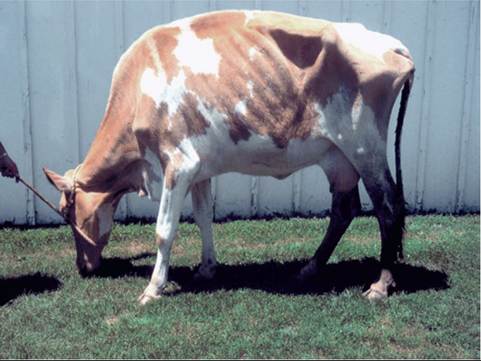
FIG. 32.140 A Guernsey cow exhibiting clinical signs of Johne’s disease, including thin body condition and fecal staining on hind end. (Courtesy Dr. Robert H. Whitlock, Kennett Square, Pa.)
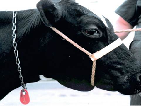
FIG. 32.141 Holstein cow exhibiting submandibular edema associated with clinical Johne’s disease, hypoproteinemia, and protein-losing enteropathy. (Courtesy Dr Robert H. Whitlock, Kennett Square, Pa.)
Goats, in particular, may become quite anemic, more than might typically be expected with anemia of chronic disease. Of course anemia, hypoproteinemia, and weight loss may be exacerbated by concurrent intestinal parasitism, which may initially obscure the diagnosis of Johne's disease. Johne's disease in farmed deer may differ in that there may be a shorter incubation period with rapid progression to clinical disease with diarrhea and emaciation. Also, postmortem examination of deer with Johne's disease may reveal caseation or calcification, or both, of mesenteric lymph nodes.22
Diagnosis
The available diagnostic tests are divided into two broad categories: (1) immune-based diagnostic tests that detect the host's antibody or cell-mediated immune response to MAP infection, and (2) organism detection tests that detect MAP organisms or MAP bacterial DNA in fecal or tissue samples.
IMMUNE-BASED TESTS. Tests of cell-mediated immunity include the intradermal Johnin test, which is similar to a tuberculin test, and in vitro lymphocyte stimulation with MAP antigens with detection of IFN-γ production. The intradermal Johnin test is generally regarded as unreliable because of both false-positive and false-negative reactions. Stimulation in vitro of peripheral blood lymphocytes with antigens of MAP, followed by detection of IFN-γ in cell culture supernatant, has shown the ability in a research setting to detect animals early in the course of MAP infection, in the eclipse phase before antibody production and fecal shedding are detectable. However, the tests are not available in a practical commercially available setting.
The most commonly applied immune tests are those that detect antibodies in serum or milk. These include the complement fixation test, the agar gel immunodiffusion test, and ELISA. The ELISAs have superior performance to the complement fixation or agar gel immunodiffusion tests when applied to serum from cattle, but some nations still list complement fixation or agar gel immunodiffusion testing as an import requirement. The ELISA is the most specific and sensitive of the commercially available antibody tests. It may be applied to serum or milk, and studies have shown comparable results when ELISA is applied to both matrices. The ELISAs are attractive because of their relatively low cost and amenability to automation, which makes testing of large numbers of samples practical and cost-effective. Many Dairy Herd Improvement Association services now offer the option to includeJohne's ELISA testing on milk samples, adding to the convenience of testing. Preservatives added to Dairy Herd Improvement Association milk samples do not interfere with milk ELISA testing.
Antibody detection tests are hindered only by the natural biological features of the disease, in that infected cattle do not ordinarily produce detectable antibodies until after fecal shedding has begun. Thus the sensitivity to detect asymptomatic cattle that are “low fecal shedders” may be as low as 15%, whereas 90% of clinically affected, “heavy shedding” cattle are positive. The ELISAs generally have high specificity (98% to 100%).23,24
ORGANISM DETECTION TESTS. Organism detection tests on fecal samples include both culture and PCR detection methods. They have the advantage over antibody-based tests in that they (1) generally allow detection earlier in the course of infection because fecal shedding usually precedes antibody detection; (2) allow quantification of the degree of fecal shedding to assess the biosecurity effect of individual animals; and (3) definitively identify MAP in a herd at the time of first diagnosis. They have the disadvantage of being more expensive than ELISA and, in the case of culture, requiring a long incubation period to obtain results. Cultures performed on solid Herrold's Egg Yolk Medium may require up to 16 weeks for incubation. Many laboratories now employ a liquid culture system that can shorten the culture time to 1 to 2 months. Commercially available real-time PCR tests for fecal samples also permit quantification of MAP shedding and have sensitivity comparable to or better than culture. The specificity of culture and PCR methods is generally regarded as 100%, but it should be remembered that when MAP-contaminated feed is consumed by uninfected cattle, the MAP organisms may pass through the gastrointestinal tract and cause a transient positive result on fecal PCR or culture. A list of USDA-approved or accredited laboratories for Johne's disease testing is available at the USDA Animal and Plant Inspection Service (USDA-APHIS) website (https://www.aphis.usda.gov/aphis/ourfocus/animalhealth/ lab-info-services/sa_approved_labs/ct_approved_labs).
TESTING SCENARIOS. Diagnostic tests should be selected on a case-by-case basis, depending on the clinical scenario, the objectives of the producer, and the inherent advantages and disadvantages of each test. For example, to confirm the diagnosis of Johne's disease in an adult cow with weight loss and diarrhea, a clinician will not have the luxury of waiting for fecal culture results before making a culling or treatment decision. Thus a test with rapid turnaround time is indicated, and both ELISA and fecal PCR would provide rapid results with a high predictive value positive (or negative). If the case in question is the index case in a herd, then organism detection would be more appropriate than ELISA, to provide a definitive confirmation that MAP is the etiologic agent because the implications of a diagnosis for the herd may be great. Postmortem demonstration of typical lesions and identification of MAP by culture of mesenteric lymph node or ileum can also be used to confirm a diagnosis.
An alternative objective might be to identify infected asymptomatic cattle in a herd that is known to be infected with MAP, for purposes of culling in an eradication program, or segregation for calving pen or colostrum management. In higher prevalence herds, serum or milk ELISA can be used if the magnitude of the ELISA result is used to interpret the results (i.e., as opposed to just “positive” or “negative”). Animals with a negative ELISA result, even if infected, are probably not shedding MAP in feces, milk, or colostrum at a significant level. Animals with “low positive results” are more probably shedding MAP in feces and might remain in the herd if production is good, but their status should be kept in mind when managing calving pens and colostrum. “High positive” ELISA results suggest heavy fecal shedding and a potential significant source of farm contamination; affected cows should be culled as soon as possible. Although more expensive, fecal culture or PCR can similarly be used to identify asymptomatic infected cattle with greater sensitivity than ELISA. Where management/ control is the objective, a low positive result will warrant appropriate calving pen and colostrum management but not necessarily culling, whereas a high positive result may warrant sooner culling. In an aggressive eradication program, any fecal culture positive result might result in a decision to cull, either immediately or at the end of the lactation. Testing can be performed on an annual, whole-herd basis, or in some systems, cows can be tested at the time of drying off. With this method, if ELISA, fecal culture with liquid medium, or fecal PCR is employed, the results will be available before calving to guide management decisions.
A third scenario is to determine whether a herd has MAP infection or to document test-negative or low-prevalence status for certification programs. One of the more cost-effective methods for this purpose, on dairy farms, is to perform fecal culture or PCR on environmental fecal samples collected from areas with high cow traffic and from the manure pit or storage lagoon. Culturing from 5 to 6 such sites has been shown to detect at least 70% of herds with infected animals.25
The USDA-APHIS Uniform Program Standards for Voluntary Bovine Johne's Disease Control Programs (VJDCP) provide testing guidelines for establishing herd infection status. Although individual states' programs may vary, the current standard establishes six classification levels. Levels 1 to 3 identify herds with low test-positive prevalence, or herds with 1 year of all-negative test results, whereas levels 4 to 6 identify herds with 2 or more years of test-negative results, with level 6 having the highest certainty of infection-free status. Herds establish classification status by whole herd (or statistically determined subset for large herds) ELISA or organism detection testing (individual samples or pooled). The reader is referred to the VJDCP Program Standards for more information about more detailed testing requirements (https://www.aphis.usda.gov/animal_health/animal_diseases/ johnes/downloads/johnes-ups.pdf).
Finally, testing may be desired to screen animals purchased as herd replacements or for use as embryo transfer recipients. Unfortunately, currently available tests are not useful for detecting infection in these animals, which are often of a young age (but the infection is rarely eliminated, and daily treatment will probably be required for the life of the animal, with relapse likely to occur when treatment is discontinued. Treatment of a clinically affected pregnant cow to salvage her fetus is not recommended because of the high likelihood of transplacental infection of the fetus. In addition, any chemotherapeutic program will involve extralabel use of antimicrobials, which in the United States must be performed in accordance with the regulations set forth in the Animal Medicinal Drug Use Clarification Act. As reliable withdrawal times are not established for many of the antimicrobials employed in treatment of Johne's disease, owners should be informed that once treatment begins, the animal or its products should never enter the human food supply chain.
The therapeutic agents most often employed include those used to treat mycobacterial infections in humans (isoniazid, rifampin, clofazimine), monensin, and aminoglycosides, and combination therapy is often recommended, at least to induce remission.26 Clofazimine, an antileprosy drug, has been used successfully by the author at a dose of 2 mg/kg q24h, alone or in combination with other agents. Improvement in fecal consistency and plasma total protein occurred within 7 to 14 days of starting treatment. However, this drug has not been available in the United States, prompting use of other combinations. Thus a typical currently employed treatment protocol, if clofazimine is not available, for cattle, goats, sheep, and camelids includes rifampin (10 mg/kg PO q24h) and isoniazid (10 to 20 mg/kg PO q24h). Again, because of its potential carcinogenicity, rifampin is not appropriate for extra-label use in animals intended for food and should thus only be used in pet animals or animals that will never enter the human food chain. Gentamicin (6.6 to 8.0 mg/kg IV q24h) for the first 5 to 10 days of treatment may hasten onset of remission, provided renal function is normal and can be monitored for signs of nephrotoxicosis. For cattle, monensin should also be recommended. Approved for its effect as a coccidiostat and for its effect on rumen microflora and digestion efficiency, momensin has also been shown to inhibit MAP growth in vitro and improve the signs and lesions of disease in experimentally and naturally infected cattle.27,28 Use of monensin as a feed additive also has the potential to play a chemoprophylactic role in prevention of MAP infections in susceptible cattle. However, monensin as a feed additive cannot be used in an extra-label fashion in the United States, so the intended animal must have a label indication for use of monensin, not solely treatment of MAP.
Prevention and Control
For herds not currently infected with MAP, prevention relies primarily on establishing a closed herd. If herd replacements must be purchased, the herd of origin should be screened as indicated earlier, and ideally have achieved level 4 or higher status in the VJDCP.
For herds that are infected, clear objectives should be established. For large production herds with high turnover, no steps may be economically warranted. For other herds, management to reduce transmission and ameliorate the economic effect of the disease may be desirable, and in other herds eradication may be the goal. Regardless of the goal, a test-and-cull strategy employed without other management changes to reduce transmission will not be successful.
Whether the goal is eradication or managing the disease to minimize losses, a risk assessment should be performed to identify potential areas of MAP transmission, especially to susceptible young stock, and a management plan developed. A plan must be individually tailored to a specific facility, but in general key control points are calving pens, calf feeding (colostrum and milk) management, and calf housing, especially as they pertain to potential exposure of calves to feces of adult infected cows, or to fecal-contaminated surfaces. Handbooks for veterinarians to assist them in performing risk assessments and management plans for both dairy and beef cattle, designed by the Johne's Disease Committee of the U.S. Animal Health Association, are available at http://www.johnesdisease.org/. On most dairy farms, control strategies focus on calving pen hygiene, early removal of calves from the calving pen after birth, providing an uncontaminated source of colostrum and milk or colostrum replacer and milk replacer, and calf housing free of contamination by feces from adult cattle. Testing in some form to identify and remove or segregate highly infectious adult cattle is usually warranted. In beef cow-calf operations, preventing transmission to calves again must focus on manure contamination of calving areas, manure contamination of cows that are nursing calves, and other possible sources of calf exposure to feces from potentially infected adult cattle. Recommendations can be adapted as appropriate for use with small ruminants and camelids, again to focus on prevention of transmission to susceptible young stock.
Vaccination has been employed with mixed results in cattle and small ruminants. In the United States, vaccination of cattle is permitted in some states, on approval by the State Veterinarian. The only licensed vaccine (Mycopar [Boehringer Ingelheim Vetmedica, St. Joseph, Mo.]) is a killed oil suspension of M. avium. The vaccine is generally regarded to reduce the incidence of clinical disease, and in some instances also reduce the level of fecal shedding, yet in other studies prevalence of fecal shedding was not affected. Vaccination should be considered as a useful adjunct, particularly in herds of high prevalence with significant losses due to clinical Johne's disease. Vaccination alone, without implementation of management changes to reduce exposure of calves to MAP, is often unsuccessful as any resistance achieved by vaccination may be overcome by heavy MAP exposure.
ZOONOTIC POTENTIAL. Traditionally, MAP was not regarded as a pathogen of humans. However, because of some similarities between Crohn's disease in humans and Johne's disease in ruminants, the possible connection between MAP and Crohn's disease has undergone further scrutiny. These studies, especially those that employ PCR methods to identify MAP in tissue samples from people with Crohn's disease, have definitively shown an association between presence of MAP in intestinal tissue and Crohn's disease.29 However, what is more difficult to determine is whether this association is causal or secondary. Crohn's disease is thought to involve an overly florid immune response to antigens in the gastrointestinal tract. Proponents of the hypothesis that MAP causes Crohn's disease believe that MAP antigens are responsible for that response, whereas skeptics believe that MAP may be present secondarily to damaged mucosa with diminished resistance to MAP colonization. Improved clinical signs in patients with Crohn's disease in response to antimicrobial therapy directed against MAP might constitute further evidence for the role of MAP in the disease.
However, results of clinical trials of antimycobacterial therapy have had mixed results with differences in opinion regarding their interpretation.30 Undoubtedly, Crohn's disease is a multifactorial disease influenced by a complex interaction of genetics, environment, cigarette smoking, and potentially infectious agents. Human exposure to MAP occurs potentially via survival of MAP in pasteurized milk or drinking water contaminated by fecal runoff from infected farms. Increasingly, regulatory agencies around the world are treating paratuberculosis as a food safety issue. More recently, an association with immune-mediated disorders such as diabetes mellitus Type 1, multiple sclerosis, and thyroiditis through molecular mimicry and dysregulation of the immune system.31 It may be difficult to scientifically establish an etiologic connection between MAP and Crohn's disease or these other disorders; however, in the author's opinion, even the potential for a role of MAP in Crohn's disease or other human conditions should motivate producers and veterinarians to control the disease in food-producing animals.