Osteochondrosis
Lameness, if any, is usually mild, except in cases with large osteochondral fragments or subchondral bone cysts. Severe cases seem to be more common in the stifle, and associated clinical signs may be present in foals as young as 6 months.
Reports in cattle indicate that young, intact male, purebred animals are most often affected. The typical clinical signs are lameness with associated joint effusion. Osteochondrosis in swine has been associated with a syndrome known as “leg weakness.” Affected animals can show a range of signs from mild lameness to difficulty rising and/or inability or refusal to mount. Evaluation of performance data from swine herds can be a useful indicator of osteochondrosis because it has been shown to have a significant effect on production traits.3,6 Production animals are often slaughtered prior to 2 years of age; thus recognizable clinical signs of the disease in cattle and swine may be apparent only in breeding animals.■ Sites of Predilection Osteochondrosis can be found in any diarthrodial joint, but sites of predilection exist in all species affected. In horses the stifle, tarsus, and fetlock are most frequently affected, and to a lesser degree the shoulder and vertebrae. In the stifle, in order of frequency, the lateral trochlear ridge of the femur, medial trochlear ridge of the femur, trochlear groove, and distal end of the patella are affected. The medial femoral condyle is the site of predilection for subchondral cystic lesions. In the tarsus, the distal intermediate ridge of the tibia, lateral trochlear ridge of the talus, and medial malleolus are affected.7 The most common manifestations in the fetlock joint are along the dorsal aspect of the sagittal ridge and the condyles of the metacarpus or metatarsus, and of the dorsal aspect of the proximal phalanx. Predilection sites in cattle are similar to those in horses, with the hock and stifle most often affected and similar distribution in the joints.
Swine show a slightly different pattern, with the medial condyle of the humerus and femur most frequently affected.■ Diagnostic Tests Animals presenting with signs of joint effusion and lameness should undergo a thorough physical examination. A compete blood cell count (CBC) indicating a septic process, along with radiography, helps differentiate osteomyelitis, septic physitis, and septic arthritis from osteochondrosis. Horses should be observed in motion and given flexion tests when possible. Regional and intraarticular anesthesia can be used to localize the site of lameness, though often the primary sign of osteochondrosis is swelling without lameness. Swine and cattle can be observed in their normal surroundings for signs of lameness. The joint in question should be radiographed, as should the corresponding joint on the contralateral limb in the tarsocrural and femoropatellar joints, because lesions are frequently bilateral. If lesions are detected in the metacarpophalangeal or metatarsophalangeal joint, the remaining three limbs should be evaluated. Joint effusion with or without lameness is more reliably seen in cases of osteochondritis dissecans (OCD) and subchondral bone cysts than in osteochondrosis.
Swine and cattle will often present with signs of lameness and mild joint effusion. Careful physical examination of the restrained animal should localize the lesion and guide appropriate radiography. As with equine patients, the lesions are commonly bilateral, and corresponding limbs should also be radiographed. Reviewing production records of finishing or breeding swine herds may be of diagnostic value, because osteochondrosis has been shown to cause significant reductions in performance and production traits.3
The manifestation of osteochondrosis may not be adequately represented by clinical signs and radiographs. Clinical signs of lameness and joint effusion have been shown to precede changes within the joint, and serial radiographs may be needed to diagnose the condition.
Radiographic abnormalities consistent with osteochondrosis of the distal intermediate ridge of the tibia and lateral trochlear ridge of the femur can return to a normal appearance by 5 and 8 months, respectively, in warmblood foals.11 Radiographs often underrepresent the severity or size of the lesion seen at surgery. Evaluation of osteochondrosis along the lateral trochlear ridge of the femur indicated that ultrasonography provided a better overall subjective assessment of the lesion (length, depth, and width) compared with radiography.12■ Pathophysiology Endochondral ossification is the process of bone formation in which cartilage scaffolds, arranged in zones, are gradually replaced by bone. It occurs at the articular/ epiphyseal and metaphyseal growth plates and at secondary centers of ossification, such as the carpal and tarsal bones. Directly beneath the articular cartilage is a zone of resting chondrocytes that divide to form the next zone of proliferating chondrocytes. These proliferative cells divide rapidly, organizing into columns perpendicular to the long axis of growth. The cells swell in the hypertrophic zone, where the columns become more organized. The chondrocytes in this zone are surrounded by increasing amounts of extracellular matrix, which becomes mineralized in the zone of calcification. These columns of chondrocytes are vascularized by metaphyseal blood vessels supplying nutrients. It is on these calcified cartilage columns that bone forms, creating the primary spongiosa, which is subsequently remodeled into mature bone.
The exact pathogenesis of osteochondrosis is unclear. The traditional theory is that the process of endochondral ossification is disrupted, resulting in areas of thickened cartilage. The deeper layers of affected cartilage do not receive adequate nutrients, resulting in necrosis of the cells and failure of proper ossification. These retained cartilage “plugs” have less structural integrity than normal cartilage and are prone to damage.
Specifically, shear forces acting on the abnormal cartilage can lead to fissure formation, which progress into fragments of cartilage and subchondral bone. When compressive forces predominate on an area of thickened cartilage, it is surmised that infolding of the cartilage plug occurs with normal endochondral ossification proceeding around it, possibly leading to the formation of a subchondral bone cyst.The traditional theory of defective endochondral ossification may be a simplistic view of a more complex, multifactorial condition. Limited reparative responses of bone and cartilage make it difficult to determine whether the origin of a lesion is developmental or traumatic. One report failed to distinguish articular cartilage differences in naturally occurring osteochondrosis versus healing osteochondral fragments.13
Computed tomography and magnetic resonance performed on fetuses and foals demonstrated that there was greater cartilage thickness in areas of joints that commonly develop OCD. More specifically, at 8 to 9 months of gestation, the lateral trochlear ridge of the femur, medial malleolus of the tibia, and distal intermediate ridge of the tibia, all OCD-susceptible sites, had the greatest percentage of cartilage compared to unsusceptible sites. Postpartum, the percentage of cartilage in the medial malleolus and distal intermediate ridge of the tibia remained high. These findings suggest that greater cartilage thickness at specific joint sites could play a role in the development of OCD.14
Arthroscopic observations of normal-thickness cartilage defects and normal subchondral bone, as well as lesions occurring preferentially at single sites at the limits of articulation, suggest causative factors other than defective endochondral ossification.15 The development and then spontaneous regression of osteochondrosis lesions in young animals suggest that the condition is a dynamic process that can be affected by numerous intrinsic and extrinsic factors, and a “window of susceptibility” may exist whereby lesions are constantly changing and result in the development of normal articular cartilage.
Alternatively, these developmental lesions may not regress, leading to osteochondrosis.16 It is currently impossible to predict how a lesion may behave while developing; thus treatment recommendations should be reserved until the lesion has been fully developed.■ Etiology The etiology of osteochondrosis is unclear and commonly described as multifactorial. Such factors as nutrition, genetics, growth rate, failure of normal vascularization, altered or excessive biomechanical forces, and hormonal influence have been implicated.
The consistent distribution of lesions at specific anatomic sites within the joint suggests trauma or regional biomechanical forces as causative factors, particularly when the lesions are bilateral or quadrilateral. It has been established in pigs that microtrauma or disruption of the metaphyseal blood vessels supplying the developing cartilage canals causes ischemic necrosis, failure of mineralization, and a retained cartilage plug.17 The same vascular pattern has been shown to be present in young foals, with the arterial supply crossing the ossification front from metaphyseal blood vessels to nourish the developing cartilage canals.18-20 Therefore such failure of vascularization, resulting in ischemic necrosis of the cartilage canal and retention of a cartilage plug, appears to play a role in the development of osteochondrosis in swine and horses.21
Nutritional influences have been evaluated. Studies have focused on dietary energy levels and mineral composition (copper and zinc). The growth rate of the animal is directly affected by energy intake and use as well as its genetic predisposition. Animals fed high-energy diets resulting in accelerated growth rates have a much higher incidence of osteochondrosis.4,22-23 More specifically, foals fed with concentrates had a higher probability of developing OCD lesions, whereas foals not receiving concentrates had a higher probability of healing from existing OCD lesions.24 In pigs, it was shown that for every 100-g increase in average daily gain during the weaning and finishing period, a 20% increase in cartilage lesions or osteochondrosis in the humeral condyle occurred.25 This is important when considering ration planning and growth rates of replacement animals, where longevity is a greater concern than in the production animal.
Low copper levels and increased zinc concentrations have both been implicated in the etiology of the disease. Low copper is thought to play a role through lysyl oxidase, a copperdependent enzyme essential in the crosslinking of collagen molecules. Increased levels of zinc antagonize copper and could work indirectly through a similar mechanism. There is conflicting evidence on the causative nature of low copper levels, and lesions seen in copper-deficient animals do not always mimic those of naturally occurring osteochondrosis. In addition, evidence suggests that neonatal copper levels exert a positive effect on the resolution of osteochondrosis lesions but are not directly involved in the pathogenesis of the disease.26
Genetics have been implicated in the etiopathogenesis of osteochondrosis in different species. A recent review article summarized the advancements in quantitative and molecular genetics in refining estimation of genetic parameters and identifying predisposing genetic loci. Whole genome scans were performed in Thoroughbred, Standardbred, French and Norwegian trotter, Hanoverian, and Dutch warmblood populations. Heritabilities were highest for tarsal osteochondrosis in Hanoverian warmbloods and Norwegian trotters, whereas in Thoroughbreds a low genetic variation seemed to be present in similar lesions. Validation studies in Spanish Purebred and Hanoverian warmblood horses corroborated osteochondrosis risk loci on horse chromosomes (ECAs) 3, 14, 27, and 29. A strong association with tarsal OCD was found for a single nucleotide polymorphism on ECA 3.27 In pigs, Landrace and Yorkshire breeds show a high frequency of osteochondrosis, whereas domestic pigs crossed with wild hogs do not develop the disease.4
Numerous hormones (e.g., insulin, somatotropin, thyroxine) are involved in endochondral ossification, and alterations of these, or their derivatives, could theoretically lead to the development of osteochondrosis. The current focus of research has been on the molecular mechanisms involved in the development of the disease. The mechanisms of normal endochondral ossification are not fully developed, further complicating an understanding of the disease process.
■ Treatment and Prognosis Treatment of osteochondrosis should take several factors into account: clinical signs, lesion severity, species, age, intended use, relative value of the animal, and owner expectations. Treatment options include both nonsurgical and surgical management.
Nonsurgical management should consist of rest, controlled exercise, and dietary evaluation.24 Dietary mineral and carbohydrate levels should be evaluated carefully, and any deficiency or excess should be corrected. Systemic NSAIDs and intraarticular medications like corticosteroids, hyaluronic acid, and polysulfated glycosaminoglycans (GAGs) can be administered, but minimal clinical evidence exists to support their use. In food-producing animals the clinician must take into consideration that these are not approved treatments and that withdrawal times may not be established.
Animals with OCD are frequently treated with arthroscopic surgery. Cases with no radiographic evidence of degenerative joint disease (DJD) before surgery and minimal articular damage at arthroscopy have a favorable prognosis. Animals that show radiographic evidence of DJD before surgery or considerable damage to the articular surfaces at arthroscopy have a less favorable prognosis. There is a site predilection for performance. Lesions in the stifle have a less favorable outcome for full athletic potential than do lesions within the tarsus. In addition, surgeon experience and age of the horse at the time of surgery also affects outcome; more experienced surgeons and older horses demonstrate improved outcomes.28 Arthroscopic approaches to the joints of cattle have been described, but the procedure is more difficult than in the horse.9 In selected cases, reattachment of large cartilage flaps with absorbable poly-p-dioxanone pins has been described.29 Dietary evaluation of surgical patients should be included and appropriate adjustments made.
Treatment of subchondral cystic lesions vary. Nonsurgical management should consist of rest, controlled exercise, and intraarticular injection of corticosteroids with or without hyaluronic acid or polysulfated GAGs. The prognosis is variable, and alternate treatments should be considered in refractory cases; these treatments include intralesional deposition of corticosteroids, arthroscopic cystic debridement, and transcystic placement of a cortical bone screw.
Arthroscopic-guided injections into the fibrous lining of the cyst have a 67% success rate in horses without preexisting secondary radiographic changes.30 Arthroscopic debridement and curettage of the cystic lesion have similar success rates, and horses younger than 3 years have a better prognosis. Packing the remaining defect with either a cancellous bone graft covered by a chondrocytic growth factor or various bone substitutes has shown promise, although it is more costly and technically challenging.29,31 Transcystic screw fixation has shown greater than 80% success rate and gained more recent acceptance as a suitable second-line treatment should intralesional injection of steroid fail.32 Treatment of osteochondrosis in food animals is generally limited by the economic value of the animal. Show animals or valuable breeding animals may be treated by any of the methods described, and the surgical options may offer a better prognosis than nonsurgical treatment. In production herds, ration evaluation and genetic predisposition may be the most important variables to address.
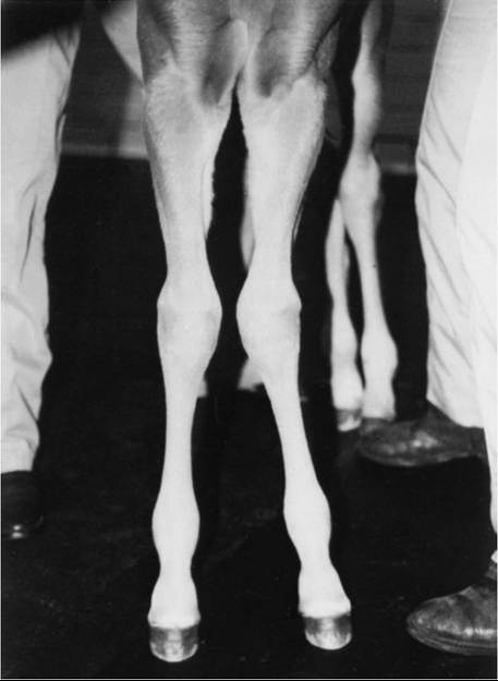
FIG. 38.1 Carpus valgus deviation.