Stomachandintestines
Rectal scrapings can lead to a diagnosis in some cases. Eosinophils can be observed in patients with eosinophilic colitis or gastroenterocolitis. Neutrophils can be observed in patients with other inflammatory disorders.
In rare cases, neoplasia can be diagnosed based on a rectal scraping. Rectal scraping is considered most useful for the diagnosis of infectious organisms, such as Histoplasma capsulatum, Cryptococcus neoformans, Prototheca, Pentatrichomonas hominis, Balantidium coli, and Entamoeba histolytica.11,12FNA cytology of masses of the gastrointestinal tract that are discovered during ultrasonographic examination and impression smears of endoscopically obtained biopsy specimens can be useful for the identification of infectious organisms or the diagnosis of neoplasia (Figures 1.101-1.104).
Facts
■ Fine-needle aspiration rarely leads to complications.
■ Tumors of the gastrointestinal tract can often be diagnosed based on cytology.
■ Degenerative hepatic disease often displays characteristic cytological features.
■ Hepatitis and hepatic cirrhosis cannot be accurately diagnosed based on cytology alone.
■ In some cases microorganisms can be identified on cytology.
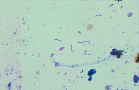
Figure 1.101:
Helicobacter infestation. Impression smear of a gastric biopsy specimen obtained from a cat. Large spiral-shaped organisms can be seen. Cytology is one of the most sensitive methods for detection of Helicobacter-like organisms.13
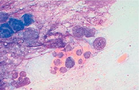
Figure 1.102:
Impression smear of an intestinal biopsy specimen in a cat. This cytology slide shows large quantities of eosinophilic granulocytes. Histopathology of this intestinal biopsy was indicative of lymphoplasmacytic enteritis.
This is a good example that cytologic and histopathologic findings of gastrointestinal specimens do not always agree.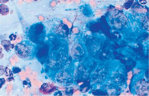
Figure 1.103:
Gastric carcinoma. Poorly differentiated epithelial cells with numerous criteria of malignancy are found on an impression smear of a gastric mucosal biopsy specimen. Neutrophilic granulocytes, few lymphocytes, and mast cells can be seen in the background.
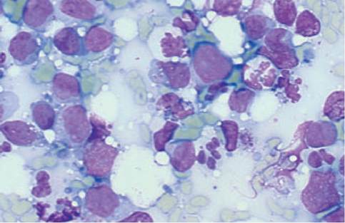
Figure 1.104:
Intestinal lymphoma. The FNA of an intestinal mass shows a homogenous population of large immature lymphocytes as well as a large number of inflammatory cells with mostly degenerated neutrophils with intra- and extracellular bacteria. Malignant lymphomas of the intestines are often contaminated with bacteria and often show large numbers of microorganisms and inflammatory cells on FNA cytology.
References
1. Hirschberger J. Organzytologie. In: Kraft W Durr UM (eds.), Klini- sche Labordiagnostik in der Tiermedizin. Stuttgart, Schattauer, 1997; 260-266.
2. Stockhaus C, Teske E. Klinische Anwendbarkeit der Leberzytologie bei Hund und Katze. Kleintierpraxis. 1997; 42: 687-701.
3. Stockhaus C, van den Ingh TS, Rothuizen J et al. A multistep approach in the cytologic evaluation of liver biopsies of dogs with hepatic diseases. Vet Clin Pathol 2002; (in press).
4. Weiss DJ, Moritz A. Liver cytology. Vet Clin North Am (Small Anim Pract) 2002; 32: 1267-1291.
5. Bjorneby JM, Kari S. Cytology of the Pancreas. Vet Clin North Am (Small Anim Pract) 2002; 32: 1293-1312.
6. Bolliger Provencher A. Cytology of the liver. Proc of the 6th ESVIM Forum. 1996; 66-67.
7. Blue JT, French TW Meyer DJ. The liver. In: Cowell RL, Tyler RD, Meinkoth JH (eds.), Diagnostic cytology and hematology of the dog and cat, 2nd ed. St. Louis, Mosby, 1999; 183-194.
8. Teske E, Brinkhuis BG, Bode P et al.
Cytological detection of copper toxicosis in Bedlington terriers. Vet Rec 1992; 131: 30-32.9. Lundquis A, Akerman M. Fine needle aspiration biopsy in acute hepatitis and liver cirrhosis. Ann Clin Res 1970; 2: 197-203.
10. Perry MD, Johnston WW Needle biopsy of the liver for diagnosis of nonneoplastic liver disease. Acta Cytol 1985; 29: 385-390.
11. Rakich PM, Latimer KS. Rectal mucosal scrapings. In: Cowell RL, Tyler RD, Meinkoth JH (eds.), Diagnostic cytology and hematology of the dog and cat, 2nd ed. St. Louis, Mosby, 1999; 249-253.
12. Baker R, Lumsden JH. The gastrointestinal tract - intestines, liver, pancreas. In Baker R, Lumsden JH (eds.), Color Atlas of Cytology of the Dog and Cat, 1st ed. St. Louis, Mosby, 2000; 177-197.
13. Kuffer-Frank M, Gerres A, Neuhaus B et al.Vergleich diagnostischer Methoden zum Nachweis von Gastric Helicobacter-like Organisms bei Hund und Katze. 9.Jahrestagung der Fachgruppe Innere Medizin und Klinische Laboratoriumsdiagnostik der Deutschen Veterinarmedizinischen Gesellschaft, Munchen, 6.-8. 3. 2000; 64-65.
1.8