Parasitic Diseases
In addition to the parasitic diseases of the liver discussed below, the liver may be the site of various Schistosoma spp. infections that can produce disease, as discussed in Chapter 8 (see Table 8.3).
A summary of parasites affecting the liver, pancreas, and related blood vessels of goats is given in Table 11.2.Stilesia and Thysanosoma Tapeworm Infections
The goat is a definitive host for the tapeworm Stilesia hepatica, which occurs extensively throughout Africa and Asia. The adult tapeworm may be found in the bile duct in large numbers. Adult S. hepatica measure 20-50 cm long and 3 mm wide. The life cycle, which is not completely elucidated, likely involves oribatid mites as an intermediate host. Eggs are passed in the feces of the goat.
This tapeworm is considered non-pathogenic in the living animal. However, large numbers of adult tapeworms may induce sufficient bile duct thickening that affected livers are condemned during meat inspection. In one study in
Table 11.2 Helminth parasites of the liver, pancreas, and related blood vessels.0
| Scientific name | Common names | Location in goat | Geographic distribution | Clinical effects |
| Fasciola hepatica | Common liver fluke | Liver parenchyma, bile ducts | Worldwide | Acute death; marked anemia, edema, emaciation |
| Fasciola gigantica | Giant liver fluke | Liver parenchyma bile ducts | Africa, Asia | Acute death; marked anemia, edema, emaciation |
| Fascioloides magna | Giant liver fluke; large American liver fluke | Liver parenchyma | North America | Acute death |
| Dicrocoelium dendriticum | Lesser liver fluke; lancet fluke | Bile ducts | Europe, Asia, North America | Marked anemia, edema, emaciation |
| Eurytrema pancreaticum | Pancreatic fluke | Pancreatic ducts, bile ducts, duodenum | Eastern Asia, Brazil | Ill-thrift |
| Stilesia hepatica | Liver tapeworm | Biliary ducts | Africa | Non-pathogenic; liver condemnation |
| Thysanosoma actinoides | Fringed tapeworm | Bile ducts, also small intestine | Western United States, South America | Non-pathogenic; liver condemnation |
a For schistosomes of the liver and related vessels see Table 8.3.
Zimbabwe, 18.75% of communal goats had their livers condemned at slaughter and 72.9% of these condemnations were due to S. hepatica (Chambers 1990). Praziquantel orally at 15 mg/kg is effective in eliminating S. hepatica in sheep, with evidence that the same dose is also effective in goats (Verster and Marincowitz 1980).
The goat is infrequently infected with Thysanosoma actinoides, the fringed tapeworm of North and South America that is commonly found in the bile ducts, pancreatic ducts, and small intestine of sheep, cattle, and deer. Clinical disease in goats caused by this tapeworm infection is poorly documented.
Echinococcosis or Hydatid Disease
Clinical hydatid disease is uncommon in goats, but hydatid cysts in liver and other tissues at slaughter are widespread and cause condemnations and economic loss.
The adult tapeworm, Echinococcus granulosus, is found in the intestine of carnivores, particularly dogs. Eggs are passed in the feces and are ingested by goats, other ungulates, or humans. In these intermediate hosts, the ingested eggs release oncospheres that enter intestinal venules or lacteals and migrate via the circulation to liver or lung. The metacestode stage, known as the hydatid cyst, develops in these organs over a period of several months. Brood capsules containing protoscolices develop within some cysts. Occasionally cysts rupture and the released protoscolices produce additional daughter cysts within the intermediate host. When dogs eat viscera containing protoscolices, these evaginate and mature to adults in the intestine in about seven weeks, completing the life cycle.
Echinococcus granulosus occurs essentially worldwide. However, there are noted areas of so-called hyperendemic- ity where the disease occurs frequently and represents a public health concern. These areas include much of northern and eastern Africa, the Mediterranean region, Eastern Europe, Central Asia, parts of China, and the cone of South America (Lightowlers 2002).
The disease also occurs in eastern Australia and the western United States, where sheep are common. Infections are likely to occur in those places where ruminant livestock and domestic and wild canids commingle.Newer studies of genome patterns indicate that there are numerous distinct strains of E. granulosus. The so-called sheep strain (G1) and the so-called camel strain (G6) affect goats (Schantz 2006). The prevalence of disease is increased when carnivore populations are high and no efforts are made to segregate domestic and wild carnivores from livestock or their grazing areas. The feeding of ruminant offal to dogs also enhances completion of the life cycle. Increases in prevalence of hydatid disease in livestock occur after rainy periods, presumably because the rain widely disseminates eggs in feces on grazing land or in water supplies.
Prevalence data of hydatid disease in goats is available from abattoir and postmortem surveys around the world. The prevalence range in endemic regions is 0.26-6.5% (Pandey 1971; Rahman et al. 1975; Naus 1982; Al-Yaman et al. 1985; Lorenzini and Ruggieri 1987). Hydatid cysts are most common in the liver, lung, and spleen, but may be found in other organs at slaughter or necropsy. In some places, lung cysts are more common than liver cysts in goats. More information on hydatid cysts in goat lung is given in Chapter 9.
Hydatid cysts may average 5-10 cm in diameter and contain a yellowish, serum-like fluid, and may have a granular inner wall containing multiple brood capsules. Hydatid “sand,” which is an accumulation of detached brood capsules, may be seen in the cyst fluid. Hydatid cysts should be differentiated from Cysticercus tenuicollis cysts, as discussed below. Cysts containing nymphs of the canid nasal parasite Linguatula serrata also have been found in the liver, lungs, and mesenteric lymph nodes of goats in Bangladesh (Rahman et al. 1980).
In the last 20 years ELISA and immunoblot assays have been developed to diagnose echinococcosis in livestock, but due to a high number of false-negative and falsepositive results, these assays remain unsuitable for surveillance of echinococcal infections (Moro and Schantz 2006).
Various imaging techniques can be used to identify hydatid cysts, including ultrasound, computed tomography (CT), and magnetic resonance imaging (MRI).There is no practical or economical treatment for hydatid cysts in ruminants. Control efforts involve disruption of the carnivore/ruminant life cycle. The principal efforts should be directed at eliminating the feeding of livestock offal to dogs, encouraging burial of livestock carcasses or viscera, and using routine anthelmintic therapy in dogs to reduce adult tapeworm populations. There has been notable progress in the development of effective vaccines for controlling hydatid cysts. Though not yet commercially available, a recombinant DNA vaccine based on the oncospheric protein EG95 has been shown to be effective in protecting cattle, sheep, and goats against hydatid disease with immunity lasting for at least one year (Dalton and Mulcahy 2001; Lightowlers 2002).
Echinococcosis is a serious zoonotic disease. Humans are infected by ingestion of tapeworm eggs shed by canids. International efforts to reduce hydatidosis, especially with regard to its zoonotic potential, have been reviewed (Moro and Schantz 2006).
Cysticercosis
Etiology
Adult Taenia hydatigena tapeworms are found in the intestine of dogs, coyotes, wolves, and other carnivores. The eggs are passed in the feces and ingested by goats, sheep, and other domestic and wild ruminants. Eggs hatch in the small intestine of the intermediate hosts and enter the blood. Upon reaching the liver, the embryos leave the portal vasculature and migrate through the hepatic parenchyma to the peritoneal cavity, causing distinct hemorrhagic tracts in the liver. The metacestode, which is the developmental phase that occurs in ruminants, is a bladderworm called C. tenuicollis. It matures over a period of five to eight
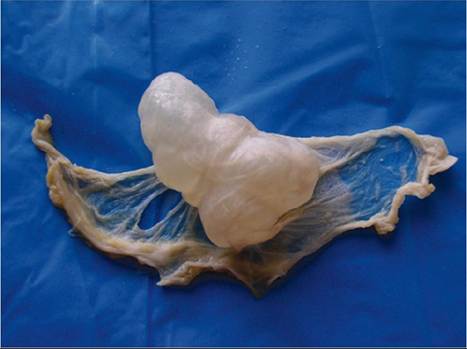
Figure 11.2 Cysticercus metacestode (Cysticercus tenuicollis) of the canid tapeworm, Taenia hydatigena, attached to the mesentery of a goat.
Source: Reproduced by permission of Dr. Jaroslaw Kaba, Faculty of Veterinary Medicine, Warsaw University of Life Sciences, Warsaw, Poland.weeks, and is found attached to the mesentery, omentum, and serosal surface of abdominal organs (Figure 11.2). Occasionally, migration out of the liver is not achieved and the cysticercus is found in the liver itself. Aberrant migrations sometimes occur, with cysticercus found in the lungs or other organs.
Epidemiology
Factors that promote the infection of goats with C. tenuicol- lis from contact with canids are similar to those described above for echinoccocosis. Cysticercosis occurs worldwide. Reported prevalence in slaughtered goats around the world ranges from 0.2% in Australia to 23.3% in Nigeria (Rahman et al. 1975; McKenzie et al. 1979; Akinboade and Ajiboye 1983; Sanyal and Sinha 1983).
Pathogenesis
The migration of embryos of T. hydatigena in the liver produces parenchymal damage and the creation of blood- filled tracts. When many embryos are migrating, acute, fatal hepatic insufficiency may occur in goats (Everett and de Gruchy 1982). Less severe migrations of embryos may still lead to liver condemnations in otherwise healthy animals, as the blood-filled tracts become fibrotic over time. Occasionally, peritonitis is associated with migration out of the liver and into the peritoneal cavity. The mature cysts in the mesentery, omentum, or liver usually do not produce any clinical disease.
Clinical Findings
Acute cysticercosis may produce signs of depression, weakness, anorexia, and possibly collapse and death caused by hepatic insufficiency. Signs consistent with pneumonia, peritonitis, and/or anemia can occur as a result of aberrant migration in the lungs and abdominal viscera, with resulting hemorrhage. Fever may be recorded if peritonitis occurs. Signs usually occur between 7 and 20 days after exposure (Pathak and Gaur 1981a). Chronic cysticercosis is usually asymptomatic.
Clinical Pathology and Necropsy
Experimentally, goats infected with eggs of T.
hydatigena showed marked elevations of ornithine carbamyltransferase (OCT) and AST 10 days after infection, but this is not specific for cysticercosis (Pathak and Gaur 1981b). Indirect hemagglutination and complement absorption tests have been used in the diagnosis of caprine cysticercosis (Varma et al. 1973, 1974). Radiography or ultrasonography can be useful in identifying hepatic or peritoneal cysts, as can CT and MRI.At necropsy, C. tenuicollis cysts may be present in the liver, but are common in the mesentery, omentum, and serosal surface of peritoneal viscera. Increased volumes of serofibrinous fluid may be present in the peritoneal cavity. Mature cysticerci have a smooth inner surface and contain only a single invaginated scolex, in contrast to hydatid cysts.
Acute cysticercosis is recognized by the presence of tubular, red, blood-filled tracts 2-4 mm in diameter in the liver, as shown in Figure 11.3. More chronic lesions may appear white as leukocytes fill the tracts and fibrosis occurs.
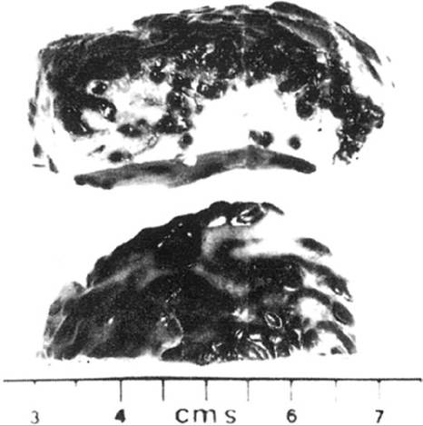
Figure 11.3 Blood-filled tracks in the liver due to migration of embryos of Taenia hydatigena. Source: Everett and de
Gruchy 1982/John Wiley & Sons, Inc.
Treatment
Praziquantel, at a single oral dose of 60 mg/kg, was reported to be 93-100% effective in removing C. tenuicollis from sheep and goats in China (Li and Li 1986).
Control
Control measures involve disruption of the carnivore/ ruminant life cycle, as described above for echinococcosis. As with echinococcosis, there has been a considerable effort to develop effective recombinant protein vaccines to control cysticercosis, with good progress reported for vaccines against Taenia ovis, but not yet specifically T. hydati- gena (Lightowlers et al. 2000)
Fascioliasis
Fascioliasis is the most common liver fluke disease of goats worldwide. In certain regions, chronic fascioliasis represents a major constraint on goat production.
Etiology
There are two Fasciola species that infect goats. Fasciola hepatica is smaller, with an adult length of 18-32 mm and a width of 7-14 mm. Fasciola gigantica is longer, with an adult length of 24-76 mm and a width of 5-13 mm.
Fasciola spp. have an indirect life cycle, with water snails of the superfamily Lymnaeidae acting as the intermediate host. More than 15 species of these snails serve as hosts for F. hepatica worldwide, and at least 13 for F. gigantica, with some overlap. When Fasciola are introduced into new areas by importation of livestock, local species of Lymnaeidae may serve as suitable, adaptive, intermediate hosts. Infected snails introduced into a new area may also act as the source of new infections.
Adult flukes reside within the biliary tract of definitive hosts such as goats and lay eggs that are passed via the bile duct to the feces. Egg production is substantial, with a mean of 303 000 eggs found in the gallbladders of infected slaughtered goats (Jimenez-Albarran and Guevara- Pozo 1977). Eggs passed in feces hatch within 10-12 days, releasing miracidia that invade intermediate host snails. Eggs hatch optimally at a temperature of 26 °C (79 °F), while temperatures must be at least 10 °C (50 °F) for mira- cidia to survive, invade snails, and develop further. Within the snail, the miracidium undergoes subsequent multiplications through sporocyst, rediae, daughter rediae, and cercariae stages. The average time from infection of snails to shedding of cercariae is 5-8 weeks, but can be as short as 21 days or prolonged as much as 10 months under adverse conditions.
When cercariae are shed by snails, they swim to a nearby plant, attach to herbage, encyst, and become infective metacercariae that are then ingested by grazing vertebrate hosts. The ingested metacercariae excyst, penetrate the intestinal wall, and migrate through the abdominal cavity to the liver. They then penetrate the liver capsule and migrate through the liver parenchyma to the bile ducts, where they mature. They reach the bile ducts about six to seven weeks after infection. Maturation within the bile ducts to egg-laying adults requires an additional two to four weeks. Adult flukes may remain in situ for months to years.
Occasionally, flukes are found in aberrant organ locations such as kidney and lungs, caused by inaccurate migration out of the intestine (Charan and Iyer 1972; Haroun et al. 1989).
Epidemiology
Fascioliasis occurs principally in domestic ruminants, but other non-ruminant livestock, numerous wild mammals, and marsupials also can be infected and may serve as reservoir hosts. Humans can contract fascioliasis from ingesting fresh plants contaminated with metacercariae.
The distribution of F. hepatica is cosmopolitan, and reports of fascioliasis in goats come from around the world (Rafyi and Eslami 1971; Jimenez-Albarran and Guevara-Pozo 1977; Chevis 1980; Quittet 1980; Vasquez- Vasquez 1980; El Moukdad 1981; Leathers et al. 1982; Bendezu et al. 1983; Anwar and Chaudhri 1984; Fernandes and Hamann 1985; Liakos 1985; Thompson 1986; Campano 1987; Wang et al. 1987; Molan and Saeed 1988). Prevalence varies considerably. No liver flukes were identified in an abattoir study in Queensland, Australia (McKenzie et al. 1979). Approximately 4% of sheep and goat livers were infected in a Mexican study (Vasquez-Vasquez 1980), while as many as 79% of sheep and goat livers inspected in Pakistan had F. hepatica (Anwar and Chaudhri 1984).
The distribution of F. gigantica is more limited, primarily to the tropical regions of Asia and Africa and to the Middle East. Caprine infection with F. gigantica has been reported from all these regions (Fabiyi 1970; Rafyi and Eslami 1971; Charan and Iyer 1972; Tager-Kagan 1978; Assoku 1981; El Moukdad 1981; Nooruddin et al. 1987). In a year-long Indian abattoir survey, 14.7% of goat livers were infected with F. gigantica and 22.3% of potentially marketable liver was condemned (Pachauri et al. 1988).
Besides a suitable snail host, the key element that regulates the occurrence of fascioliasis is a suitable combination of moisture and temperature that allows persistent surface wetness on pasture for the snail and the free-living stages of the parasite to thrive. The relationship of infection rate to weather conditions is sufficiently well known that meteorologic calculation of increased fascioliasis risk is done to determine the need for timing of prophylaxis (Soulsby 1982). These forecasting techniques were first applied in Europe, but are now being applied elsewhere, including North America (Malone and Zukowski 1992), South America (Fuentes et al. 1999), and Africa (Yilma and Malone 1998).
Low-lying, poorly drained, persistently marshy, or swampy areas in warm climates grazed by ruminants are ideal for completion of the life cycle of Fasciola spp. Water sources, however, do not need to be permanent. Intermittent flooding of fields by overflow of streams or frequent irrigation can facilitate infection, because snails can burrow into mud and survive temporary dry periods. Irrigation canals and ditches can be an important point of infection for goats and sheep in dry regions where fresh herbage and water may only be available at certain periods of the year along the snail-infested irrigation channels.
In temperate regions, infection of snails typically peaks in late spring and early summer, leading to peak pasture contamination with infective metacercariae in late summer and early fall. Peak incidence of clinical disease in ruminants is then seen in late fall and winter. Where winters are not extremely cold or excessively dry, some snails and eggs survive winter and recommence development in early spring, so that a less significant cycle of infection with metacercariae occurs on pastures in late spring and early summer as well. In tropical regions, metacercariae can be present all year long, and there is no well-defined season for clinical outbreaks in ruminants. Grazing is not a prerequisite for infection if stabled animals are fed fresh green herbage contaminated with metacercariae, though feeding fresh cut grass to penned goats in zero-grazing systems is often promoted as a means of reducing gastrointestinal parasitism due to nematodes.
Goats, like sheep, do not develop as strong an immunity to Fasciola infection as cattle, and remain largely susceptible to reinfection. However, some immunity does develop, as evidenced by reduced size and numbers of flukes recovered from repeated challenge infections (Reddington et al. 1986; El Sanhouri et al. 1987; Haroun et al. 1989).
Pathogenesis
There is little or no damage produced during migration of metacercariae through the intestinal wall or the peritoneal cavity. The potential for damage begins when metacercar- iae penetrate the liver capsule and start to migrate through the parenchyma. When large numbers invade simultaneously, as may happen in the early fall, a traumatic hepatitis ensues and is recognized clinically as acute fascioliasis. When moderate numbers invade over an extended period of time, then subacute fascioliasis can occur. Fatal acute fascioliasis in goats can be produced by 1000 infective metacercariae, and severe subacute fascioliasis by 200. F. gigantica is more pathogenic to goats than F. hepatica, and goats may be more susceptible to F. gigantica than are sheep (Ogunrinade 1984).
In acute fascioliasis, there is marked disruption of liver parenchyma with extensive hemorrhage. The liver capsule may rupture, leading to severe intra-abdominal bleeding and death. Even without fatal hemorrhage, animals may die within a few days as a result of hepatic insufficiency from parenchymal damage. In the subacute form, invading metacercariae still produce migratory tracts, hemorrhage, and necrosis within the liver and lead to a more prolonged clinical picture of hepatic insufficiency. Extensive fibrosis occurs in the liver during subsequent attempts at healing.
Acute fascioliasis is not commonly seen in goats and the reason is not clear. It may be related to feeding behavior, because goats are not intensive grazers and may not ingest sufficient numbers of metacercariae in short periods to produce severe acute reactions.
Chronic fascioliasis occurs after the arrival and maturation of flukes in the bile ducts. Persistence of adult flukes in the biliary tree results in hyperplastic cholangitis, with ongoing loss of plasma proteins caused by increased permeability of the biliary mucosa. Feeding activity and movement of flukes within the biliary tree produce chronic blood loss. Under natural circumstances of exposure, individual animals may simultaneously experience acute, subacute, and chronic fascioliasis.
In sheep, infectious necrotic hepatitis, or black disease, is a common complication of fascioliasis. In these cases, C. novyi organisms already present in the liver suddenly proliferate in the anaerobic environment of necrotic tissue created by migrating fluke larvae. The release of toxins by proliferating bacteria produces an acute, fatal toxemia. The prevalence of this disease in goats is poorly documented, but it is considered to be a serious potential risk in areas of Australia and New Zealand where goat populations have expanded into traditional sheep-raising areas (Pauling 1986). In Australia, black disease has been documented in Angora goats. It occurs seasonally between February and June in well-conditioned goats between 9 months and 4 years of age. It is presumed that goats are less frequently affected than sheep because of their browsing habits. The prevalence in goats increases when they are forced to graze in wet areas during drought, because C. novyi is a soil-borne organism (King 1980). Black disease was also confirmed in goats in the Sudan, where 18 normal-appearing animals in a herd of 435 were found dead overnight. At necropsy, all the goats were heavily infested with F. gigantica and the presence of C. novyi type B was established (Hamid et al. 1991). Where there is concern of the risk of black disease, goats can be vaccinated against C. novyi, which is included in many of the commercially available, multivalent clostridial vaccines.
Clinical Findings
Acute fascioliasis, though uncommon, may present as sudden death or as progressive weakness, depression, anorexia, and pallor lasting up to three days and resulting in death. Before death, pain may be elicited by deep palpation over the liver. Subacute fascioliasis may show similar signs for as long as several weeks. Black disease usually presents as sudden death, with no other signs except some possible frothing at the mouth and nostrils.
Chronic fascioliasis is the most common presentation. The history may include a period of depression, poor appetite, lethargy, and weight loss for one month or longer. In prolonged cases, some animals have diarrhea. There may also be a complaint of decreased milk production in milking does. Physical findings include poor body condition, rough haircoat, pale mucous membranes, and tachycardia. Intermandibular edema, or bottle jaw, frequently occurs in long-standing cases and is highly suggestive of chronic fascioliasis.
Clinical Pathology and Necropsy
Acute fascioliasis is most often diagnosed at necropsy. In subacute disease, the hemogram may document normochromic anemia, eosinophilia, and possibly neutrophilia. Hypoalbuminemia and increases in serum concentration of liver-related enzymes may be noted in the chemistry profile.
Normocytic or macrocytic anemia, hemoglobinemia, hypoalbuminemia, and eosinophilia can be expected in chronic fascioliasis. Total protein may be in the normal range as a result of accompanying hypergammaglobulinemia. Increases in liver-specific enzymes in the blood occur less reliably in chronic fascioliasis, although increases in GDH and OCT have been reported to occur even when flukes are present only in the biliary tree (Treacher et al. 1974).
In chronic fascioliasis, fluke eggs can be found in the feces of goats. Sedimentation methods for fecal examination are preferred to flotation methods. Eggs are yellowcolored, ovoid, and operculated at one end. Eggs of F. gigantica are slightly larger than eggs of F. hepatica. Where rumen flukes occur, their eggs must be differentiated from those of liver flukes.
Because acute fascioliasis occurs during the prepatent period of infection, detection of ova in feces is not a useful aid for identification of early infections with F. hepatica or F. gigantica. This inability to detect prepatent infections has spurred a good deal of research in the area of serodiag- nosis over the last two decades. The main focus has been to isolate and characterize parasite antigens suitable for use in serodiagnostic tests such as ELISA to detect anti-fasciola antibodies in infected animals. One category has been the so-called excretory-secretory antigens of Fasciola spp., including cathepsins. These antigens have been derived directly from flukes and purified, and more recently obtained through use of recombinant DNA technology. ELISA assays using such antigens have been able to detect circulating IgG against F. hepatica and F. gigantica as early as one week post infection (Paz-Silva et al. 2005), but more commonly three to seven weeks post infection (Cornelissen et al. 2001; Mezo et al. 2003; Yadav et al. 2005).
However, there are still some constraints with serodiag- nostic tests. Though it is becoming less common as antigen purification techniques improve, cross-reactivity still may occur with other helminths sharing similar antigens, notably paramphistomes. Also, detectable antibodies persist even after effective treatment of fascioliasis, making interpretation of test results difficult. A commercial ELISA test kit for identifying prepatent fascioliasis in cattle and sheep is currently available in Europe, Australia and New Zealand.
Necropsy in acute fascioliasis demonstrates a swollen, traumatized, friable liver with multiple perforations of the capsule and subcapsular hemorrhage. Fibrin tags may be present on the liver surface and blood accumulated in the peritoneal cavity. Migrating flukes are not easily seen on cut surface of the liver parenchyma, but may be visualized by shaking a piece of liver in a pan of water and looking for the larvae to settle out at the bottom.
When black disease complicates acute fascioliasis, signs of clostridial toxemia may be evident. These include blackening of the subcutaneous blood vessels observed as the skin is reflected, serosanguinous pericardial and peritoneal effusions, and patchy areas of liver necrosis with reddish borders. These reddened edges are likely sites for isolation of C. novyi.
In chronic fascioliasis the carcass is emaciated, subcutaneous edema may be present, and organs are pale. The liver may be cirrhotic and irregularly nodular and the capsule opaque in areas. On cut surfaces, there is extensive fibrosis of the parenchyma. Bile ducts are thickened and fibrosed, but mineralization is uncommon. Upward of 100 adult flukes may be found in the cystic biliary ducts and gallbladder. They may be expressed by squeezing on cut sections of liver. Flukes have also been found in the pancreatic duct of goats at necropsy (Leathers et al. 1982).
Diagnosis
Acute fascioliasis is one of many potential causes of sudden death in goats, as discussed in Chapter 16. Definitive diagnosis is by necropsy. Chronic fascioliasis is most easily confused with gastrointestinal nematodiasis, and, in practice, concurrent nematode and trematode infections are common in goats. Schistosomosis, as discussed in Chapter 8, can also present similarly to chronic fascioliasis. When chronic weight loss is the major complaint or presenting sign, a wide variety of other diseases needs to be considered, as discussed in Chapter 15.
Treatment
The choice of therapy largely depends on the stage of infection requiring treatment (Boray 1985). Carbon tetrachloride, hexachloroethane, bromsalans, oxyclozanide, niclofolan, and albendazole are only effective on mature flukes, 10 weeks of age or older. Nitroxynil and clioxanide are moderately effective in flukes from 8-9 weeks of age and highly effective against mature flukes. Brotianide and rafoxanide are most effective against flukes 8 weeks and older, but only have 50-90% effectiveness against flukes 6-7 weeks of age. Clorsulon is effective against all flukes 2 weeks of age and older. Triclabendazole and diam- phenethide are effective against flukes from 1 day of age through maturity. This information is summarized in Table 11.3.
Other considerations in drug selection include safety, cost, regulatory concerns, and lactation and pregnancy status. The earliest available treatments, namely carbon tetrachloride and hexachloroethane, are themselves hepatotoxic and can cause mortality in goats. These products may still be available in some developing countries where liver flukes exist. Though inexpensive, the use of these products should be discouraged. Bromsalans, which are substituted salicylanilides, can be toxic when combined with benzimidazoles, and should be used cautiously in goats. Diamphenethide is expensive and may be cost prohibitive for widespread use in commercial flocks. Only albendazole and clorsulon are available for treatment of fascioliasis in the United States and only albendazole is specifically approved for use in goats. The use of flukicides in lactating goats is problematic. Few products are specifically approved for use in lactating animals of any species, and withholding times for the same products may vary between countries. As a rule of thumb, if flukicides are used that are approved for other lactating species but not goats, then milk should be discarded for at least the time recommended for the other species, or for seven days, whichever is longer.
Specific doses for goats are established for a number of flukicides for use against F. hepatica. Albendazole orally at 15 mg/kg was 95.9% effective against adult flukes and produced no signs of toxicity, even when given at 75 mg/kg (Foreyt 1988a). In France, it is marketed for goats at a dose of 7.5 mg/kg orally. Diamphenethide given orally at 150 mg/ kg removed 87.5% of juvenile and adult flukes. Treated animals included pregnant does and no adverse reactions were observed (Wang et al. 1987). In another goat study, the drug was 84% effective against 1-week-old larvae and 97% effective against 3-week-old larvae (Hughes et al. 1974).
Table 11.3 Summary of information on flukicides used in goats.
Withholding Effective against times (days)
| Flukicide | Class of drug | Reported doses (mg/kg bw) | Immature flukes 1-6 weeks old | Immature flukes 6-10 weeks old | Adult flukes >10 weeks | Meat | Milk |
| Triclabendazole | Bezimidizole | 5-15 PO | + | + | + | 28 | X |
| Diamphenethide | Aromatic amine | 150 PO | + | + | ± | 7 | X |
| Clorsulon | Sulfonamide | 7 PO | ± | + | + | 8 | 4 |
| Closantel | Salicylanilide | 10-20 PO | - | ± | + | 28-42 | X |
| Rafoxanide | Salicylanilide | 7.5 PO | - | ± | + | 28 | X |
| Brotianide | Salicylanilide | 7.5 PO | - | ± | + | 21 | X |
| Clioxanide | Salicylanilide | 20 | - | ± | + | ? | ? |
| Nitroxynil | Substituted phenol | 15 SC | - | ± | + | 30-60 | X |
| Albendazole | Benzimidazole | 7.5-15 PO | - | - | + | 14 | 3 |
| Niclofolan | Substituted phenol | 0.8 SC, 2-4 PO | - | - | + | ? | ? |
| Oxyclozanide | Salicylanilide | 15 PO | - | - | + | 28 | X |
| Bromsalans | Salicylanilide | 20 | - | - | + | ? | ? |
| Bromophenophos | Organophosphate | 16.5 PO | - | - | + | 21 | 7 |
| Hexachloroethane | Chlorinated hydrocarbon | 200-300 | - | - | + | ? | ? |
| Carbon tetrachloride | Chlorinated hydrocarbon | 80 PO | - | - | + | ? | ? |
Note: Doses in bold italic are reported in the literature specifically for goats. Other doses are those reported for sheep because goat-specific information was not found in the literature. Similarly, very little goat-specific information is available for meat and milk withholding times following use of these flukicides. Times given are for cattle and/or sheep and come from European and Australian reports. They are to be used as general guidelines. Regulatory authorities should be contacted in individual countries to determine if there is goat-specific information available on the use and withholding times for these products.
-, not effective; ±, variably effective, tending to be more effective at the older range of the category; +, effective; ?, no information found; bw, bodyweight; PO, orally; SC, subcutaneously; X, not suitable for use in lactating animals.
Oral Iriclabendazole completely eliminated ova shedding in chronically infected goats at a dose of 5 mg/kg (Wolff et al. 1983). It is commercially marketed in drench or bolus form in some countries for oral use in goats at a dose of 10 mg/kg. In sheep, it has been shown that the lower dose kills flukes only as young as 2 weeks of age, whereas the higher dose is effective on flukes as young as 1 day of age.
Niclofolan was 90% effective against F. hepatica when given subcutaneously at a dose of 0.8 mg/kg in goats, with no adverse effects (Girardi et al. 1979). In China, it is used in goats at 2-4 mg/kg given orally (Weng 1983). Closantel effectively eliminated F. hepatica in goats at an orally administered dose of 10 mg/kg (Lee et al. 1996), but a dose of 20 mg/kg was required for 100% elimination of F. gigan- tica (Yadav et al. 1995).
Rafoxanide was reported to be 100% effective against F. hepatica in goats at a dose of 7.5 mg/kg orally (Campos Ruelas et al. 1976). Nitroxynil was 89% effective against 6-week-old larvae at a subcutaneous dose of 15 mg/kg (Hughes et al. 1973). Closantel, rafoxanide, and nitroxynil are also effective against Haemonchus contortus in goats.
Clorsulon given to goats orally at a dose of 7 mg/kg reduced fluke numbers and fecal egg counts by 98% (Sundlof et al. 1991). Oral bromophenophos at 16.5 mg/kg eliminated all adult F. gigantica in goats, but was only 50% effective against immature forms. Doubling the dose had little additional effect (Qadir 1979, 1981).
In many regions of the world where goats are kept, access to veterinary products may be limited and local treatments are employed. In a controlled study in Sudan, oral administration of extracts from either the local shrub Albizia anthelmintica or the local tree Balanites aegyptiaca showed reduction in liver fluke counts relative to untreated controls of 95.5% and 93.2%, respectively (Koko et al. 2000).
In most cases, drugs and dosages effective against F. hepatica are effective against F. gigantica. Acute and subacute fascioliasis are best treated with either diampheneth- ide or triclabendazole. All animals at risk should be treated whether they are showing signs at the time of first diagnosis or not. The prognoses in acute and subacute fas- cioliasis are poor.
The prospects are much better in treating chronic fascio- liasis. Any of the drugs effective against mature flukes can be used in these cases. However, if drugs not effective against immature forms are used, then animals should be retreated at an interval appropriate for that drug to eliminate subsequently maturing larvae. Early response to treatment can be demonstrated by eliminating ova shedding. It may take one month or longer for erythrocyte and protein parameters to return to normal in treated goats.
In the last 20years, parasite resistance to anthelmintics has emerged as a major challenge to treatment and control of parasitic diseases. The problem of resistance has been most pronounced in relation to treatment of gastrointestinal nematode parasites and is discussed further in Clhrptor '1(0 However, there have also been reports of anthelmintic resistance in liver flukes. The main problem, reported from a number of countries, is with triclabendazole, but failures of Irealmenl with closantel have also been reported from Australia.
Because triclabendazole is effective against flukes at all stages of development, it has become the most widely used drug for fascioliasis, and this has contributed to the development of resistance. Resistance may first be recognized as a failure for the drug to kill the youngest stages of the fluke. This may be manifested as the reappearance of fluke eggs in the feces sooner after treatment than normally expected. Gradually, as resistance grows stronger, more mature flukes survive treatment as well (Abbot et al. 2004).
There are no in vitro tests for evaluating resistance to flu- kicides (Coles et al. 2006). When resistance is suspected, other flukicides should be used. Because most other fluki- cides are not effective against young flukes, the treatment may need to be at a more frequent interval than when using triclabendazole.
Abbot et al. (2004) discourage the use of combination fluke and worm products because they can lead to off-target selection for resistance to broad-spectrum anthelmintics in nematodes, or fasciolicide resistance in F. hepatica. However, in the case of closantel/benzimidazole combinations, there may actually be synergistic activity that can enhance their activity against resistant F. hepatica (and H. contortus) and slow the emergence of resistance to either class of compound.
Control
The objective of control is to limit fluke burdens in grazing animals. This is accomplished mainly by strategic use of flukicides and by keeping grazing animals out of environments likely to contain infective metacercariae from associated snail populations. Snail control is another tool, but may be difficult to achieve.
Pasture contamination by metacercariae is a dynamic process largely dependent on weather conditions. When calculated, the meteorologic index can be helpful in determining the amount of tactical control necessary to avoid clinical disease and production loss on a seasonal basis (Urquhart et al. 1988). At the minimum, in temperate regions or arid zones, flukicide treatments should be administered strategically once in the early spring before turnout to pasture using an adult flukicide. Goats should be retreated in the autumn to kill developing and adult flukes picked up over the grazing season. An anthelmintic that kills immature and mature flukes should be used.
In tropical settings, particularly when year-round grazing occurs, treatment every 8-10 weeks with triclabenda- zole or diamphenethide may be indicated. This facilitates destruction of all ages of immature flukes within the prepatent period to eliminate egg production while controlling both acute and chronic fascioliasis in the goat. Egg production can also be controlled by using rafoxanide or closantel at five- or six-week intervals, but the risk of acute fascioliasis is still high because immature larvae are not killed. Such intensive control measures are effective in reducing pasture burdens and may not have to be repeated in subsequent years. When goats are part of mixed grazing systems, all potential hosts need to be treated simultaneously. When flukicides are used frequently, the potential for anthelmintic resistance should be monitored by fecal examinations for fluke eggs.
Limiting access of goats to snail populations is an adjunct to the use of flukicides. High-risk areas such as low-lying marshy portions of grazing land, drainage ditches, irrigation channels, or stagnant ponds should be identified and drained or fenced off when possible. Cutting back vegetation at the edges of standing water makes the environment less amenable to snails. High-risk situations should also be identified, such as intensified grazing by animals near water after drought or at the end of the grazing season when forage is in short supply. Housed goats should not be offered freshly cut herbage likely to be contaminated with metacercariae.
When mild winters and warm, moist summers promote high snail populations, the application of molluscicides to snail habitat could be considered. Copper sulfate in solutions of 1 : 100 000 to 1 : 5 000 000 or as a powder at 10-35 kg/ ha is effective in destruction of snails and their eggs (Soulsby 1982). Applications in spring kill overwintered snails, and applications in summer and fall kill newly developed snails. Goats are less susceptible to copper toxicity than sheep, but caution should still be taken in turning goats out on copper-treated pastures before a dilution by rain has occurred. There are environmental concerns related to the use of molluscicides, which may be toxic to fish and other species. Check local regulations before use. Biologic control strategies using ducks and competitive non-host snail populations have also been applied for snail control, with some success.
FascioLoidosis
Fascioloides magna, also known as the large American liver fluke, is a more important cause of morbidity in goats than is fascioliasis in certain parts of North America and Europe.
EtioLogy and Pathogenesis
Adult F. magna are larger than F. hepatica and F. gigantica. The length is 23-100 mm and the width 11-26 mm. They are ovoid and pink-colored. Eggs are not found in goat feces, for reasons described below.
This fluke has an indirect life cycle. The intermediate hosts are snails of various genera, including Fossaria, Galba (Lymnaea), Pseudosuccinia, and Stagnicola. These snails tolerate a wider range of habitat, temperature, and moisture conditions than the snail hosts of Fasciola spp. The normal definitive hosts are members of the family Cervidae, including various deer, elk, and moose. Abnormal hosts include the large Bovidae such as bison, yak, and cattle, as well as goats and sheep.
The adult fluke resides in the bile ducts of definitive hosts and lays eggs that are passed to the outside in the feces. Eggs hatch after four weeks and miracidia invade the intermediate host snails. Development to cercariae takes an additional seven to eight weeks. Voided cercariae encyst on vegetation. These encysted metacercariae are quite resistant to desiccation. When consumed by definitive hosts such as deer, the metacercariae penetrate the intestine, migrate through the peritoneal cavity, invade the liver, migrate through the parenchyma, and finally form encapsulated cysts. These cysts communicate with the biliary tree so that mature flukes can pass eggs out through the bile. The prepatent period is 30-32 weeks.
In cattle, a similar pattern is followed, except that cysts rarely communicate with the biliary tree. Eggs, then, are not passed and the life cycle is not completed. Adult flukes reach maturity in 32-44weeks and the cysts become thickwalled and fibrotic.
In goats and sheep, migrating larvae rarely form cysts. Instead, they continue to wander aimlessly through the liver parenchyma, causing considerable damage and hepatic dysfunction. Peritonitis and hemorrhage into the abdominal cavity can also contribute to disease severity. Even a single wandering fluke can be potentially fatal to goats. Clinical disease usually occurs three to six months after exposure (Foreyt and Leathers 1980). Ova are rarely if ever passed in the feces of infected goats, so microscopic examination of feces does not aid in the diagnosis.
Epidemiology
F. magna is native to North America and is concentrated particularly in the Great Lakes region, the Gulf coast, and the Rocky Mountains and Pacific Northwest, including western Canada. The parasite was introduced into Europe with game deer and has become well established, particularly in Eastern Europe, including Italy, Germany, Austria, Slovenia, the Czech Republic, Slovakia, and Hungary.
Goats become infected with F. magna when they feed in areas also inhabited by deer or elk (wapiti) and intermediate host snails. Though the risk is increased when these areas are marshy, the requirement for standing water is not as strict as for fascioliasis. In the Great Lakes region of North America, small ruminants become infected in late August or September and clinical disease is usually seen in January and February.
CLinicaL Findings
The most frequent presentation is sudden death. There are no descriptions of subacute disease in goats. In sheep, animals so affected show depression, pallor, weakness, anorexia, and abdominal pain that is accentuated by palpation over the liver.
CLinicaL Pathology and Necropsy
Animals examined before death may have increases in serum concentrations of liver-related enzymes. Necropsy reveals extensive liver damage, characterized by necrosis and numerous hemorrhagic tracts throughout the liver parenchyma (Figure 11.4). These tracts are characteristically black in color due to accumulation of black iron
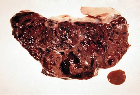
Figure 11.4 Fascioloides magna fluke and cross section of a necrotic goat liver through which it migrated. Source: Courtesy of Dr. M.C. Smith.
porphyrin pigment. The pigment may also be seen in mesenteric nodes. Though rare, cysts containing live or dead flukes may be found in the livers of goats (Olsen 1949).
Diagnosis
Diagnosis is based on a history of sudden death and characteristic lesions in the liver at necropsy Differential diagnosis must include acute fascioliasis, cysticercosis, toxic hepatitis, and, in younger animals, haemonchosis and coccidiosis. Other causes of sudden death are reviewed in Chapter 16.
Treatment
Treatment is difficult because clinical disease is caused by immature flukes that can induce serious or fatal illness when present even in small numbers. This means that useful drugs must approach 100% efficacy against immature flukes. Albendazole administered orally to goats at 15 mg/ kg was reported to be 99% effective against flukes 8 weeks of age or older (Foreyt and Foreyt 1980). The use of albendazole during the first months of pregnancy may carry some risk to the fetus.
Other flukicides have not been evaluated specifically for F. magna infection in goats. Closantel was 95-98% effective in sheep at a dose of 15 mg/kg orally or 7.5 mg/kg intramuscularly (Stromberg et al. 1985). Clorsulon was not effective against immature F. magna in sheep at a single oral dose of 15 mg/kg (Conboy et al. 1988), but was 92% effective against 8-week-old larvae in sheep at a dose of 21 mg/kg (Foreyt 1988b). Oral rafoxanide at 15 mg/kg has been reported to be effective in red deer (Rajsky et al. 2002). Of these various anthelmintics, only albendazole is approved for use in goats in the United States and it is only approved for F. hepatica.
Control
Control of fascioloidiasis is complicated by the need to control wild animal definitive hosts and an ecologically diverse, intermediate host snail population. In endemic areas where cases have occurred, intensive management without pasturing should be considered for dairy goats. This is obviously difficult for extensively managed fiberproducing goats. Avoidance of marshy pastures and attempts to fence off deer may be helpful. When exposure cannot be controlled, reduction of fluke loads in goats can be undertaken. In northern climes, prophylactic treatment with albendazole is administered after the first killing frost and repeated one month later to help reduce clinical losses.
Lancet Fluke Infection
Dicrocoelium dendriticum, the lancet fluke, generally produces a chronic form of liver fluke disease in goats that is less severe than that produced by Fasciola spp. The fluke is widely distributed around the world.
Etiology
D. dendriticum is a small liver fluke measuring 6-10 mm in length and 1.5-2.5 mm in width. It has an indirect life cycle involving two intermediate hosts: terrestrial snails and ants. There are 40 species of snails from nine families that may serve as primary intermediate hosts, but all secondary host ant species are of the subfamily Formicinae (Boray 1985). Goats, sheep, and cattle are the common definitive hosts, but other livestock species and wild mammals and humans can be infected.
Adult flukes reside in the bile ducts and excrete eggs to the outside in feces. Eggs are ingested by snails, where they develop into cercariae. Cercariae are expelled by snails in slime balls and are ingested by ants. Infectious metacercar- iae develop in the ants and are neuropathogenic, causing ants to remain paralyzed on herbage during peak times of livestock grazing. Infected ants are ingested by grazing livestock and the young flukes excyst from metacercariae in the small intestine of the definitive host. In contrast to the Fasciolidae, these immature flukes do not migrate through the peritoneal cavity and liver parenchyma. Instead, they enter the hepatic biliary system directly through the common bile duct via the small intestinal lumen. The prepatent period for D. dendriticum is 8-12 weeks.
Epidemiology
D. dendriticum infection is common in North America, Europe, Asia, North Africa, and the Middle East. Prevalence in goat populations is reported to be as high as 45% in endemic areas (Manas-Almendros et al. 1978). It is less common in South America and is not found in Australia, New Zealand, and much of Africa. Dicrocoelium hospes, a cattle fluke of West and East Africa, has been reported in goats in Niger and Nigeria (Tager-Kagan 1979; Schillhorn van Veen et al. 1980).
Numerous habitats are potentially contaminated with D. dendriticum because there are multiple snail hosts, terrestrial ant populations are widespread, and many wild mammals serve as reservoir hosts. Grazing areas near forests are particularly high-risk sites. The fluke eggs are also resistant to desiccation and freezing. Hibernating ants can maintain metacercarial infections, so that grazing stock are at risk from the beginning of spring grazing.
Pathogenesis
These flukes are less pathogenic than the Fascioloidae because they do not migrate through liver parenchyma. D. dendriticum infection, which may involve thousands of flukes per goat, can cause progressive inflammation of the entire biliary tree and potentially lead to fibrosis, biliary cirrhosis, and hepatic insufficiency.
Clinical Findings
D. dendriticum may be a subclinical infection or produce a disease syndrome similar to chronic fascioliasis. Affected goats have a history of weight loss. They appear thin and depressed and may show signs of anemia and hypoproteinemia, including pallor and intermandibular edema.
Clinical Pathology and Necropsy
Identification of lancet fluke eggs in the feces of goats is the most reliable means of diagnosis. The dark brown, opercu- lated eggs are much smaller than those of F. hepatica or F. gigantica and can be found by both fecal flotation and sedimentation methods. D. dendriticum infection may produce elevated serum levels of GGT and AP caused by biliary inflammation. Anemia and hypoproteinemia are evidenced in the hemogram.
At necropsy, adult lancet flukes are found in the bile ducts. In early cases there is thickening of the biliary ducts. In severe, chronic infections, biliary fibrosis, cirrhosis, and scarring of the liver surface may be noted. The comparative liver pathology of D. dendriticum and F. hepatica in goats has been described (Rahko 1972).
A paramphistome parasite of the bile duct of buffalo and cattle in Asia, Gigantocotyle explanatum, sometimes is found in the bile duct of goats (Upadhyay et al. 1986). It should be differentiated from D. dendriticum, which has a similar appearance.
Diagnosis
Lancet fluke disease must be differentiated from chronic fascioliasis, gastrointestinal helminthiasis, paratuberculosis, and other causes of chronic wasting, as discussed in Chapter 15.
Treatment
In France, albendazole is marketed for use against the lancet fluke at 15 mg/kg orally, with a precaution against use during the first three months of pregnancy. Thiophanate is marketed at 50 mg/kg orally, with a milk withdrawal time of three days. Diamphenethide at 220-330 mg/kg orally is highly effective, with no toxic side effects noted (Devillard and Villemin 1976). Drugs reported to have an efficacy of 90% or greater in sheep include thiabendazole at 200 mg/ kg, fenbendazole at 100 mg/kg, praziquantel at 50 mg/kg, and netobimin at 20 mg/kg (Boray 1985; Sanz et al. 1987). Note that some of these doses exceed the routine doses used in helminthiasis. Brotianide was found to be ineffective in goats at oral doses as high as 22.5 mg/kg (Shahlapour et al. 1986).
Control
Control measures are complicated by the existence of two intermediate hosts, and strategic anthelmintic therapy may be the only practical control method. In endemic areas in temperate regions, goats should be treated in the autumn to reduce adult fluke burdens before the winter feeding season begins. In tropical and subtropical areas, year-round, repetitive treatments timed within the prepatent period of the flukes reduce fluke burdens and pasture contamination.
Pancreatic Flukes
Eurytrema spp., or pancreatic flukes, produce mostly subclinical infections in goats. The presence of Eurytrema eggs in feces may confuse the diagnosis of the more clinically important lancet fluke, D. dendriticum.
Eurytrema pancreaticum is widely distributed throughout Asia and also in Brazil. The fluke has a similar life cycle to D. dendriticum, with different snails serving as primary intermediate hosts, and grasshoppers instead of ants serving as secondary intermediate hosts. The definitive hosts are goats, sheep, cattle, buffalo, and humans. The young flukes ascend the pancreatic duct, but occasionally mature in the bile duct or duodenum. This fluke is wider and spinier than D. dendriticum. The prepatent period is 11-15weeks. Other species of Eurytrema reported from goats include Eurytrema cladorchis in China and Nepal (Chongti and Tongmin 1980; Mahato 1987) and Eurytrema coelomaticum from Taiwan and Brazil (Shien et al. 1978; Fernandes and Hamann 1985).
Pancreatic fluke infestations are usually subclinical, but heavy infections may contribute to emaciation. A case of sudden death in a cachexic adult goat in Nepal was caused by rupture of the gastroepiploic vein that contained E. cladorchis (Mahato 1987).
At necropsy, pancreatic flukes may be found in the duodenum and bile ducts, but most commonly in the pancreatic ducts. These ducts may show catarrhal inflammation and thickening and the pancreas itself may show focal areas of atrophy and fibrosis. The severity of lesions increases with the number of flukes present (Shien et al. 1979).
Praziquantel given two consecutive days at 20 mg/kg orally is effective against Eurytrema spp. in goats (Kono et al. 1986). Triclabendazole (Kono et al. 1986), niclofolan (Weng 1983), and nitroxynil (Suh 1983) are not effective.
More on the topic Parasitic Diseases:
- References
- Smith Mary C., Sherman David M.. Goat Medicine. 3rd edition. — Wiley-Blackwell,2023. — 976 p., 2023
- CLASSIFICATION
- Classification of cases: Sheep
- Eosinophilic enteritis (EE)
- Smith Bradford P., Van Metre David C., Pusterla Nicola (eds.). Large Animal Internal Medicine. Part 2. 6th edition. — Elsevier,2020. — 2279 p., 2020
- References
- Acquired Torticollis
- Edema Is a Clinically Noticeable Excess of Interstitial Fluid
- CARDIOVASCULAR DISEASES