Physical Assessment
Numerous factors influence a foal's body weight at birth, including breed, gender, gestational age, and intrauterine environment. Estimates of body weight for newborn Thoroughbred foals for the purpose of drug calculation range between 40 and 55 kg, although many healthy foals may well exceed this range.
Birthweight of Thoroughbred foals was positively correlated with mare body condition score.11 The average daily weight gain for healthy foals is breed dependent but is typically between 1.3 and 1.7 kg/day for Thoroughbredtype foals.12 Daily weight gain in hospitalized foals is determined by disease, calorie intake, and a lack of exercise and is almost always less than that of healthy, active foals.The degree of physical development should be considered in relation to the estimated gestational age. This assessment should be made in light of the variable gestational length in horses. Typically gestational length is calculated from the time of insemination through birth, a figure that overestimates the true gestational period by as much as 7 days. The mean gestational period in Thoroughbreds is around 340 to 342 days with 95% of mares foaling between 327 and 357 days.13 A number of factors influence this period, including breed, gender, and time of year. Colts on average have a gestational length that is approximately 1.5 to 2.5 days longer than that of fillies; they are slightly heavier, slower to stand, and have a heavier placenta.14 Mares that conceive early in the breeding season have pregnancies that may be up to 10 days longer than those that become pregnant late in the season.13,15 The role of dam, sire, and the dam's sire in determining gestational period has been suggested in some but not all breeds.16
Physical characteristics of immaturity include low birth weight, small body size, short and shiny hair coat, doming of the head, periarticular laxity, and droopy ears.
Foals with a shortened gestational period and signs of physical immaturity are termed premature, while foals that are physically immature in the face of an appropriate gestational length are termed dysmature. Foals born after 356 days should be regarded as post-term. This is distinguished from foals that are post-mature, a condition of increased morbidity as a consequence of failing placental function.16 Post-mature foals tend to have a lean and lanky physical appearance.Rectal Temperature
Rectal temperature in normal foals ranges between 99° F and 102° F (37.2° C and 38.9° C) during the first 4 days after birth.2 The upper end of the normal range, which is around 1° F (0.6° C) above the upper end of the adult range, is strongly influenced by environment and exercise. Temperature can be variable in foals with systemic sepsis; in the early stages of sepsis the rectal temperature is commonly within the normal range or mildly elevated. Foals in septic shock will usually have a low rectal temperature, and those with localized infection will often be afebrile.
Ears
The ears of a normal foal are typically erect at the time of birth. Drooping of the ear pinnae is most commonly associated with prematurity or dysmaturity and, as such, is often accompanied by other physical signs of immaturity, such as small body size, short silky hair coat, and a domed head. Examine inside the ear pinnae for evidence of dermal hemorrhages. These can be a manifestation of systemic sepsis (see Chapter 17) or thrombocytopenia caused by disseminated intravascular coagulation (DIC) or alloimmune thrombocytopenia.
Eyes
The neonatal foal may not be able to close its eyelids completely (lagophthalmos), has reduced production of tears, and lacks a menace response. The menace response is a learned one and is usually acquired in the first 14 days of life. The pupil is oval-shaped, and pupillary light reflexes are present from birth but are slower. Other normal ocular findings include persistence of the hyaline artery remnants, the blood supply to the developing lens, and a pale pink to red optic disc with smooth margins.
Newborn foals also have very prominent lens “Y” sutures. Lens suture lines are apparent in most foals and can persist into adulthood. An ophthalmic survey of healthy Standardbred foals conducted within 48 hours of birth reported abnormalities in 36% of foals.17 Most were acquired and included retinal and subconjunctival hemorrhages; these had resolved in all foals by 1 (retinal) and 2 weeks (subconjunctival) of age. All foals in that study had hyaloid system remnants of various degrees.17 In an ophthalmic survey of older foals (mean age of 9.4 weeks), suture lines were observed in 95% of animals.18 The anterior suture was predominately Y-shaped, but the authors reported that posterior sutures were more variable, from “Y-shaped to sawhorse, to stellate.” Posterior lens sutures are reportedly more difficult to recognize in Thoroughbred foals. The suture patterns are not consistently symmetric between eyes. Prominent lens sutures can be confused with congenital cataracts. Congenital cataracts are amenable to surgery in the absence of uveal tract inflammation, a normal retina, and an appropriate demeanor. Other abnormalities that can occur include atresia of the nasolacrimal system, dermoids, retinal dysplasia, and optic nerve hypoplasia.The neonatal foal may have significant corneal ulceration without tearing, blepharospasm, or photophobia due to reduced corneal sensitivity when compared with older foals and adult horses.19 Consequently, special attention is required to monitor for ocular disease. It is recommended to fluorescein stain the cornea daily in sick recumbent neonatal foals. The combination of lagophthalmos, recumbency, and seizure predisposes foals with encephalopathy to a high incidence of corneal ulceration.
Entropion (inward rolling of the eyelid margin) is a common ocular problem of newborn foals. This usually occurs as a complication of dehydration and/or malnutrition and may lead to corneal abrasion and ulceration.
Treatment is placement of vertical mattress sutures or staples to evert the offending lid while the primary condition is corrected. An alternative strategy is to inject procaine penicillin G subcutaneously into the lower lid to cause temporary eversion. It is extremely rare that surgery is required to correct entropion.Examine the conjunctiva and sclera for signs of hemorrhage. This is commonly associated with birth trauma, and affected foals should be monitored closely for development of more classical postasphyxia complications. These hemorrhages resolve by week 2 of postnatal life.17 Episcleral injection (engorgement and proliferation of scleral vessels) is a prominent feature of systemic sepsis. Iridocyclitis is usually associated with systemic sepsis and can include hyphema, hypopyon, and fibrin. These changes are not uncommon in foals with in utero-acquired infection and may be present at birth. Iridocyclitis is also seen in older foals with Salmonella or Rhodococcus equi infection.
Oral Cavity
Oral mucous membranes in healthy newborn foals should be pink and moist and have a capillary refill time (CRT) of 1 to 2 seconds. Bright pink to red mucous membranes, accompanied with injected scleral vessels and hyperemia of the coronary bands, are consistent with the early stages of sepsis. During this early hyperdynamic phase the CRT may be shorter than normal. Darkening of mucous membranes with a delay in the CRT may indicate sustained sepsis. In severe cases this is accompanied by petechial or ecchymotic hemorrhages.
Icteric mucous membranes are most commonly associated with systemic sepsis but are also a feature of neonatal isoeryth- rolysis, liver disease, internal hemorrhage, meconium retention, or infection with equine herpesvirus type 1. Pale membranes suggest anemia and can occur with external umbilical cord hemorrhage, internal hemorrhage from torn umbilical vessels, fractured ribs, or hemorrhage within gastrointestinal or urinary tracts.
Pallor with icterus would normally indicate neonatal isoerythrolysis or internal hemorrhage. Petechial or ecchymotic mucosal hemorrhages suggest advanced sepsis when associated with darkened membranes and a prolonged capillary refill time, or alloimmune thrombocytopenia when the membranes are pink (Fig. 16.1). Blue to gray discoloration of the membranes is consistent with hypoxemic circulatory shock. Overt cyanosis is uncommon and should prompt close examination of the cardiovascular and respiratory systems.In term foals the central incisors usually erupt during the first 5 to 7 days of postnatal life. The middle incisors rise between 4 and 6 weeks of age, but the corner incisors do not erupt until around 6 to 9 months. In miniature horses and ponies the eruption of the middle and corner incisors is delayed at 4 months and 12 to 18 months, respectively. The 12 temporary molars are present at birth or erupt within the first week of life. These teeth are replaced by permanent premolars at 2.5, 3, and 4 years of age. The three molar teeth do not erupt until 1, 2, and 3.5 years of age for molars 1, 2, and 3, respectively.
Dental problems are uncommon in foals with the exception of those associated with facial deformities such as maxillary prognathism, mandibular prognathism, and campylorrhinus. Maxillary prognathism (parrot mouth; bird face; overbite;
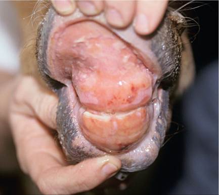
FIG. 16.1 Spontaneous mucosal hemorrhages and ulceration in a foal with alloimmune neonatal thrombocytopenia.
brachygnathism; brachygnathia) describes the condition where the mandible is shorter than the maxilla, producing an overjet or overbite. The condition may affect the incisors, cheek teeth, or both, and is the most common congenital oral malformation of foals. The majority of foals with maxillary prognathism suffer little impact to their quality of life, and the defect simply represents a cosmetic problem. In moderate to severely affected foals the lack of incisor contact results in excessive growth of the lower teeth and secondary soft tissue trauma to the palate caudal to the upper incisors.
The premaxilla and incisive bones tend to bend forward and downward as growth continues due to the lack of an opposing force, normally provided by the mandible. This can lead to difficulty in grazing. Mandibular prognathism (sow mouth; monkey mouth; brachygnathia superior; underbite) describes the condition where the maxilla is shorter than the mandible, producing an underbite. The condition occurs much less commonly than the maxillary equivalent and is most frequently seen in pony and small horse breeds. The defect in its most severe form can be associated with deformity of the nasal passages and nares, and there may be difficulty in breathing and an inspiratory stridor. Mandibular prognathism is commonly associated with congenital hypo- thydroidism.20 Campylorrhinus lateralis (wry nose, wry face) describes the condition where the premaxilla and nasal septum are deviated laterally. It may occur singularly or in combination with other deformities, such as wry neck, cleft palate, and maxillary or mandibular prognathism. If the deviation is severe, the foal may have great difficulty in sucking from the mare. There may also be problems with breathing. In most foals the deviation is mild and represents a simple but obvious cosmetic defect. Close attention must be paid to dental wear as maxillomandibular malocclusion occurs commonly.Cleft palate is an uncommon congenital defect of foals that results from incomplete fusion of the lip and/or secondary palate during the early gestational period. Previous reports estimate the incidence between 0.1% and 0.2% of all horse births.21 Almost all clefts occur in the secondary palate, the horizontal partition dividing oral and nasal cavities. The secondary palate includes all of the soft palate and most of the hard palate, with the majority of defects restricted to the soft palate. The basis of this congenital defect is not known but could include a heritable component and/or exposure of the developing fetus to infection, toxins, or nutritional disturbances. The diagnosis is usually straightforward and centers on an appropriate history and signalment. Confirmation is through palpation using the third finger of an upturned hand or by direct inspection of the oral cavity. It is important that the hand is clean before direct palpation. Endoscopic examination of the nasal passages or through the oral cavity under anesthesia may be required to detect small defects. Oral palpation is recommended for all sick neonatal foals before commencing potentially costly treatment. A cleft in the secondary palate permits the oral contents to pass directly into the nasopharynx and nasal passages. Consequently, the most common complaint is nasal regurgitation of milk immediately after sucking. As the foal matures, feed particles will also appear at the nares. Foals with small palate defects may have intermittent milk drainage from the nostril and consequently escape diagnosis during the neonatal period. Rarely, cases are initially diagnosed in adult horses. Most will develop an aspiration pneumonia, which can be difficult to identify in its early stages due to immaturity of cough receptors. Surviving foals will typically be poorly grown and ill-thrifty. The presence of milk at the nares after feeding does not definitively indicate that a cleft is present and is more commonly associated with pharyngeal/ laryngeal weakness or incoordination. The tongue should be examined for evidence of candidiasis, appearing from small white plaques up to a diffuse tan coloration of the tongue. The condition can be local or systemic and is common in debilitated foals and those that have been on chronic antibiotic therapy. In severe cases the condition will cause reduced nursing and salivation.
Thyroid Gland
Hypertrophy of the thyroid gland (goiter) can occur in response to deficient or excess dietary iodine. One of the more common reasons for thyroid gland enlargement in neonatal foals is excess iodine supplementation during pregnancy.22 There are reports of neonatal goiter when seaweed was incorporated into the diet of broodmares or when pellets were fed to mares that had been formulated with excessive iodine.23,24 Dysmature foals with thyroid hyperplasia and concurrent musculoskeletal problems have been identified in Western Canada and northwest United States.25,26 The syndrome results in hypothyroidism and may be related to the feeding of diets that are high in nitrate or low in iodine to mares during pregnancy. Thyroid hormone is an important cofactor in maturation of the respiratory system, and hypothyroidism has been linked to respiratory failure in a newborn foal.27 In older foals enlargement of the thyroid has been associated with dietary iodine deficiency and low circulating levels of T4.22 Newborn foals have baseline T3 and T4 levels that are considerably greater than that of adult horses.28,29 These levels decline over the first 12 days after birth.
Neck and Back
A number of congenital anomalies affect the alignment of the vertebral column. These include atlantoaxial malformations, scoliosis, kyphosis, lordosis, and combined anomalies, such as kyphoscoliosis. Atlantoaxial malformations may occur with or without cervical scoliosis or signs of spinal cord compression. Arabian foals are most commonly affected, and a familial predisposition has been suggested.30 There may be palpable abnormalities of the atlas and axis and altered head carriage. The diagnosis is confirmed using radiography where a range of abnormalities is seen, including atlantooccipital fusion, hypoplasia of the dens, and axis malformation. Prognosis is poor as many have signs of ataxia and paresis involving all four limbs at birth. Some animals may have normal neurologic function. Foals with severe kyphosis or kyphoscoliosis often have underlying malformation of the thoracic vertebrae.31
Gastrointestinal System
Most foals display abdominal straining within a few hours of birth and pass most of the meconium within the first 1 to 4 hours after delivery, but passage may be delayed without concern for up to 12 hours. Differentiating straining to defecate from straining to pass urine can be difficult in some foals. Typically foals straining to pass feces will have an arched appearance to their hindquarters (Fig. 16.2). A change in the color and consistency of the feces from a dark brown to black pasty material to a lighter brown, less tenacious material indicates that the meconium has been passed. The routine administration of an enema is performed commonly in healthy newborn foals in an attempt to reduce the straining associated with passage of meconium. A variety of methods are used, with commercial glycerine phosphate-based products the most common. These are relatively easy to administer, although care should be taken to avoid direct trauma to the rectal mucosa with the applicator tip. Many veterinary practices probably use gravity enemas using 400 to 800 mL of warm soapy water delivered through a soft tube, such as a canine stomach tube or male urinary catheter. Acetylcysteine retention enemas are not administered routinely, but rather in foals with resistant meconium impactions.
Auscultation of the abdomen can provide an indirect assessment of intestinal contractility. The technique should be interpreted with caution as the presence of borborygmi does not equate to propulsive motility or ingesta transit. The changes
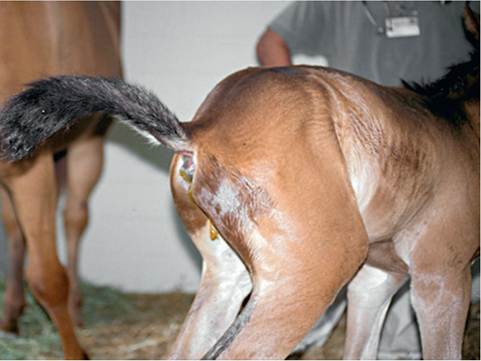
FIG. 16.2 A foal demonstrating a classical hunched appearance while straining to pass feces.
in gastrointestinal sounds over time may be more useful in determining a diagnosis. For example, a silent abdomen that progresses to a hyperactive one could be indicative of inflammatory intestinal disease, whereas an abdomen with normal sounds that changes to silent could be consistent with a surgical lesion. Percussion and ballottement are used to differentiate abdominal distention caused by gas or fluid accumulation. A tympanic sound on percussion is consistent with intraluminal or free peritoneal gas, and ballottement can detect waves of free abdominal fluid. Progressive abdominal distention, irrespective of the underlying cause, can be monitored using a soft measuring tape repeatedly positioned over small clipper marks. Gastric distention can cause a “sprung rib,” a swelling that projects beyond the 18th rib.
Firm structures can be detected using transcutaneous abdominal palpation, assuming the foal is suitably relaxed. It is possible to palpate bowel impacted with meconium, fecoliths, intussusceptions, the urinary bladder, or enlargement of the internal umbilical remnants caused by infection or hemorrhage. The external umbilicus should also be palpated for enlargement or herniation, and, in colts, the inguinal rings and scrotum should be assessed for bowel herniation. These are typically soft and fluctuant swellings that are easily reduced when the foal is placed on its back and are managed by manual reduction and strapping. Incarceration is rare, but bowel integrity can be assessed using ultrasound; devitalized bowel lacks motility and has thickened and edematous wall. In some colts the intestine can protrude through a rent in the parietal vaginal tunic and migrate subcutaneously in what is described as a direct inguinal hernia32 (Fig. 16.3). This is often associated with preputial swelling and requires surgical intervention.
Digital rectal examination is indicated in a colicky foal or in any foal where meconium passage has not been observed. Distal meconium impaction is typically apparent on rectal examination, but high impactions are not. Intestinal atresia should be suspected if there is no history of fecal passage and there is clear or white mucous on the glove.
Passage of a nasogastric tube is an often-overlooked diagnostic procedure in colicky neonatal foals. Removal of gastric fluid can reduce or eliminate colic signs, as well aid in the localization of disease. Gastric distention may also be present in sick neonates showing no signs of colic. The condition occurs with primary gastric outflow problems or mechanical or functional small intestinal obstruction. Functional obstruction caused by small intestinal ileus occurs more commonly in neonates secondary to a range of diseases including hypoxic
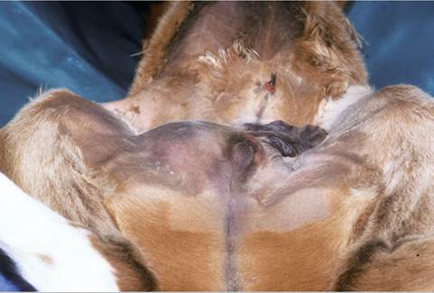
FIG. 16.3 A dissecting scrotal hernia before surgical repair.
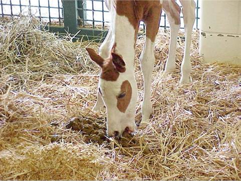
FIG. 16.4 Coprophagy is an important behavior in establishing hindgut flora.
ischemic syndrome, sepsis, prematurity, enteritis, electrolyte derangements, and overfeeding.
Coprophagy is observed in normal foals from birth through 5 to 6 months of age.33-35 Most foals demonstrate coprophagy by 7 days of age (Fig. 16.4). The consumption of feces is not driven by hunger, and foals have a selective preference to consume feces of their dams. The most likely basis for coprophagy is as a mechanism to populate the intestinal tract with bacteria, fungi, and protozoa essential for digestion of a herbivorous diet. Coprophagy precedes the passage of protozoa in foal feces and the development of the syndrome known as “foal heat diarrhea.” This is a benign diarrheal disease of newborn foals and should not require treatment.
Abdominal Ultrasound
Ultrasound is a key component in the examination of a foal abdomen. Numerous conditions are recognized on ultrasound, including uroperitoneum, bacterial peritonitis, hemoperitoneum, strangulating intestinal obstructions, meconium impaction, gastric enlargement, enteritis, necrotizing enterocolitis, and hernias. The foal can be scanned in lateral recumbency or standing; the latter is preferred as thickened or heavy structures will move to the dependent region of the abdomen, gas-filled structures will migrate dorsally, and visualization of peritoneal fluid is improved. Clipping is not essential if the hair is dampened with alcohol. The stomach is identified in the mid to ventral abdomen on the left side between the 6th and 12th intercostal spaces and lies medial to the spleen. In contrast to adults, the stomach is commonly in contact with the ventral body wall during the first week of life and the contents are readily imaged.36 Gastric wall thickness is typically around 2.0 mm during the first week of life. The duodenum is readily located in normal foals on the right side of the abdomen between the ventral and caudal aspect of the liver and the dorsal margin of the right dorsal colon, as well as ventral to the caudal pole of the right kidney. The remainder of the small intestine is also easily identified throughout the abdomen and should have a wall thickness less than 2.5 mm. The large intestine is differentiated from the small intestine by increased luminal diameter, bowel sacculations, and greater likelihood of gas. The colon wall should be less than 2.8 mm during the first week of life. The neonate with acute colic should be assessed with ultrasound for small intestinal intussusception. These are readily identified, are typically jejunojejunal, and are often located in the ventral abdomen when the foal is standing. They have a characteristic appearance with the intussusceptum surrounded by fluid and the intussuscipiens creating a “target” sign. Some will resolve spontaneously, but most require surgical reduction. Meconium impactions have an echogenic ball or log appearance when in the small colon or a more varied appearance when present in the large colon, a hypoechoic to hyperechoic mass that may be surrounded by fluid.37 Thickening of the wall with intramural gas echoes is consistent with a strangulated obstruction or necrotizing enterocolitis. Abdominal hemorrhage results in a classical “smoke swirl” appearance to the peritoneal fluid.
Abdominal Radiography
Radiography has been displaced as the principle imaging modality of the foal abdomen but still can provide essential information.38 Contrast radiography remains an important technique to assess gastric outflow problems, intestinal transit, intestinal atresia, and high meconium impaction. The abdomen of most neonatal foals can be captured on a 35 cm ? 43 cm cassette. A grid should be used to minimize film fogging. For a standing foal weighing around 50 kg, using a 400-speed film-screen system, a grid with a 10:1 grid ratio, and a focal spot-film distance of 100 cm, the appropriate technique should be in the range of 80 to 90 kV (peak) and 15 to 20 mA. This technique can be reduced slightly lateral projections in the recumbent foal. The beam should be centered on the last rib. For upper gastrointestinal contrast studies, 5 mL/kg of 30% wt/vol barium suspension is administered through a nasogastric tube; ideally foals are fasted for 4 hours before the procedure and should not be sedated. Barium typically empties rapidly from the stomach, allowing examination of the stomach and duodenum on initial films. The contrast should clear the small intestine by 3 hours and be completely cleared by 36 hours. The cecum should be visible by 1 to 2 hours post administration. A barium enema can be used to evaluate the lower intestinal tract, specifically intraluminal obstruction and atresia. Sedation can be used as the procedure is not defining a dynamic problem. A 30-French Foley catheter with a 30-cm3 balloon is inserted 6 to 10 cm into the rectum, the cuff is inflated, and 300 to 500 mL of 30% wt/vol barium sulfate suspension are infused. Volumes up to 20 mL/kg have been described; colonic or rectal rupture is a rare but fatal complication of the procedure. Computed tomography (CT) is an underused tool in the assessment of the foal abdomen, with most applications involving examination of limbs, head, or neck.39
Abdominocentesis
Peritoneal fluid can also be evaluated by abdominocentesis. The technique has a greater complication rate in neonates, and ultrasound should be used to identify pockets of fluid for sampling. Most complications arise due to a combination of hypodermic needles, a moving patient, and a relatively thin bowel wall. If no fluid is identified, then the procedure should not be performed. For small volumes it is recommended to use a blunt, small bovine teat cannula; for larger volumes a hypodermic needle can be used. Peritoneal fluid nucleated cell counts are lower in foals than in adult horses.40 Nucleated cell counts above 1.5 ? 109∕L may be considered abnormal in foals up to 4 months of age,41 although another study suggested a value of 3.0 ? 109∕L as a cutoff for foals younger than 75 days of age.42 Cytologic evaluation is similar to adults with a predominance of large mononuclear cells (macrophages and mesothelial cells) with smaller numbers of lymphocytes and neutrophils. The relative percentage of neutrophils (15%) may be less than that seen in normal adult peritoneal fluid. The protein concentration of foal peritoneal fluid is similar to that of adults, below 20 gm/L, and therefore values above this are considered abnormal. An increased total nucleated cell count, increase in the percentage of neutrophils with cell degeneration, and intracellular and extracellular bacteria would be expected in foals with septic peritonitis. This would be accompanied by a low pH (2.7 mmol/L).40
Cardiovascular System
Physical assessment of the cardiovascular system includes examination of visible mucous membranes, palpation of peripheral arterial pulses, assessment of extremities for warmth, detection of limb or ventral edema, and cardiac auscultation. The heart rate in newborn foals in the minutes after birth is influenced by vagal tone and varies between 36 and 80 bpm. As the foal becomes active and struggles to stand, the heart rate will increase to 120 to 150 bpm before stabilizing in a range of 80 to 100 bpm over the first week of life.43 Tachycardia is an important mechanism in neonates to improve cardiac output and blood pressure, and it is expected during periods of asphyxia and in response to hypovolemia, sepsis, anemia, or hypoxemia. A sustained increase in heart rate may also accompany pain or stress or reflect a primary cardiac problem. Bradycardia can be due to hypothermia, electrolyte abnormalities, or hypoglycemia.
Cardiac arrhythmias are rarely recognized but occur in most foals during the immediate postpartum period with supraventricular premature contractions and sinus arrhythmia predomi- nating.44,45 Other arrhythmias include supraventricular (atrial) tachycardia, atrial fibrillation, second-degree atrioventricular block, ventricular premature contractions, ventricular tachycardia, and idioventricular rhythm. These rhythm disturbances typically only last for 5 minutes and in most foals have disappeared by 15 minutes of age. Rarely they may persist up to 2 hours of age. The more severe arrhythmias are associated with a lower arterial oxygen concentration, suggesting a role for hypoxemia in the pathophysiology of these events. Arrhythmias that persist beyond this timeframe should be investigated by electrocardiogram. Causes include electrolyte abnormalities, persistent hypoxemia, and sepsis. Foals with sustained and rapid ventricular tachycardia need treatment with intravenous quinidine, gluconate, lignocaine, or propranolol.46
Cardiac murmurs are expected in the early neonatal period. A patent ductus arteriosus (DA) can result in a continuous machinery murmur with a point of maximal intensity high over the left heart base in the third intercostal space.46,47 These are typically audible up until 3 to 4 days of age.48 As the DA begins to close, the systolic component of the murmur often remains audible. Low-intensity, left-sided systolic murmurs are commonly heard through the first months of postnatal life. Some may be associated with flow across the foramen ovale, but most are likely due to turbulence in the great vessels created during left ventricular ejection.46 Others have suggested that flow through the DA may persist for weeks to months.49 Loud murmurs that persist beyond 7 days of age or any murmur that is accompanied by signs of cardiac disease, such as poor growth, cyanosis, lethargy, or exercise intolerance, should be referred for echocardiography. Auscultation of both sides of the thorax is important as right-sided cardiac murmurs are a feature of several congenital cardiac anomalies. Occasionally serious cardiac defects may not be associated with audible murmurs. Although most congenital cardiac diseases are detected in foals or young horses, some defects do not become apparent until the horse is required to perform or may occur simply as incidental findings in older horses.
Distal extremities should be free of edema and warm to the touch. Limb edema can occur for several reasons, but most commonly secondary to asphyxia or in response to inappropriate fluid therapy in oliguric or anuric foals. Limbs that are cool or cold are poorly perfused and will typically have weak arterial pulses. However, the relationship between peripheral pulse pressure and hypotension is not consistent; hypotension is not always associated with weak pulses and, conversely, weak pulses do not always indicate hypotension.50 Arterial thrombosis should be suspected if a single limb is cold, although an aortic thrombosis could result in decreased perfusion to both hind limbs.51 Pulse quality can be assessed in the distal limbs using the great or dorsal metatarsal arteries on the lateral aspect of the hind cannon bone. In the forelimbs the brachial artery can be palpated at the level of the medial collateral ligament of the elbow joint. Several methods are available to assess arterial blood pressure. Direct blood pressure monitoring is ideal because it is accurate and continuous, but it is invasive and difficult to maintain and can be complicated by vessel thrombosis or sepsis. More commonly, blood pressure is monitored indirectly using oscillometry or Doppler-based devices over the coccygeal, brachial, or great metatarsal arteries. An infant-sized inflatable cuff (bladder 52 mm) placed snuggly around the base of the tail yields data that are comparable to mean arterial measurements taken directly from a catheterized great metatarsal artery in a day-old foal weighing between 44 and 69 kg.52 The technique tended to underestimate systolic and overestimate diastolic values, although the differences were not large. The mean arterial pressure of a healthy Thoroughbred foal using an oscillometric device was reported to be 95, 100, and 101 mm Hg at days 1, 2, and 3, respectively.53 Other noninvasive methods to measure cardiac output, such as volumetric echocardiography, have shown good correlation with more robust but invasive techniques in anesthetized foals. The question remains if this correlation will be sustained in critically ill foals.
Respiratory Tract
In newborn foals the nasal passages are examined for meconium staining and, if present, the passages should be suctioned using a dose syringe and soft catheter or other portable or fixed suction device. The early stages of pulmonary disease are difficult to recognize, as cough and nasal discharge are typically absent. Immature cough receptors also contribute to a lack of awareness of ongoing milk aspiration into the major airways. Auscultation of the lung fields can also be misleading in young foals. Fluid sounds are normally present immediately after birth, as are end-inspiratory crackles due to simple atelectasis of the dependent lung during or after lateral recumbency. These crackles should resolve when the foal has been standing for several minutes. Conversely, foals with bronchopneumonia may have minimal abnormal lung sounds until the pathology becomes severe. In the absence of advanced diagnostics the clinician must rely on clinical signs, such as restlessness and agitation, increased respiratory rate and depth, or respiratory distress, to diagnose lower airway disease.
The normal response after delivery is an increase in minute ventilation, with elevations in both rate and tidal volume. The respiratory rate is usually rapid for the first hour of life, around 60 to 80 breaths/min, but should decline to a steady rate of 30 breaths/min thereafter in normal foals. Persistent elevations in the respiratory rate can be caused by a range of pulmonary and extrapulmonary causes. Pulmonary causes include meconium aspiration, bacterial or viral pneumonia, atelectasis due to recumbency, congenital pulmonary disease, rib fracture or dislocation, or pleural effusion. Nonpulmonary causes include fever, pain, excitement, exercise, brain disease, a compensatory response to metabolic acidosis, or idiopathic or transient tachypnea syndrome. An increase in the effort of breathing may be the first sign of respiratory disease. As the disease progresses, the foal can develop paradoxical thoracic wall motion due to muscular fatigue where the thoracic wall paradoxically moves inwards during inspiration and outwards during expiration. This is more commonly seen in premature foals with lung disease and is a sign of impending respiratory failure. Another sign of failure is an expiratory “grunt”; foals will keep their glottis closed until the end of the breathing cycle in order to maintain sufficient pressure in the respiratory tract to prevent airway collapse.
Respiratory rhythm should be regular in awake foals but may be irregular in normal foals during rapid eye movement (REM) sleep. This can also include body and limb movements and vocalization. Irregular or erratic breathing patterns in conscious foals, or in foals that are in coma or semicoma, are consistent with central nervous system disease or, less commonly, metabolic or electrolyte disorders, hypothermia, or prematurity or dysmaturity. Patterns include periodic breathing, ataxic breathing, tachypnea, hypopnea or intermittent apnea, and apneustic breathing. Hypopnea or apnea in foals with central nervous system disease may result in severe respiratory acidosis, prompting the use of respiratory stimulants or mechanical ventilation. Doxapram administered as a constant rate infusion was more effective than oral caffeine at reducing the arterial carbon dioxide concentration (PaCO2) in affected foals.54
Respiratory distress or stertorous breathing can be caused by several uncommon conditions in the newborn foal. A range of congenital cardiac or respiratory tract malformations should be considered if abnormalities are present at or shortly after birth. Those involving the respiratory system include stenotic nares, choanal atresia, subepiglottic cysts, collapsing trachea, and lung lobe agenesis. Tearing of the diaphragm during birth with herniation of abdominal contents can also result in immediate respiratory distress. Similar distress can be seen with a dorsally displaced soft palate caused by neurologic or anatomic dysfunction. Soft fluctuant swelling in the parotid region could be consistent with guttural pouch tympany. The condition is more commonly seen in older foals but has been reported in the early neonatal period, often associated with pharyngeal/laryngeal weakness and altered ostia function. This may also result in milk entering the pouch. There is a reported predisposition in fillies and certain breeds, including Arabians and Paints.55
Localized or widespread crepitus of the subcutaneous tissues could be consistent with air leakage from respiratory tissues, most commonly from ruptured pulmonary bullae.56 The thoracic wall should be palpated for evidence of rib shaft fractures or costochondral dislocation. Thoracic trauma and rib fracture occur most commonly as a complication of the birthing process. They are identified by observation of thoracic wall symmetry and synchrony, palpable crepitus, and localized edema. In addition, there may be an audible “click” on auscultation of the thorax. Affected foals may grunt on expiration and prefer to lie on the unaffected side. Diagnosis is confirmed by ultrasound or radiographic examination. On ultrasound examination there is disruption of the cortical surface and associated soft tissue edema. Management includes recognition and treatment of potential complications and avoidance of further thoracic trauma. Potential clinical complications of rib fracture include hemothorax, lung laceration and pneumothorax, and pericardial/myocardial puncture.
Arterial Blood Gas Collection and Analysis
Due to the limitations of physical examination in accurately assessing the respiratory system, arterial blood gas (ABG) analysis is commonly performed at referral practices during the initial assessment. It is a sensitive indicator of respiratory function.57 The sample is usually collected from the dorsal metatarsal artery. Alternative sites include the brachial artery, located at the level of the medial collateral ligament of the elbow joint, or the carotid artery, but hematoma formation is a common sequel to aspiration from this site. The sample will remain useful for up to 90 minutes in a capped plastic syringe at room temperature. Contamination of the sample with room air will artificially increase the arterial oxygen concentration (PaO2) and decrease the PaCO2. Interpretation of blood gas values of venous blood can be deceptive and should be restricted to evaluation of metabolic conditions and not pulmonary gas exchange.
Normal ABG values for neonates of different postnatal and gestational ages are presented in Table 16.2. Interpretation of the ABG sample involves consideration of the amount of struggling and position of the foal during sample collection. Foals that require considerable restraint to obtain a sample probably do not require sampling. Lateral recumbency can reduce the PaO2 by as much as 30 mm Hg. Supplemental oxygen affects ABG values; a flow rate of 10 L per minute (lpm) delivered by nasal insufflation increased the PaO2 from 84 ± 6 mm Hg to 298 ± 69 mm Hg in healthy, full-term newborn foals.58 This flow rate approximates to a fraction of inspired oxygen (FiO2) of 1.0.59 If the respiratory rate is rapid and the depth shallow, supplemental oxygen will be diluted by room air because of the large quantity of room air entering the upper respiratory tract, and the concentration of alveolar oxygen will be less than expected. A general guide for supplemental oxygen use is to set an initial flow rate of 5 lpm. Oxygen should be humidified if delivered at high flow rates or for long periods of time.
Hypoxemia (PaO2 < 70 mm Hg) can occur with hypoventilation, ventilation/perfusion mismatching, or right-to-left shunting of blood.57 The two most common respiratory-derived derangements are hypoxemia with normocapnia or hypocapnia and hypoxemia with hypercapnia. Treatment of the former involves supplemental oxygen. The latter is usually indicative of severe respiratory disease but could reflect primary hypoventilation, as may occur in cases of botulism. Acute hypercapnia is associated with a more substantial drop in blood pH and may lead to circulatory collapse and coma, particularly if accompanied by hypoxemia. Chronic exposure to elevated CO2 permits adaptation and subtle clinical effects. The change in pH is less dramatic because of enhanced bicarbonate reabsorption in the proximal tubules of the kidney, a process that begins within 6 to 12 hours and is maximal by 4 days. Hypercapnia can be exacerbated by fever or the administration of carbohydrates or bicarbonate. The latter is often clinically relevant and highlights the danger of giving large amounts of sodium bicarbonate to foals with pulmonary disease, potentially taking a primary metabolic acidosis and creating a more severe respiratory or mixed acidosis. Hypercapnia is also an expected response to metabolic alkalosis.60
Airway sampling either by transtracheal aspiration (TTA) or bronchoalveolar lavage is rarely performed in sick neonates, as the added stress of the procedures on an already compromised neonate can have fatal consequences. The TTA procedure is more likely to cause additional complications in foals with high ventilation rates, including pneumomediastinum and subcutaneous emphysema.
Pulmonary Imaging
Radiography can provide additional information that can assist in establishing a diagnosis, assessing the type and distribution of disease, and monitoring the response to treatment.38 Radiographs are particularly useful in the diagnosis of rib shaft fractures, pneumothorax, pleural effusion, and diaphragmatic hernia. Radiographic changes associated with pulmonary disease tend to lag behind clinical changes, and it is difficult to differentiate atelectasis from pneumonia. Infiltrate tends to settle or move ventrally as the foal becomes ambulatory. Unfortunately, capturing diagnostic thoracic images in the field is difficult because of the amperage of portable machines and the limitation in plate size. The relatively low amperage results in long exposure times, which in a poorly compliant patient with high respiratory rate leads to motion artifact and a false interpretation of disease. Images tend to be limited to standing or recumbent lateral projections as positioning for ventrodorsal views can further compromise the cardiopulmonary system. Pleural and parenchymal diseases have a similar ultrasonographic appearance in newborn foals to that seen in adults. Anechoic pleural fluid can be seen with some infectious processes, in foals with uroperitoneum, or with some congenital anomalies. Echogenic or cellular fluid is seen with other infectious processes, and in hemothorax cases there will be characteristic “smoke whirls” present.
■ TABLE 16.2
Normal Blood Gas Values for Foalsa
| Age Group | Arterial | Venous | |||||||
| O2 (mm Hg) | CO2 (mm Hg) | pH | Base Excess | HC-3 (mEq/L) | O2 (mm Hg) | CO2 (mm Hg) | pH | HC ; (mEq/L) | |
| Immediate post foalingb | 40-50 | 52-60 | 7.2-7.3 | +2 | 24-26 | — | — | — | — |
| 2 hoursb | 68 ± 10 | 49 ± 2 | 7.37 ± 0.01 | +4 | 26 ± 2 | 42 ± 2 | 56 ± 2 | 7.33 ± 0.01 | 28 ± 2 |
| 4-12 hoursc | 75 ± 5 | 47 ± 2 | 7.39 ± 0.01 | +6 | 28 ± 2 | 42 ± 2 | 52 ± 2 | 7.38 ± 0.01 | 30 ± 2 |
| 24 hoursc | 81 ± 6 | 48 ± 2 | 7.4 ± 0.01 | +6 | 28 ± 2 | 42 ± 2 | 52 ± 2 | 7.38 ± 0.01 | 30 ± 3 |
| 1-3 daysc | 90 ± 6 | 148 ± 2 | 7.4 ± 0.01 | +6 | 28 ± 2 | 43 ± 2 | 52 ± 2 | 7.38 ± 0.01 | 29 ± 2 |
| 4-14 daysc | 86 ± 5 | 45 ± 2 | 7.41 ± 0.01 | +6 | 28 ± 1 | 38 ± 2 | 53 ± 2 | 7.38 ± 0.01 | 31 ± 2 |
| Prematureb birth | 39 ± 5 | 55 ± 4 | 7.27 | -.274 ± 1 | — | — | — | — | |
| (320-330 days’ | 52 ± 4 | 48 ± 3 | 7.33 | -.33 | 25 ± 1 | — | — | — | — |
| gestation)—1 hour | bgcolor=white> | ||||||||
aLateral recumbency.
b Data from Rose RJ, Rossdale PD, Leadon DP: Blood gas and acid-base status in spontaneously delivered, term-induced and induced premature foals. J Reprod Fertility 32:521, 1982.
cData from Stewart JH, Rose RJ, Barko AM: Response to oxygen administration in foals: effect of age, duration and method of administration on arterial blood gas values. Equine Vet J 16:329, 1984.