Physical Examination
With the animal properly restrained, the physical examination can now progress to specific palpation, auscultation, and percussion. Obtaining a sample of urine for urinalysis is of great value if incorporated into the physical examination; it is easy to perform with the use of dipsticks such as N-Multistix (Bayer AG, Leverkusen, Germany).
Stroking the perineal region can aid in eliciting urination in the female bovine; however, even this is futile if the animal is apprehensive. Consequently, it is recommended that this be done first, while the patient is still fairly relaxed. In the male, elicitation of urination is slightly more difficult and requires massaging of the preputial orifice. Another method is to wash the outside of the prepuce with warm water, but this is less successful. In the female sheep and goat, stroking the perineal area can be attempted, but positive results are rarely achieved. A method that has been reported but which causes the animal great stress is to prevent it from■ TABLE 1.1
Normal Values for Temperature in the Ruminant1-3
| Animal | Degrees Celsius | Degrees Fahrenheit |
| Cattle | ||
| Adult | 38-39 | 100.5-102.5 |
| Calf | 39-40.5 | 101.5-103 |
| Sheep | ||
| Adult | 39-40 | 102-103.5 |
| Lamb | 39.5-40.5 | 102.5-104 |
| Goat | ||
| Adult | 38.5-39.5 | 101.5-103.5 |
| Kid | 39-40.5 | 102-104 |
breathing until urination is stimulated.
In male or castrated male sheep and goats, gentle massage of the prepuce sometimes results in urination. If that fails, the breath-holding technique can be used. Because of the extreme stress associated with this induced hypoxic state, the author cautions against using this technique and further recommends that it not be attempted on patients that are severely compromised because of any disease process. (See Chapter 22 for further information on interpretation of the urinalysis.)Body temperature is then measured with a rectal thermometer. Normal values for each species are given in Table 1.1. There are no absolute values, and the upper and lower limits should be adjusted as needed to account for ambient temperature and housing. For example, if the ambient temperature is greater than 37.5° C (100° F), a body temperature of 39.5° C (103° F) may still be considered normal for the adult bovine, especially if the animal is not allowed access to shade. When body temperatures approach 41° C (106° F), as a result of high ambient temperatures, heat stroke may occur. Keep in mind that the animal tries to maintain its body temperature within these normal limits, and marked deviation from the norm would be indicative of a disease process. A markedly elevated temperature is seen in acute, severe inflammatory processes. Pathologic lowering of the body temperature is seen in disorders that cause an inhibition of metabolism such as postparturient paresis, neonatal hypoglycemia, the end stages of a chronic disease, or severe septicemia resulting from gram-negative bacteria. There is a normal diurnal variation in body temperature of as much as 0.5° to 1° C (1° to 2° F). In the female there can also be a slight increase in temperature in the days preceding estrus. Neonates are poor thermoregulators and often have a normal body temperature that is 0.5° to 1° C (1° to 2° F) higher than that of adults.
In the evaluation of the thoracic and abdominal cavities, the initial step is the ballottement of the abdomen on the right side.
Hearing “sloshing” sounds while performing this bal- lottement of the abdomen would be presumptive evidence of intraabdominal fluid. An increase in fluid being sequestered intraabdominally could be related to some degree of intestinal or ruminal stasis or associated with an increase in peritoneal fluid, as with peritonitis or ruptured bladder. Ballottement can also reveal whether any firm mass such as a fetus, impacted abomasum, abscess, or tumor is located in the abdomen. In goats the abdominal fat pad is quite prominent and tends to obscure any significant finding on ballottement. Deep palpation of the paralumbar fossa can sometimes reveal masses in this region, including lymphomas, fat necrosis, or abscesses. In goats, lambs, and calves, two hands are used to deeply palpate the abdomen; the normal freely movable left kidney is usually readily palpable. On the right side an enlarged or painful liver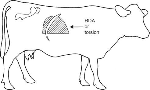
FIG. 1.4 Schematic representation of the area of the gas ping percussed in association with a right displaced abomasum (RDA) or abomasal torsion.
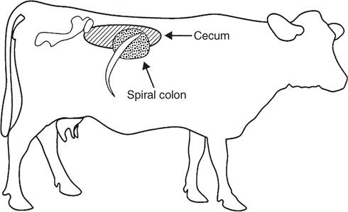
FIG. 1.3 Schematic representation of areas of gas pings elicited by percussion of the cecum and spiral colon.
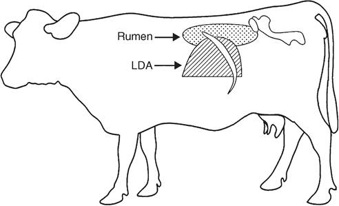
FIG. 1.5 Schematic representation of the area of the gas ping percussed in association with a left displaced abomasum (LDA) or gas ping in the rumen.
or kidney can be noted. Palpation of an abnormal swelling or firmness, especially with the elicitation of pain, indicates a problem that must be further evaluated.
The spinal column and ribcage are then palpated; the presence of fractures, enlargement of the costochondral junctions, or the elicitation of pain is noted. Enlargement or fractures of the costochondral junctions are commonly seen in young animals with deficiencies of calcium, copper, or vitamin D.
Auscultation with concurrent percussion by snapping the finger against the thoracic and abdominal walls is the next procedure. Gas trapped within abdominal viscera elicits a “pinging” sound that can be heard with the stethoscope. Localization of these gas pings to certain areas within the abdomen is helpful in determining which alimentary structure is involved (Figs. 1.3 to 1.5). If the cecum is enlarged and gas filled, an abdominal ping can be heard. This can extend caudally to the tuber coxae and cranially through the paralumbar fossa and under the ribcage on the right side (see Fig. 1.3). The diameter of this area can be variable and range from 6 inches (15 cm) in a cecal displacement to 3 feet (1 m) horizontally in cecal torsions. Spiral colon pings are generally localized to the right dorsocranial paralumbar fossa and rarely extend farther forward than the tenth intercostal space. They tend to be round areas 10 inches (25 cm) or less in diameter centered high under the last rib (see Fig. 1.3). Although commonly found in sick cattle that are anorectic, these spiral colon pings have no specific diagnostic significance and can be seen in normal cattle occasionally. Gas pings associated with a rightsided displacement or torsion of the abomasum can extend as far cranially as the ninth intercostal space and caudally into the paralumbar fossa (see Fig. 1.4). The diameter of displacements is usually 18 inches (45 cm), whereas that of torsions can be up to 3 feet (1 m). In cases of abomasal volvulus, the animal is usually exhibiting other systemic signs such as increased heart rate, dehydration, depression, scleral injection, and mild colic. In simple right-sided displacements or dilations of the abomasum, the only significant finding may be the small gas ping localized to the abomasum in a cow with depressed appetite and decreased milk production.
On the left side, gas pings can be noted as originating from the peritoneum, the rumen, or a left displaced abomasum (LDA).
The auscultation of a gas ping that is primarily localized to the dorsal aspect of the paralumbar fossa and auscultable on both sides of the spinal column would be indicative of a pneumoperitoneum. The extent of these pings can be from the thoracolumbar junction caudally to the retroperitoneal space. Pings associated with ruminal tympany occupy the whole of the paralumbar fossa and can extend dorsally to the spinal column but generally do not extend over to the right side (see Fig. 1.5). LDA results in a gas ping that is localized, easily outlined, and approximately 12 to 18 inches (30 to 45 cm) in diameter. Caudal extent of the displacement is generally the 13th rib; however, it can extend into the paralumbar fossa, in which case the outline of the abomasum can be easily palpated. The LDA should ping over the 11th rib on a line from the hip to the elbow (see Fig. 1.5). Rumen gas associated with a left-sided ping will rarely ping at this location. Identification of a fluid line within the displacement can aid in diagnosis and is accomplished by balloting the left paralumbar fossa while auscultating the area of the gas ping concurrently, a process known as succession. LDA often gives a pitch that changes in tone as it is percussed, as a result of movement of the rumen behind the abomasum. Often with LDA, intermittent gas bubbling or “sloshing fluid” sounds are heard. Rumen gas can be further differentiated from gas trapped in an LDA by rectal palpation of the rumen. One can also differentiate rumen gas from that trapped in an LDA by passage of a stomach tube into the rumen. Blowing into the rumen yields obviously auscultable sounds unless an LDA is present, in which case the sounds are muffled as the practitioner listens over the area of the ping. Performing a rumen or abomasal tap, the Liptac test, can further differentiate whether the ping originates from an LDA or the rumen. Fluid collected from an LDA would have a pH of less than 4, whereas that of the rumen should be 6 or higher, although cattle suffering from ruminal acidosis, or “grain overload,” can have a ruminal pH approaching 4.The rumen is examined by both auscultation and palpation. It should have a doughy texture with a small gas cap in its
■ TABLE 1.2
■ TABLE 1.3
Normal Resting Heart Rates (Beats/Min) for Adult and Young (sounds can vary over the different areas of the thorax or can be found singularly over the entire lung field. Significant pulmonary pathology may be present in ruminants without any auscultatable abnormalities. Total absence of lung sounds ventrally indicates pleural effusion or pulmonary abscessation with loss of airways. When ventral consolidation of the lung occurs, airway sounds are transmitted well and easily heard ventrally as pipestem sounds similar to those heard over the trachea, whereas percussion reveals a marked increase in ventral lung density. The trachea should also be auscultated. Inspiratory dyspnea and stridor are usually the result of extrathoracic obstructions to airflow (nose, pharynx, larynx, extrathoracic trachea). Pneumothorax can also result in loss of auscultatable airway sounds, which may be absent dorsally or entirely over the entire side if the lung has collapsed completely.
Place the middle finger in the intercostal space and slap the finger with the opposite hand or use a tablespoon and rubber hammer to accomplish percussion of the chest wall to determine the ventral lung border. Percussion is most useful in goats and calves. In sheep the wool precludes effective use of the technique, and in adult cattle the chest wall is often too thick to effectively evaluate changes in percussion tones. The chest is percussed in a dorsal-to-ventral direction, moving caudal to cranial on the chest wall. A change in resonance is noted when the ventral border is reached; a line demarcating this change in resonance is the junction of the diaphragm to the thoracic wall. In the ruminant this line should be described by joining a point at the junction of the 11th rib and the epaxial musculature dorsally to a point at the middle of the 9th rib, then cranially to the point of the olecranon. The cranioventral portion of the percussed thorax is dull because of the heart field (≈3 inches [7.5 cm] above the olecranon in adult cattle; 1 to 2 inches [2.5 to 5 cm] in calves, sheep, and goats). Finding an increased area of dullness in the cranioventral lung field associated with harsh lung sounds would be an indication of lung consolidation as seen in Pasteurella pneumonia. Pulmonary emphysema (atypical interstitial pneumonia) should be considered when the lung field is larger than expected, the animal is dyspneic, and airway sounds are minimal, whereas a ventral border that is markedly elevated and in a straight line could be an indication of pleural effusion. In this case auscultation would reveal decreased lung sounds ventrally and possibly the presence of pleural friction rubs. Acoustic percussion of the lung field can extend into the chest only to a depth of 2 to 2 and 12 inches (5 to 6 cm); therefore lesions within the thoracic cavity that lie deeper than this cannot be percussed.
The next step in the physical examination is to assess for evidence of pain in the ventral portion of the abdomen and thorax. This can be accomplished through the use of the withers pinch test or by ballottement of the xiphoid region. The withers pinch test involves auscultating the trachea while the withers are simultaneously squeezed and pushed ventrally. Painful lesions result in the ruminant resisting normal ventral movement of the spine and/or emitting a grunt or holding its breath when this test is performed. Ballottement of the xiphoid is also done while the trachea is auscultated. The xiphoid region is pushed with a knee or struck with a closed fist, and an elicitation of a grunt would indicate pain in this region. The examiner can then ballotte the remainder of the ventral abdomen and thorax to classify the lesion as localized or diffuse. If localized, the region affected should be identified. It should be noted that the animal may kick the examiner during the xiphoid ballottement test; thus proper precautions should be taken.
The subcutaneous abdominal veins are assessed next and palpated along their length for the presence of thickened walls, distention, or pulses. Distention and pulses may be abnormal if they correlate with other clinical evidence of right-sided heart failure. Thickening of the wall or evidence of thrombosis is often a consequence of faulty intravenous injection of irritating substances or of injuries that result in hematomas or abscesses.
If the animal is lame or has postural abnormalities, the feet and legs are palpated next. If the animal is uncooperative, it may be necessary to sedate and/or cast it. If sedation is necessary, it should be performed at the end of the physical examination. Care should be exercised for obvious reasons. The examination consists of comparing one foreleg with the other and then comparing both with the expected norm. The same procedure is followed for the hind legs. Abnormalities in the shape of the claw may be hereditary or may be caused by nutritional deficiencies, poor leg conformation, poor housing, or as a sequela to laminitis. The coronary bands should be palpated for evidence of pain or increased heat. An attempt should be made to pick up all four feet individually and observe the soles and interdigital regions for necrotic areas, areas of bruising or swelling, draining tracts, or presence of foreign bodies. The fetlock, carpal and tarsal, stifle, and elbow joints are all easily accessible and should be examined for swelling, tenderness, edema, heat, instability, and crepitation. Each joint should be tested over its full range of motion, and any elicitation of pain should be noted. Physical findings should be consolidated into a decision as to whether the joint problems are infectious or traumatic. Sheep or goats with acute polyarthritis most commonly have mycoplasma or chlamydial infection. Chronic joint pain in goats (often with soft tissue thickening and enlargement caused by synovitis) is frequently attributable to caprine arthritis encephalomyelitis. Conformation of the legs should be analyzed because this could contribute to a joint or foot problem. The pelvic girdle does not lend itself to extensive examination; however, one can note symmetry or asymmetry and further evaluate during the rectal examination. Fractures of the tuber coxae, subluxation of the sacroiliac junction, acetabular fractures, fractures of the head or neck of the femur, and dislocation of the coxofemoral joint may all be diagnosed by evidence gained during palpation and observation of the pelvic area. The dislocated or fractured limb is frequently shorter than the normal opposite limb. The tail can also be examined for evidence of fractures, paresis, or paralysis.
The perineal region is examined by noting the external condition of the genitals and rectum. Anal sphincter tone can be assessed, and the presence of vaginal discharge can be noted. In males the testicles, spermatic cord, and epididymis should be palpated for the presence of nodules or areas of fibrosis. The testicular circumference can be measured and compared with what is expected for age and breed. These measurements are noted in Tables 1.4 and 1.5. The perineal part of the penis can be palpated for the presence of hematoma or swelling and pain (cellulitis or abscess). In the female the supramammary lymph nodes can be felt at the attachment of the udder to the perineum. Enlargement of these nodes occurs with mastitis or lymphosarcoma. The udder is palpated for the presence of fibrotic areas, commonly seen secondary to staphylococcal mastitis or associated with Trueperella pyogenes (formerly called Arcanobacterium [Actinomyces] pyogenes) abscesses. The presence of a swollen quarter or quarters with pain and heat may be associated with mastitis caused by gram-negative bacteria. Cold damp areas of skin on the udder that are discolored, necrotic, and possibly sloughing are evidence of gangrenous mastitis. In lactating animals, milk should be present in each quarter
■ TABLE 1.4
Expected Values for the Scrotal Circumference of the Bull at Different Ages4
| Age (Months) | Scrotal Circumference (cm) |
| 12-14 | bgcolor=white>30-34|
| 15-20 | 31-36 |
| 21-30 | 32-38 |
| >31 | 34-39 |
■ TABLE 1.5
Expected Values for the Scrotal Circumference of the Ram at Different Body Weights5
| Body Weight (kg) | Scrotal Circumference (cm) |
| within 1 second. Using other factors such as sunken eyes, dryness of mucous membranes, abnormal heart rate, and degree of illness, an estimation of percent dehydration is made. Dehydration is first noted clinically when the animal is approximately 5% dehydrated, and death occurs at 12% to 15% dehydration. The larynx should be palpated for enlargement and the presence of pain. The trachea should also be palpated for the presence of fractured tracheal rings, collapsed areas, and pain on palpation. The jugular veins are examined for the presence of distention and pulses. Thrombosed veins should also be noted because this condition may alter the desired course of therapy and often prevents placement of a catheter. The venous stasis test is performed on the jugular by holding off the vein in the midcervical region. Normally the vessel fills above the point of occlusion and remains collapsed below. With restricted venous blood flow, as seen in cases of right-sided heart failure, a positive stasis test can result. In these cases, the vessel below the occluded point fails to collapse or takes a prolonged time to do so and a jugular pulse is frequently present. The final step in the physical examination is rectal palpation. This cannot be accomplished in small ruminants, so this section is directed primarily toward the bovine. The pelvic area is evaluated for the presence of retroperitoneal abscesses or fractures of the pelvic bones. The left kidney is more easily palpated than the right kidney because of its location within the abdominal cavity. Both kidneys are located to the right of midline, as the left kidney is displaced by the rumen and is located caudally to the right kidney. The kidney should be palpated for overall size and shape, which is normally lobulated in cattle, and the kidney can be gently squeezed to determine if pain is evident. The rumen should be palpated and the findings compared with those noted on percussion and auscultation. If there is any evidence of intraabdominal gas pings, an attempt should be made to palpate the suspected organ. This can provide information that may be helpful in determining therapeutic or diagnostic directions. The rectal palpation can reveal the presence of masses that were not palpable externally, such as fat necrosis or tumors. Adhesions and evidence of peritonitis can also be palpated rectally, and attempts should be made to localize them to a specific area of the abdomen to establish potential cause. The preiliac (or internal iliac) lymph nodes are located by sweeping the hand along the craniodorsal face of the ilium. These nodes normally have the size and shape of a walnut. Enlargements are noted with lymphosarcoma, peritonitis, and severe limb inflammation. The lymph nodes of the aortic bifurcation are small and not easily palpated. Their ability to be palpated would be evidence of abnormal enlargement. Palpation of the genital tract is directed toward establishing size, shape, and presence of abnormalities. These contents could be normal, as with a pregnant animal bearing a fetus, or abnormal, as seen with a pyometra. If it is determined that a cow is not pregnant, the ovaries and oviducts should be palpated for structures and abnormalities. In the bull, particular attention should be paid to the prostate, seminal vesicles, and bulbourethral gland. Diagnostic imaging using an ultrasound probe in conjunction with rectal palpation can add to the information already gathered during the physical examination to further refine the problem and aid in prognostication and development of the therapeutic plan. Proper manipulation of the probe can allow for imaging the kidney, uterus, bladder, and rumen wall. Abnormal structures noted on palpation of the abdominal cavity can also be imaged. The accessory sex glands in the male can also be visualized.
More on the topic Physical Examination:
-
Veterinarian -
|