Poisonous Plants
Tam Garland
Plant Poisoning Considerations
The need for forage and therefore risk of toxicosis in both ruminants and Equidae is well established. Significant economic losses have been attributed to poisonous plants in regions and countries that rely on grasslands for raising livestock.
Studies in the western United States suggest that plant poisonings adversely affect 3% to 5% of rangeland cattle, sheep, goats, and horses.1 In China, poisonous grassland species of plants are responsible for an estimated $12.3 million of all livestock losses, including loss of performance and mortality from 2000 to 2006.2 The estimated price of plant poisonings in Australia is $100 million (Australian dollars) per year.3,4Based on these statics, poisonous plants pose significant dangers to all large animal species. Poisonings can occur for a variety of reasons. Toxic plants may be consumed inadvertently when baled into hay or processed into silage. Consequently, the animal may be unable to avoid the plant. However, hungry or thirsty animals are much more prone to eat plants that may otherwise be avoided. Naive animals, those unaccustomed to the surroundings, are more likely to consume plants that are unfamiliar to them.
The severity of toxicity may be affected by a variety of factors acting on the plant, such as season (summer versus winter) or moisture (either excessive or drought conditions). Severe heat may affect the toxicity of plants. The type of soil may be an influencing factor in the toxicity of the plants as well as the micronutrient levels in the soil. For example, some soils may be high in such nutrients as selenium, and some plants are selenium accumulators.
Investigation of possible plant poisoning on the affected premises include limited availability of forage as evidenced by animal observation and body condition scores; drought affecting plant growth, availability, and potentially high concentrations of toxins such as nitrates; recent storms having downed trees or fences or even a lightning strike; feed changes; inappropriate disposal of garden, shrub, or tree trimmings; and administration of herbal products for medicinal value.
The level of toxins within a given plant species is often variable, even in samples obtained from the same pasture. Toxicity may be more pronounced in specific growth phases of the plant species, during times of stress or rapid growth, or after drying. Endophyte or fungal infections of the plant may also be important, either as the sole or an additional source of toxicity. These factors can be important in the timing and choice of plant specimens to identify, as well as assay for toxicity.Individual animal factors play a role in both quantity of plant consumed and expression of clinical signs, thus making investigations more challenging. More than one type of toxic plant may have been ingested, which may lead to a wider variety of clinical signs. An individual animal may develop a taste for a plant being avoided by others in their herd or flock or be forced to eat it due to feed competition and low social order. Herbicide application may also enhance the palatability of plant species such as water hemlock.5 Recent fertilizer application may increase nitrate concentrations in both plants and water supplies. Some plants can create a wide range of clinical signs depending on the amount consumed, either acutely or chronically. Research done on specific plants and toxins, and applied to feeding trials, has demonstrated that differences in metabolism, GI flora, and genetics may have a role in disease expression. Yet another variable is interaction between plant toxins and medications. Consumption of hay with two separate species of nightshade (Solanum spp.) has recently been identified as a factor in the development of clinical signs of ivermectin toxicosis in horses.6
Any suspicion of plant poisoning in an individual or group of individuals in a herd or flock should trigger a diligent investigation. The first step is a complete history of the affected individuals, the herd or flock, and the premises. In addition to routine health questions, emphasis should be placed on current feed, and whether any changes have been instituted; weather factors possibly affecting feed and water access or quality; fertilizer use on forage sources; differences in the management of affected and unaffected animals; and the potential for feeding errors or inadvertent exposure to garden refuse.7 If the history yields evidence to strengthen the suspicion of plant poisoning, it may be necessary to immediately remove access to the suspected source of the clinical problems.
If multiple animals have been affected, determining a timeline of events and cases may be helpful in evaluating the history and determining the cause.The second step in the investigation should be thorough examination of affected animals to identify and characterize clinical abnormalities and lesions. Low body condition scores, particularly if accompanied by a strong appetite, should raise clinical suspicion of consumption of normally unpalatable plants, or lack of appropriate nutrition. Pregnant animals should be evaluated for signs of potential abortion. Quick access to a veterinary diagnostic laboratory, an animal poison control center, or an available online database where clinical signs are entered to yield potential diagnoses may help veterinarians quickly sort through potential differential diagnoses for the animal(s).8 Symptomatic treatment is often the first line of medical response if the etiology has not been determined or a specific antidote is not available or approved for use in a particular species, especially a food animal species. Stress of treatment, particularly for ruminants, must be weighed against the benefits. Activated charcoal administration remains advisable for most acute toxicoses, if the animal can withstand the stress of administration. In monogastrics, stomach lavage followed by instillation of a slurry of activated charcoal in water may be the best option for acute cases. However, in ruminants this may be more challenging, and a rumenotomy for evacuation of contents and instillation of activated charcoal could be considered. In herd situations, rumenotomies on multiple animals may not be feasible or affordable. The cost of treatment of an animal may exceed the value of the animal, and treatment considerations for large animals and the associated cost should be discussed with owners or caregivers prior to treatment. A limited number of specific antidotes are described for a few plant poisonings, but their use may be restricted by cost and the need to avoid food residues.
Enhanced excretion of toxins through laxatives or manipulation of urine or rumen pH may be applicable in specific situations where the toxic plant or principle has been confirmed.Postmortem examination of affected animals should take place as soon after death as possible to maximize the opportunities to identify lesions and an etiology. In some plant poisonings, a specific odor may be noted, such as a bitter almond smell in cyanide poisoning (e.g., Prunus spp.), an acetone smell with white snakeroot poisoning, and a mousy aroma due to poison hemlock ingestion.5
Samples should be taken of rumen or stomach contents to look grossly and microscopically for recognizable plant fragments or seeds. An aliquot of the ingesta should be collected and labeled and a second sample frozen as soon as possible for toxicologic analysis. This is especially necessary when the possible toxin is unknown. Depending on the history and the timeline, more ingesta samples may be warranted from other segments of the digestive tract, especially the abomasums of calves. If the animal is a calf, collect contents of the abomasum in a similar fashion. Intact seeds may be identifiable in the distal intestinal tract or manure.
Samples of aqueous humor, amniotic fluid, and fetal stomach fluid for nitrate concentrations should be considered if clinical signs are compatible with a particular etiology. If the animal has died within a few hours, blood samples for hematologic, chemistry, and toxin profiling may be rewarding. Tissue samples are important for characterizing histologic lesions to confirm a plant toxicity or alternative etiology. All grossly affected tissues should be sampled and a piece of liver, both fresh and frozen, submitted for toxicologic analysis. If neurologic signs were observed, the brain should be removed with proper barrier precautions.7
Identification of the plants, foodstuffs, or water responsible for clinical signs or deaths is important for guiding treatment and further control of an offending agent.
Inspection of the feed, including grain, hay, and pasture, may be necessary when dealing with suspected plant intoxications or suspected mycotoxins. Mycotoxins may be visible as moldy bits in the feed. However, the presence of mold does not ensure a mycotoxin, just as the absence of mold does not ensure the absence of mycotoxins (see the Mycotoxins section later). Several samples of the feed should be obtained for both toxic plant and mycotoxin testing. Remember as well that grasses can contain mycotoxins such as ergot. Walking the fence line looking for known toxic plants and evidence of grazing them is necessary for completeness. Samples of plants in question or unknown to you should be collected, especially if there is evidence of grazing.8 Fresh plants may be placed in plastic bags, chilled, and shipped chilled overnight to a diagnostic lab. Additional samples can be pressed between newspaper sheets or brown paper with a board or heavy cardboard with some type of weight (e.g., books, gym weights) on top to keep them flat. The plants should remain under the weight for several days. Modern technology (smart phone) can be used to take photos of the plants and email or text them immediately to the toxicologist at the diagnostic lab. A telephone conversation with the toxicologist may assist you in finding the offending plant more quickly, perhaps even while you are on site. For identified plants known to have variable toxicity, such as larkspur (Color Plate 54.1) or selenium- accumulating plants, multiple samples should be taken of the same species, concentrating on areas where the plants have been visibly browsed or grazed.9 Resources for plant identification include toxicology texts, the U.S. Department of Agriculture (USDA) plant identification website, the USDA Poisonous Plant Research Laboratory website and its publications, other country-specific websites, and university or extension service plant experts.10-16 In the United States, plant samples can be sent to the USDA Poisonous Plant Research Laboratory or Texas A&M's Veterinary Medical Diagnostic Laboratory. Other veterinary diagnostic laboratories may be able to identify the plants. Your diagnostic laboratory of choice may send the sample to one of these other labs, or you may need to request that it be sent to one of these laboratories.Common plant poisonings often trigger a recognizable pattern of clinical signs and disease progression. These are reviewed below in more detail, with emphasis on diagnostic features, treatment options, and preventive measures. Less frequent causes of livestock morbidity and mortality are listed in tables, grouped by predominant clinical signs. Highlights of newly described toxicities that have the potential to harm multiple animals are presented in this chapter section.
Sudden Death
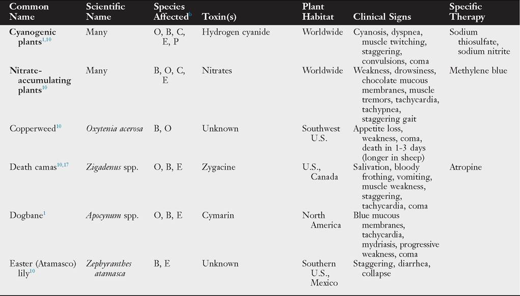
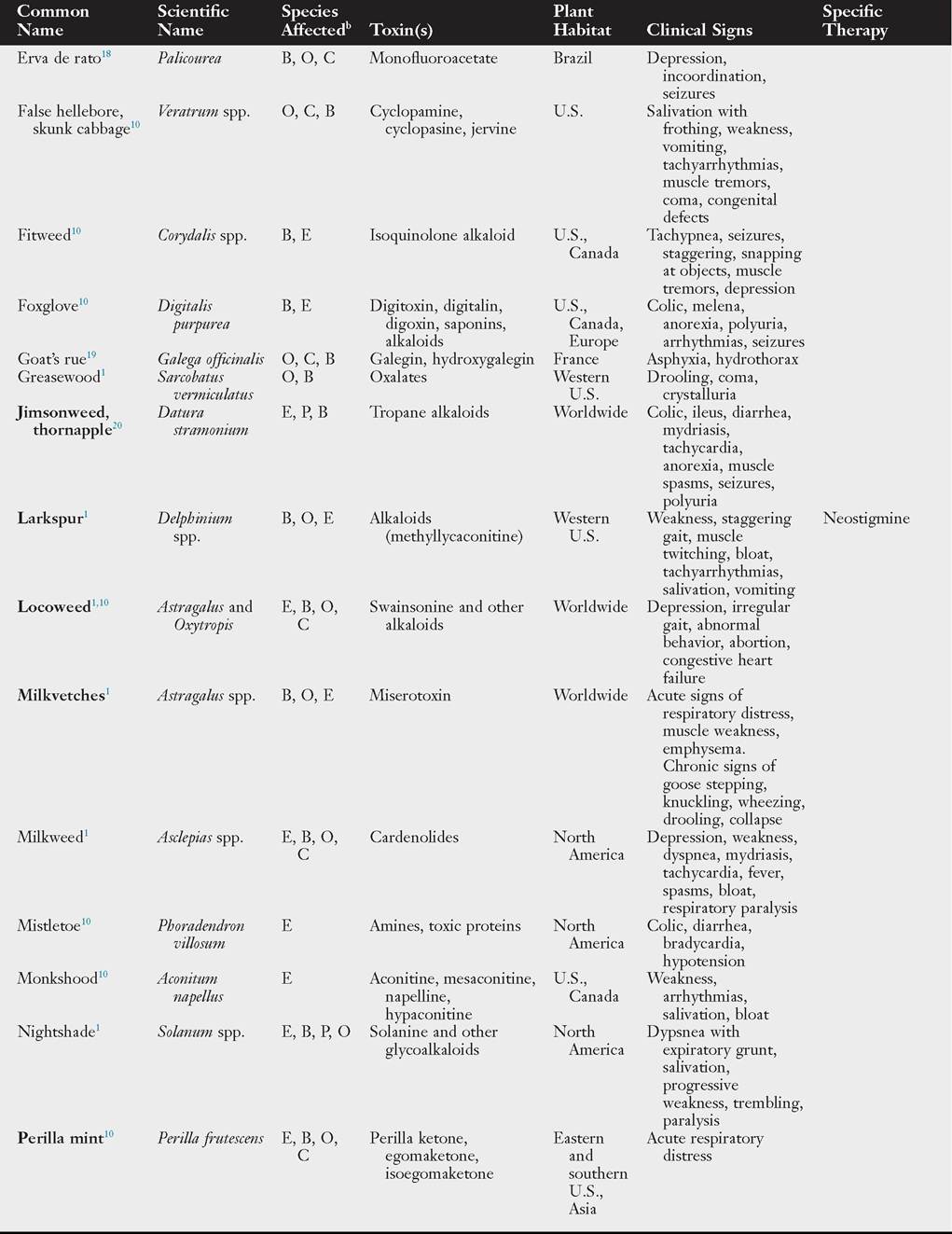
Poisonous plants, chemical toxins, lightning strikes, and infectious diseases are causes to consider when investigating sudden death in multiple animals. Numerous plant species have the potential to kill multiple animals within minutes to hours (Table 54.2).
■ TABLE 54.2
Plants With Potential to Cause Sudden Death in Large Animalsa
■ TABLE 54.2
Plants With Potential to Cause Sudden Death in Large Animals—cont'd
■ TABLE 54.2
Plants With Potential to Cause Sudden Death in Large Animals—cont'd
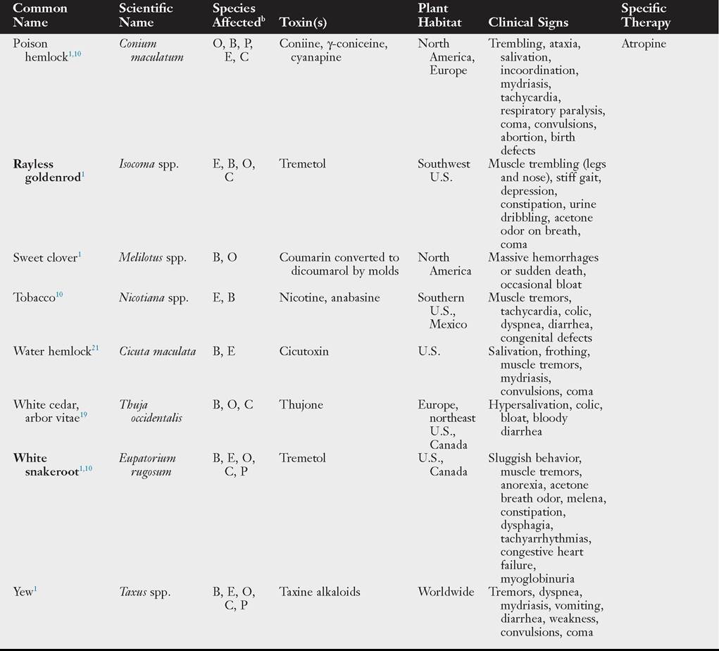
aPlant names in bold are discussed in more detail in the text.
bSpecies listed in relative order of toxicity importance: B, Bovine; C, caprine; E, equine; O, ovine; P, porcine.
Several groups of plants deserve greater discussion because of the frequency with which they are incriminated in the death of livestock and horses. All produce a range of clinical signs influenced by the amount consumed, time period of consumption, concentration of toxins, animal species, and in some instances, accommodation of ruminal microbes.
Larkspur (Delphinium spp.)
Larkspur (Color Plate 54.1) is considered one of the most important plant poisonings of range animals in the United States, second only to locoweed in its impact.10 Cattle are most susceptible, but sheep and horses can be affected. The toxicity varies with the specific plant, stage of growth, amount eaten, and time period over which it is consumed. Numerous toxic diterpenoid alkaloids have been identified in the nine Delphinium spp., the most often cause of poisoning in cattle. Tall larkspur (Delphinium barbeyi) is considered the most problematic.1 These alkaloids cause a curare-like blockade of neuromuscular junctions, by reversibly binding to nicotinic acetylcholine receptors. Larkspur consumption may also enhance the toxicity of another range plant, death camas (Zigadenus spp.).17
Death varies from sudden to 3 hours post larkspur consumption. Clinical signs include excitability, muscle tremors, stiffness, staggering, and a basewide stance.10 Cattle may assume a kneeling position before collapsing. Bloat, regurgitation (ruminants), constipation, mild GI irritation, venous congestion, and aspiration pneumonia may be observed. There are no pathognomonic postmortem changes. A definitive diagnosis is usually reached by identifying pieces of larkspur in the upper GI tract.
If treatment is possible, bloat should be immediately addressed to relieve thoracic compression and reduce the chances of aspiration; however, the stress of treatment may hasten death. Intravenous (IV) physostigmine has been advocated at dosages of 0.04 to 0.08 mg/kg, repeated as needed over several hours.10 Neostigmine at 0.04 mg/kg is slightly less effective. Neither drug is approved for use in food animals. An affected animal may be mistaken for a grass tetany case; however, treatment with magnesium exacerbates the effect of larkspur’s toxins at the neuromuscular junction and should be avoided.
Prevention of larkspur toxicity in range cattle is challenging and requires knowledge of when the plants are most toxic and most palatable, particularly if other forage is limited. For example, early spring grazing of cattle in pastures with tall larkspur should be curtailed as soon as the flower stalks begin to elongate. Sheep are more tolerant and can be used to reduce the plant’s density. Aversion to eating larkspur has been created with lithium chloride administration, a strategy that may be worthwhile on some ranches.22 Larkspur reduction may not be cost-effective with herbicides, leading investigators to use insect control with the larkspur myrid (Hoplomachus affiguratus).10
Cyanogenic Plants
Toxicity induced by consumption of cyanogenic plants produces sudden death, along with the characteristic signs of bright red mucous membranes and bright red venous blood.
Clinical signs are rarely observed but include obvious respiratory distress, rapid breathing, bloat, salivation, muscle twitching, staggering, mydriasis, cardiac arrhythmias, and convulsions.10 Cyanide blocks molecular oxygen transfer in cytochrome oxidase systems in mitochondria causing tissue anoxia.23 Ruminants are more susceptible than monogastrics because rumen microorganisms readily release cyanide from the cyanogenic glycosides. Acute, sublethal cases generally recover within an hour. Toxicities have also been reported in alpacas.24 Chronic cyanide toxicity has been associated with sorghum consumption in pregnant horses and cattle. The foals and calves are born with arthrogryposis. Animals may also develop neurologic damage to the brain and spinal cord resulting in weakness, ataxia, and urinary incontinence. Low doses of cyanogenic glycosides can be goitrogenic.10
Cherry red venous blood may be observed in fresh postmortem examinations. In ruminants, a bitter almond odor may be detected in rumen contents.10 Tracheal, pulmonary, and muscle congestion may be observed, along with hemorrhages in multiple organs. Cyanide ion content may be measured in refrigerated samples of heart, brain, or skeletal muscle or rumen contents. Blood samples for analysis should be kept in airtight containers at 4° C until analyzed. A sensitive field test for use on forage samples or rumen content has been developed using alkaline picrate-treated filter paper strips.11
A number of plants have varying levels of cyanogenic glycosides, including the following trees, shrubs, crops, and grasses: acacia, African star grass, apple, arrow grass, Bahia grass, bamboo, Bermuda grass, birdsfoot trefoil, bracken fern (Color Plate 54.2), cassava, chokecherry, Christmas berry vetch, corn, elderberry, eucalyptus, flax, flowering quince, hydrangea, Indian grass, Johnson grass, lima bean, mountain mahogany, poison suckleya, reed canary grass, service berry, sorghum, South African daisy, star grass, Sudan grass (Sorgum spp.), tall manna grass, Texas queen’s delight, and white clover.10,23 Seeds of apples and crabapples (Malus spp.) and the seeds, as well as leaves of the Prunus spp. (bitter almond, cherry, chokecherry, apricot, peach, nectarine, plum, mock orange), may have large amounts of cyanide. Drying as hay or incorporation into silage generally decreases the toxic potential of the forage plants; most are not routinely grazed or browsed. Early growth phases of the plants are the most toxic. Poisoning is most likely if animals eat a large quantity in a short period of time. Wilting, frost, drought, or other damage to the plant enhances toxicity.1 Toxicity is measured as cyanogenic potential. Plant material containing more than 20 mg per 100 g (200 ppm) cyanide potential is reason for concern, but this varies between species.23 Cyanogenic potential is significantly reduced by properly curing forages for hay or ensilage.
If the diagnosis is made rapidly, some cattle and sheep may be saved by IV or intraperitoneal (IP) injection of 20 mL of a 10% solution of sodium thiosulfate mixed with 10 mL of a 10% solution of sodium nitrite.1 However, this treatment is not approved for use in food animals. An alternative treatment has been described for ruminants using only a 30% to 40% thiosulfate solution IV at a dose of 25 to 50 g/100 kg of body weight.11 Administration of a gallon of vinegar diluted in 3 to 5 gallons of water administered by stomach tube to help acidify the rumen has also been recommended.10 Preventive measures are largely focused on limiting access to highly cyanogenic plants and trees, particularly during rapid early-growth phases, and planting varieties with lower cyanogenic potential.
Nitrate Toxicity
Many plants and some water sources contain harmful concentrations of nitrates, which can cause a wide range of clinical signs from sudden death to congenital defects in ruminants, camelids, and monogastrics. Examples of plants known to accumulate nitrates are listed in Table 54.3. When photosynthesis is impaired by shade or prolonged cloudiness or excessive nitrogen is applied to plants (e.g., fertilizer), nitrate accumulation may reach dangerous levels. Other triggers for nitrate accumulation include drought, frost, hail, and herbicide application. Nitrates are water soluble and can leach from harvested forages, occasionally causing toxicosis by elevating concentrations in the lowest bales of a rained-on haystack.25
In ruminants, ingested nitrates are converted to nitrites and ammonium in the forestomachs. Nitrates can cross the placenta. If excessive nitrates are ingested, absorbed nitrites can trigger oxidation of the ferrous iron in hemoglobin, producing methemoglobin. Chocolate brown mucous membranes and blood are characteristic findings. Vasodilation and hypotension may result, leading to hypoxia, dyspnea, exercise intolerance, death, fetal stress and subsequent abortion, stillbirths, and/or weak neonates.27 In bulls, sperm abnormalities and degenerative changes within the testes have been reported.28 If the animals are anemic or hypoxic for any other reason, methemoglobinemia will have even greater consequences. At postmortem examination, a brownish cast of the tissues due to the methemoglobinemia may be apparent.
Congenital hypothyroidism in equine neonates is most often caused by insufficient iodine intake by the broodmare but can also be caused by excessive nitrate intake. Multiple animals in the herd may be affected.29,30 Signs of high nitrate intake may not be observed in the broodmare. Abortion may result, or more commonly a dysmature foal is produced after prolonged gestation. The affected neonates have varying degrees of signs but classically include mandibular prognathism, flexural deformities of the forelimbs, secondary rupture of the common digital extensor tendon just above the carpus, poorly ossified cuboidal bones, and poor immune function. If recognized at birth, these foals can survive to be useful animals if treated intensively with splints to address the flexural deformity, very limited weightbearing until the cuboidal bones ossify, and measures to reduce the likelihood of infection. If an affected foal is allowed to bear weight before full ossification of the cuboidal bones in the hocks and carpi, angular limb deformities will likely result. The syndrome can be prevented by avoiding high nitrate forage or water sources and ensuring that there is adequate iodine in
■ TABLE 54.3
Plants With Potential to Accumulate Nitrates
| Type | Common Name | Scientific Name | Reference(s) |
| Crops | Barley | Hordeum spp. | 25 |
| Beets | Beta vulgaris | 10 | |
| Corn | Zea mays | 10, 25 | |
| Flax | Linum spp. | 10 | |
| Kale | Brassica oleracea | 10 | |
| Oats | Avena spp. | 24, 25 | |
| Pearl millet | Pennisetum typhoides | 10, 25 | |
| Rape | Brassica napus | 10 | |
| Rye | Secale cereale | 10, 25 | |
| Soybeans | Glycine max | 10, 25 | |
| Sunflower | Helianthus annuus | 10 | |
| Turnips | Brassica rapa | 10 | |
| Wheat | Triticum aestivum | 10, 25 | |
| Forages | Alfalfa | Medicago sativa | 10, 25 |
| Barnyard grass | Echinochloa spp. | 10 | |
| Button grass | Dactyloctenium radulans | 26 | |
| Johnson grass | Sorghum halepense | 10, 25 | |
| Sudan grass | Sorghum vulgare | 10, 25 | |
| Sweet clover | Melilotus spp. | 10 | |
| Tall fescue | Lolium arundinaceum | 25 | |
| Weeds | Canada thistle | bgcolor=white>Cirsium arvense10 | |
| Cheese weed, mallow | Malva spp. | 10 | |
| Dock | Rumex spp. | 10, 25 | |
| Field bindweed | Convolvulus arvensis | 10 | |
| Fireweed | Kochia spp. | 25 | |
| Goldenrod | Solidago spp. | 10 | |
| Jimsonweed | Datura stramonium | 10 | |
| Kochia (summer cypress) | Kochia scoparia | 10, 25 | |
| Lambsquarters | Chenopodium spp. | 10, 25 | |
| Nightshades | Solanum spp. | 10, 25 | |
| Pigweed | Amaranthus spp. | 10, 25 | |
| Ragweed | Ambrosia spp. | 10 | |
| Russian thistle | Salsola kali | 10 | |
| Smart weed | Polygonum spp. | 10 |
the mare's diet. Confirmation of nitrate toxicity is based on demonstration of elevated levels in forages, weeds, water, or body fluids such as aqueous humor, fetal stomach fluid, and amniotic fluid.1,10 In ruminants, plants containing more than 1.5% nitrates (measured as KNO3) dry weight may be lethal. Sublethal effects are seen from feed containing 0.5% to 1.5% nitrates. Seeds are usually unaffected by high nitrates in other plant parts. Ruminants can adapt to higher nitrate feeds if their introduction is gradual. Reproductive effects in ruminants are linked to forage concentrations greater than 5000 ppm on a dry matter basis. In adult ruminants, signs of methemoglobinemia are seen with forages containing 2000 to 10,000 ppm nitrates on a dry matter basis. Water with more than 200 ppm is excessive for pregnant ruminants; 30 ppm is the recommended limit for broodmares.10,29,30 In nonpregnant animals, a range of 445 to 1000 ppm in water is considered dangerous for livestock.10,26
Treatment of nitrate toxicity in ruminants is restricted by the paucity of approved drugs. Methylene blue administration is prohibited in food animals in the United States for at least 180 days before slaughter but, if permitted in other areas, may be given IV at a rate of 4 to 15 mg/kg body weight as a 2% to 4% solution.10 In acute ruminant cases, the adjunct therapy of the administration of either cold water or vinegar to slow nitrate reduction, and mineral oil to hasten passage of the ingested source has been used, provided the animal can withstand the stress of intubation.10
Forages known to have high nitrate potential should be tested prior to feeding livestock, particularly if fertilizer or poultry litter has been used on the forage. High-nitrate forages can potentially be ensiled to reduce the nitrate content by as much as 30%.26 Nitrate levels are highest at night and in early morning, and this knowledge can be applied to the timing of pasture access and hay cutting. If high-nitrate forage is harvested, the nitrate content will not dissipate on curing. Gradual introduction of high-nitrate forage into the ruminants' diet will permit rumen microflora to adapt, but this should preferentially begin with nonpregnant adult animals to minimize the reproductive effects. Monensin in the diet may change the threshold for toxicity and probiotics containing Propionibac- terium spp. reduce nitrates, which may also be useful.10
Neurologic Signs
■ TABLE 54.4
Plants Associated With Neurologic Signsa
| Common Name | Scientific Name | Species Affectedb | Toxin(s) | Plant Habitat | Clinical Signs | Specific Therapy |
| Blown grass, Pacific bent grass3 | Agrostis avenacea | B, O | Corynetoxins in nematode | Australia, New Zealand, southwest U.S. | Incoordination, weakness, muscle weakness, rocking stance, high-stepping, collapse, seizures, abortion | |
| Bracken fern10 | Pteridium aquilinum | E, P, B, O | Ptaquiloside | Worldwide | Weight loss, progressive incoordination, depression, tremors, crouching stance with arched neck (equine), blindness (sheep), aplastic anemia (ruminants), thrombocytopenia, bladder and gastrointestinal cancer | Thiamine |
| Buttercup10 | Ranunculus spp. | B, E | Ranunculin | North America, Europe | Salivation, depression, blindness, hematuria, oral ulcers, diarrhea, colic | |
| Caltrop or carpetweed10 | Kallstroemia spp. | B, O, C | Unknown | Southwest U.S. | Hindlimb weakness, knuckling, seizures, posterior paralysis | |
| Castor bean19 | Ricinus communis | E, B, O, C, P | Ricin | Worldwide | Bloody diarrhea, tenesmus, colic, trembling, incoordination, seizures | |
| Coyotillo10 | Karwinskia humboldtiana | B, O, C, E, P | Unknown | Southwest U.S. | Hypersensitivity, muscle tremors, abnormal high-stepping gait, diminished reflexes, progressive paralysis | |
| Fitweed10 | Corydalis spp. | B, E | Isoquinolone alkaloid | U.S., Canada | Tachypnea, seizures, staggering, snapping at objects, muscle tremors, depression | |
| Golden chain tree10 | Laburnum anagyroides | E, B | Cystisine | Southern U.S. | Incoordination, muscle tremors, mydriasis, excitement, seizures, coma, vomiting, diarrhea, paralysis | |
| Hairy cat’s ear, flatweed32 | Hypochaeris radicata | E | Unknown | U.S., Europe, Australia | Bilateral stringhalt, depression, aggression | Phenytoin |
| Hairy vetch10 | Vicia villosa | B, E | Unknown | U.S., Europe | Excitement, slobbering, oral ulcers, nasal discharge, cough, stiffness, anorexia, weakness, seizures, swelling of the head and neck, pruritic dermatitis, diarrhea, abortion, myocardial necrosis | |
| Halimium33 | Halimium brasiliense | O | Unknown | Brazil, Uruguay | Transient seizures when excited, muscle tremors, ventroflexion of neck or opisthotonus, nystagmus, tetanic spasms, postictal ataxia | |
| Horse | Aesculus | B, O, C, | Aesculin, | U.S., | Muscle tremors, incoordination, | |
| chestnut, buckeye10 | hippocastanum | E | fraxin, alkaloids | Europe | depression or excitability, hopping gait, dorsomedial strabismus (ruminants), gastroenteritis (monogastrics) | |
| Horse nettle10 | Solanum carolinense | B, E | Solanine | U.S., Canada | Falling when excited, incoordination, anorexia, emaciation, rough coat, constipation, mouth inflammation | Physostigmine |
| Horsetail or scouring rush10 | Equisetum spp. | E, B, O | Aconitic acid, palustrine, thiaminase | Worldwide | Weight loss, staggering, muscle tremors or rigidity, paraparesis, tachycardia, diarrhea, coma | Thiamine |
| Jamaican nettle tree34 | Trema micrantha | B, C, E | Trema toxin, ammonia | Americas | Apathy, blindness, recumbency, paddling, coma (hepatic encephalopathy) | |
| Jimmyweed10 | Haplopappus heterophyllus | B, E | Tremetol | Western U.S., Mexico | Depression, trembling, weakness, knuckling, seizures, paralysis, congestive heart failure, acetone breath smell |
■ TABLE 54.4
Plants Associated With Neurologic Signs—cont'd
| Common Name | Scientific Name | Species Affectedb | Toxin(s) | Plant Habitat | Clinical Signs | Specific Therapy |
| Jimsonweed, thornapple35 | Datura spp. | E, P, B | Tropane alkaloids | Worldwide | Colic, ileus, diarrhea, mydriasis, tachycardia, anorexia, muscle spasms, seizures, polyuria | |
| Johnson grass10 | Sorghum halepense | B, O, E | Hydrocyanide | Worldwide | Posterior ataxia, urinary incontinence, cystitis, weight loss | |
| Kochia1 | Kochia scoparia | B, O, E | Thiaminase, nitrates, oxalates | Worldwide | Ataxia, muscle spasms, depression, blindness, photosensitization | Thiamine |
| Locoweed36 | Astragalus and Oxytropis spp. | E, B, O, C | Swainsonine (endophyte produced) | Worldwide | Abnormal behavior, abortion, reduced fertility, skeletal malformations, weight loss | |
| Locust trees19 | Robinia spp. | E, B, O, C | Robinin, robitin | North America, Europe | Diarrhea, anorexia, weakness, posterior paralysis, mydriasis, dyspnea, arrhythmias | |
| Lupines1 | Lupinus spp. | O, B | Piperidine, quinolizidine | U.S., Canada | Salivation, frothing, depression, dyspnea, loss of muscle control, convulsions, coma, birth defects | |
| Mescal bean, Eve’s necklace | Sophora spp. | B, O, C, E | Cystisine | Southwest U.S., Mexico | Incoordination, violent muscle tremors, falling, stiffness, respiratory paralysis | |
| Milkvetches10,36 | Astragalus spp. | B, O, E | Miserotoxin | North America | Acute and chronic—acute: respiratory distress, muscle weakness, emphysema; chronic: goose-stepping, knuckling, wheezing, drooling, posterior paresis, collapse | |
| Milkweed36 | Asclepias spp. | E, B, O, C | Glycosidic cardenolides | North America | Depression, weakness, dyspnea, ataxia, seizures, arrhythmias | |
| Morning glory3 | Ipomoea spp. | B, O, C, E | Calystegines | Worldwide | Ataxia of hindlimbs, staggering | |
| Nightshade10 | Solanum spp. | B, O, C, E | Solanine | Worldwide | Fatigue, muscle tremors, head tremors, seizures, bloat, congestion of lungs, heart, spleen | Physostigmine |
| Oak10 | Quercus spp. | B, E, O, P | Gallotannins | North America, Europe | Incoordination in horses along with weakness, ileus, mouth ulcers, mucosal discoloration, patchy sweating, abnormal feces, colic, dark urine | |
| Paspalum 37 grasses3 | Paspalum spp. | B, E | Ergot alkaloids | Worldwide | Staggers, muscle tremors, seizures, abortion, gangrene | |
| Poison hemlock1 | bgcolor=white>Conium O, B, P, E | Coniine, γ-coniceine | North America, Europe | Trembling, ataxia, salivation, incoordination, mydriasis, tachycardia, respiratory paralysis, coma, convulsions, birth defects | Atropine | |
| Russian knapweed1 | Centaurea repens | E | Sesquiterpene lactones | Western U.S. | Twitching of lips, tongue flicking, involuntary chewing, weight loss, emaciation, poor prehension, incoordination, muscle tremors, facial paralysis | |
| Ryegrass38 | Lolium perenne | B, O, E | Lolitrem B | Worldwide | Muscle tremors, fasciculations, truncal swaying, basewide stance, slowed movement, limb edema | |
| Sagebrush10 | Artemisia spp. | E | Unknown | Western U.S., Mexico | Abnormal behavior, ataxia, sage aroma in breath and manure | |
| Sneezeweed1 | Hymenoxys spp. | O, B, E | Sesquiterpene lactones | North America | Equine: weakness, incoordination, foaming at the mouth, seizures; ruminants: chronic vomiting, emaciation, stiffness | |
| Squirrel corn, Dutchman’s breeches10 | Dicentra spp. | B, E | Isoquinoline alkaloid | North America | Muscle tremors, seizures, extensor rigidity, dyspnea, pain |
■ TABLE 54.4
Plants Associated With Neurologic Signs—cont'd
| Common Name | Scientific Name | Species Affectedb | Toxin(s) | Plant Habitat | Clinical Signs | Specific Therapy |
| Sudan grass10 | Sorghum sudanense | B, O | Hydrocyanide, nitrates | Worldwide | Posterior ataxia, urinary incontinence, cystitis, weight loss | |
| Yellow | Gelsemium | E, B, O, | Gelsemine, | Eastern | Weakness, hypothermia, mydriasis, | |
| jessamine10 | sempervirens | C, P | gelseminine | U.S., Mexico | seizures, extensor rigidity, coma | |
| Yellow star thistle1 | Centaurea solstitialis | E | Sesquiterpene lactones | U.S., Argentina, Australia | Twitching of lips, tongue flicking, involuntary chewing, weight loss, emaciation, poor prehension, incoordination, muscle tremors, facial paralysis |
aPlant names in bold are discussed in more detail in the text.
bSpecies listed in relative order of toxicity importance: B, Bovine; C, caprine; E, equine; O, ovine; P, porcine.
sagebrush.10 Pregnant animals may abort or give birth to neonates with neurologic signs. In cattle and sheep, swainsonine- induced damage to the lungs and hearts may contribute to high mountain disease, characterized by right-sided heart failure.10
Nitro-toxins are the most common toxins found in the milkvetch species of Astragalus and are primarily responsible for ruminant herd and flock problems. The toxic principles are β-D-glycosides converted to 3-nitro-1-propanol (misero- toxin) in the rumen and subsequently changed to the toxic compound 3-nitropropionic acid in the liver. Signs of acute toxicosis include weakness, tachycardia, respiratory distress, coma, and death. Chronic consumption results in neurologic signs beginning with weakness, incoordination, fetlock knuckling, and goose stepping, progressing to paralysis and death. Respiratory emphysema, as well as incoordination and weakness, may be observed in both acute and chronic cases, particularly in sheep.
Chronic ill-thrift is attributed to chronic locoweed consumption. Research in food animals has demonstrated a number of effects attributed to swainsonine, including reduced production of growth hormone and thyroxine, decreased intestinal absorption of nutrients, and reduced immune response to vaccinations. These can lead to poor weight gain and increased frequency of infections.10
Diagnosis of locoism is based on clinical signs and confirmed by measurement of swainsonine and/or α-mannosidase in serum. Vacuolation of peripheral lymphocytes may be evident in some cases. Vacuolation of many tissues, particularly brain and liver, may be seen histologically after finding little gross change at necropsy. Treatment is removal from locoweed forage.10
Locoweed is usually not palatable but will be eaten if more enticing forage is not available. For example, many locoweed species start growth early in the spring, attracting livestock less interested in dormant grasses. This period of preference is influenced by rainfall, so the impact of locoweed and milkvetch species will vary from year to year even within a region. Prevention is challenging if alternative forages are not available in the spring. One innovative approach is a conditioned food aversion model that combines feeding fresh-picked locoweed to naive animals with lithium chloride administration by stomach tube. The treated animals form a strong and lasting (>3 years) aversion to locoweed following a single dose but must be pastured separate from nonaverted animals on locoweed areas to prevent social facilitation from extinguishing the aversion.22
Musculoskeletal Disease Attributed to Plant Poisoning acute, nonexertional, severe, often fatal rhabdomyolysis in pastured horses and a donkey in the United States and Canada.43 The syndrome of atypical myopathy seen in horses, donkeys, and zebras in Europe may also have this underlying cause because outbreaks have been linked to ingestion of maple leaves (Acerpseudoplantanus) covered with European tar spot (Rhytisma acerinum)4 Box elder trees are also found in Europe. In addition, European researchers have demonstrated a link between atypical myopathy and Clostridium sordellii toxin.45 In both syndromes, one or multiple animals may be affected, typically on a premises having the equids for a portion of the day on a pasture with sparse grass surrounded by or with trees, which, along with any seeds or fruiting bodies, are often knocked down after a storm or high winds.43,46 The toxic principle identified in box elder seeds is hypoglycin A, a branched-chain amino acid, which is metabolized to methylene cyclopropyl acetic acid (MCPA), a potent inhibitor of multiple acyl-CoA dehydrogenases. Lack of these enzymes damages mitochondria and impairs lipid metabolism within muscle cells, leading to lipid accumulation, thus limiting the muscle cell's energy supply and causing cell death.43
Clinical signs include muscle weakness, recumbency, and myoglobinuria.43 Other reported signs include dysphagia, esophageal obstruction, or colic-like signs. In atypical myopathy, myocardial damage may be detected with electrocardiography (ventricular premature depolarizations, prolonged QT intervals); echocardiography (abnormal ventricular wall motion); and elevated serum troponin concentrations.47 Histologically, the change in both diseases is marked myofibril degeneration with intramyofiber lipid accumulation in skeletal, respiratory, and cardiac muscles. Additional confirmation of this etiology for rhabdomyolysis can be obtained at specialized laboratories by demonstration of increased urinary organic acids (specifically ethylmalonic and methyl succinic acids and glycine conjugates) and abnormal patterns of serum acylcarnitines.48 Other toxic etiologies to be considered for muscle disease are white snakeroot, rayless larkspur, and monensin toxicity.
Treatment of affected cases is symptomatic but should include attention to establishing a positive energy balance. Case fatality rate is high despite treatment.43 Preventive measures should focus on provision of sufficient forage to deter horses from consuming box elder seeds, rapid removal of downed box elder branches after high winds, and planting less toxic trees as windbreaks.
Cardiovascular and Hematologic Plant Toxicities
Ingestion of plants containing toxins affecting myocardial function may result in a wide variety of signs, including sudden death. Consumed plants most noted for producing myocardial effects are those containing cardiac glycosides. Cattle are most frequently affected. These include the pasture plants milkweed (Asclepias spp.), dogbane or Indian hemp (Apocynum cannabinum), blue-eyed grasses (Sisyrinchium spp.), and a number of plants possibly more likely to be found in gardens than pastures: foxglove (Digitalis spp.), hellebores (Helleborus spp.), hyacinth (Hyacinthium spp.), kalanchoe (Bryophyllum spp.), lily of the valley (Convallaria majalis), oleander (Nerium spp.), periwinkle (Vinca major), pheasant's eye (Adonis microcarpa), spindle tree (Euonymus spp.), squill (Drimia or Urginea spp.), and star of Bethlehem (Ornithogalum spp.). Digestive signs of hemorrhagic enteritis with colic may also be seen.10 Gousiekte is a syndrome of heart failure and sudden death affecting sheep and cattle in South Africa 4 to 6 weeks after ingestion of the pavetamine- containing Rubiaceae (coffee) family of plants. Six plants in the genera Fadogia, Pavetta, and Vangueria have been implicated in significant losses. New research suggests that toxin production in the plants may be linked to their infection by Burkholderia spp. of endophytic bacteria.49 The toxin inhibits myocardial protein synthesis, resulting terminally in dilated cardiomyopathy and congestive heart failure.50
Four important plants with toxins that damage the myocardium and cause mortality in large animals are yew (Taxus spp.), avocado (Persea americana), death camas (Zigadenus spp.), and summer pheasant eye (Adonis spp.). Avocado poisoning in horses produces edematous swelling of the head and neck and subsequent respiratory distress, which may progress to congestive heart failure. Likewise, goats may develop signs of heart failure. The udder is affected by avocados in female horses, cattle, and goats, creating a noninfectious mastitis.10 Summer pheasant eye consumed in hay caused signs of colic and death in horses, and pigs have died after consuming a diet containing Adonis seeds.51
Signs of anemia and hemorrhage are linked to ingestion of several groups of plants that cause methemoglobinemia, hemolysis, or thrombocytopenia. Ingestion of wilted maple leaves or bark (Acer spp.) (Color Plate 54.6), most often red maple, results in oxidative damage to erythrocytes and acute Heinz body anemia or methemoglobinemia in horses and zebras. Weakness, tachycardia, tachypnea, icterus, and hemoglobinuria may occur. In severe cases, laminitis, colic, or abortion may occur.52 In ruminants, nitrate-accumulating plants that cause methemoglobinemia should be considered (see Table 54.3). Wild onions and garlic can produce hemolytic anemia, most often observed as a clinical problem in cattle.10
Plants containing coumarin, such as sweet clover (Melilotus spp.), have the potential for endogenous coumarin to be converted to dicoumarol if cut plants become moldy. Signs ranging from subtle to death may be observed, particularly in cattle, depending on the amount of dicoumarol present and any trauma to the animal causing bleeding. Transfusion and vitamin K1 administration may be necessary in moderate to severe cases.10
Thiaminase enzymes in bracken fern (Pteridium aquilinum) and horsetail (Equisetum spp.) produce a thiamine deficiency most often causing neurologic signs in horses. Other signs associated with bracken fern ingestion are due to alternate toxins, particularly ptaquiloside, a sesquiterpene glycoside responsible for detrimental effects on the bone marrow and neoplastic changes in the bladder and upper GI tract. The cancerous changes in the urinary tract may manifest as enzootic hematuria in cattle. Retinal changes resulting in blindness are reported in sheep. The toxins are transmitted through milk. Protamine sulfate administration may counteract the effects on blood coagulation. In horses, bracken fern may also produce colic.10,36
Digestive and Hepatic Plant Poisonings
A plethora of plants have the potential to induce pathology in the digestive tract and liver. Plants causing significant impact in large animals are listed in Table 54.5. The effects of a plant may be mechanical or chemical irritation, altered motility, or altered cell function. Depending on the toxin, plant species, animal species, and stage of intoxication, a single species of plant may produce a wide range of signs, even within the same animal. Clinical signs may include excessive salivation or slobbering, anorexia, dysphagia, colic, vomiting or regurgitation in ruminants, bloat, constipation, diarrhea, and melena. Excessive salivation may result from injury due to plant awns, irritant substances in the plant, mold infection of the plant (e.g., red clover and slaframine production), and inability to swallow. Sources of plant awns include grasses (e.g., foxtail, needle, squirrel tail, bristle, sandbur, medusahead rye, prairie threeawn, tanglehead, tickle grass), cacti (e.g., prickly pear), nettles, and many seed pods.10,55 Substances irritating to mucous membranes are found in a number of plants and may also produce signs of diarrhea in the more distal digestive tract.
■ TABLE 54.5
Plants Producing Gastrointestinal (GI) Clinical Signsa
| Common Name | Scientific Name | Species Affectedb | Toxin(s) | Plant Habitat | Clinical Signs | Specific Therapy |
| Pyrrolizidine alkaloid plants10 | See Table 54.6 | P, B, E, C, O | Pyrrolizidine alkaloids | Worldwide | Loss of appetite, lethargy, diarrhea, constipation, icterus, photosensitization | |
| Autumn crocus10’16 | Coltbicum autumnale | B, E, P | Colchicine, colchiceine | Europe | Salivation, dysphagia, colic, abdominal pain, diarrhea, tenesmus, melena, cardiorespiratory failure, congenital defects | |
| Bitterweed1 | Hymenoxys odorata | O, B | Sesquiterpene lactone | Southwest U.S. | Weight loss, poor appetite, vomiting, bloat, abdominal pain, stiff gait (lambs) | |
| Box or boxwood10 | Buxus sempervirens | E, B, O, | Alkaloids | Worldwide | Colic, melena, respiratory failure | |
| Broomweed’ snakeweed1 | Gutierrezia spp. | O, C, E, B | Saponins | Western U.S., Mexico | Diarrhea, constipation, rough hair coat, anorexia, abortion, photosensitization | |
| Buffalo bur10 | Solanum rostratum | E, B, O, C | Solanine alkaloids and glycoalkaloids | U.S. | Trauma to mucosal surfaces; tiredness; muscle tremors; bloat; congestion of lungs, heart, liver, spleen | Physostigmine |
| Buttercup10 | Ranunculus spp. | B, E | Ranunculin | North America, Europe | Salivation, depression, blindness, hematuria, oral ulcers, diarrhea, melena, colic | |
| Castor bean10 | Ricinus communis | E, B | Ricin | Worldwide | Bloody diarrhea, tenesmus, colic, trembling, incoordination, seizures | |
| Chinaberry10 | Melia azedarach | P, B, O, C, E | Alkaloids, saponin | China, southeast U.S. | Colic, vomiting, constipation, hemorrhagic diarrhea or muscle tremors, ataxia, seizures, tetraparesis | |
| Coffeeweed10 | Cassia spp. | B, O, C, E, P | N-methylmorpholine and anthraquinone glycoside | Eastern, southern U.S. | Diarrhea, anorexia, staggering gait, myoglobinuria, renal failure, liver failure (horse) | |
| Colorado rubberweed (Pingue)1 | Hymenoxys richardsonii var. floribunda | O, B | Sesquiterpene lactone | Western U.S. | Weight loss, poor appetite, vomiting, salivation, GI stasis, bloat, abdominal pain, weakness, depression, stiff gait | |
| Corn cockle10 | Agrostemma githago | B, O, P, E | Githagenin | U.S., Europe | Colic, diarrhea | |
| False hellebore’ skunk cabbage10 | Veratrum spp. | O, B, C | Steroids | U.S., Canada | Salivation, colic, diarrhea, respiratory failure, abortion, malformed neonates | |
| Foxtail grass10 | Hordeum jubatum | E | None | U.S., Canada | Foreign body-induced ulceration in ears, eyes, neck, face, mouth; colic; impaction |
■ TABLE 54.5
Plants Producing Gastrointestinal (GI) Clinical Signs—cont'd
| Common Name | Scientific Name | Species Affectedb | Toxin(s) | Plant Habitat | Clinical Signs | Specific Therapy |
| Hairy vetch10 | Vicia villosa | B, E | Unknown | U.S., Europe | Excitement, slobbering, nasal discharge, cough, stiffness, anorexia, weakness, seizures, swelling of the head and neck, pruritic dermatitis, hair loss, diarrhea, abortion, photosensitivity | |
| Horse nettle10 | Solanum carolinense | B, E | Solanine | U.S., Canada | Anorexia, emaciation, rough coat, constipation, mouth inflammation, falling when excited | Physostigmine |
| Jimsonweed, thornapple35’53 | Datura spp. | E, P, B | Tropane alkaloids | Worldwide | Colic, ileus, diarrhea, mydriasis, tachycardia, anorexia, muscle spasms, seizures, polyuria | |
| Kentucky coffee tree10 | Gymnocladus dioica | B, O, E | Cystisine | Eastern U.S., Canada | Vomiting, colic, diarrhea, hypotension, bradycardia, seizures, muscle paralysis | |
| Lantana10 | Lantana camara | B, O, C, E | Lantadene A and B | Worldwide | Photosensitization, anorexia, constipation, gastroenteritis | |
| Laurel10 | Kalmia spp. | E, O, B, C | Andromedotoxin | Northern U.S., Canada | Colic, nasal discharge, salivation, odontoprisis, bloat, bradyarrhythmias, collapse, coma | |
| Leafy spurge10 | Euphorbia spp. | B, E | Unknown | North America | Blistering of lips, tongue, skin, GI irritation, photosensitization | |
| Lobelia10 | Lobelia spp. | B, O, C | Lobeline | U.S., Mexico | Excessive salivation, vomiting, diarrhea, mydriasis, coma, ± oral and corneal ulcers | |
| Locust trees36 | Robinia spp. | E, B, O, C | Robinin, robitin | North America, Europe | Diarrhea, anorexia, weakness, posterior paralysis, mydriasis, dyspnea, arrhythmias | |
| Marsh marigold10 | Caltha palustris | B, O, E | Anemonin | Northern U.S., Canada | Ulceration of skin, oral cavity, salivation, diarrhea, colic, nervousness | |
| Mesquite10 | Prosopis glandulosa | bgcolor=white>E, B Arabinose | Western U.S. | Colic, salivation, rumen or intestinal obstruction (phytobezoars), tremors, anemia, emaciation | ||
| Nightshade10 | Solanum spp. | B, O, C, E, P | Solanine | U.S. | Fatigue, muscle tremors, mydrasis, bloat, colic, watery diarrhea, congestion of lungs, heart, spleen | Physostigmine |
| Oak10 | Quercus spp. | B, E, O, P | Gallotannins | North America, Europe | Incoordination in horses along with weakness, ileus, mouth ulcers, mucosal discoloration, patchy sweating, abnormal feces, colic, dark urine |
■ TABLE 54.5
Plants Producing Gastrointestinal (GI) Clinical Signs—cont'd
| Common Name | Scientific Name | Species Affectedb | Toxin(s) | Plant Habitat | Clinical Signs | Specific Therapy |
| Oleander10 | Nerium oleander | B, E | Oleandrin, neriine | Southern U.S., Mexico, Mediterranean | Tachycardia, severe gastroenteritis, diarrhea, abdominal pain, sweating, weakness | |
| Persimmon10 | Diospyros virginiana | E | Tannins | Southern U.S. | Obstruction due to phytobezoars, colic, weight loss | |
| Pokeweed10 | Phytolacca americana | B, O, C, P, E | Saponins, oxalates, alkaloids | Eastern U.S. | Oral irritation, excessive salivation, vomiting, colic, bloody diarrhea, depression | |
| Privet10 | Ligustrum vulgare | E, B | Ligustrin, ligustron | U.S., Canada | Diarrhea, severe colic, hypotension, renal failure | |
| Rape10 Red clover10 | Brassica napus Trifolium pratense | B, E | Glucosinolates Slaframine from mold infection | North America U.S. | Colic, bloat, diarrhea, pulmonary emphysema (cattle), icterus, anemia, goiter Slobbers, bloat, stiffness, diarrhea, blindness, abortion, laminitis | |
| Rhododendron10 | Rhododendron maximum | B, O, C, E | Andromedotoxin | U.S., Canada | Colic, nasal discharge, salivation, odontoprisis, bloat, bradyarrhythmias, collapse, coma | |
| Rosary pea10 | Abrus precatorius | E, C | Abrin | Southeast U.S. | Anorexia, abnormal temperature, ulcers, incoordination, lung congestion, liver and urinary damage | |
| Sesbania or bladderpod10 | Sesbania spp. | B, O, C | Saponins | North America | Severe hemorrhagic diarrhea, abomasitis, renal and hepatic necrosis | |
| Sneezeweed1 | Hymenoxys hoopesii | O, B, E | Sesquiterpene lactone | North America | Vomiting, stiff gait, poor appetite, weakness, coughing ± aspiration, pneumonia, emaciation, ascites | |
| Tobacco10 | Nicotiana spp. | E, B | Nicotine | Southern U.S., Mexico | Muscle tremors, tachyarrhythmias, colic, dyspnea, diarrhea, congenital defects (blindness, staggering, forelimb collapse, coma at high doses) | |
| Tung or candle nut tree10 | Aleurites spp. | B | Saponins | China, southeast U.S. | Anorexia, atonic rumen, watery diarrhea, emaciation | |
| Yellow wood3 | Terminalia oblongata | B, O | Tannins | Australia | Nervous signs (sheep). Colic, photosensitization, icterus, dark urine, stiffness, nephrosis (cattle) | |
| Yellow bristle grass10 | Setaria lutescens | E | None | U.S., Canada | Ulcers in mouth, digestive tract | |
| Yerba de pasmo54 | Baccharis pteronioides | B, O, E | Trichothecenes | Southwest U.S., South America | Anorexia, hemorrhagic enteritis |
aPlant names in bold are discussed in more detail in the text.
bSpecies listed in relative order of toxicity importance: B, Bovine; C, caprine; E, equine; O, ovine; P, porcine.
Bracken fern (P aquilinum) (Color Plate 54.2) has long been recognized as a cause of enzootic hematuria and bladder cancer but more recently has been linked to upper digestive tract squamous cell carcinoma in cattle grazing pastures with high bracken content in conjunction with bovine papillomavirus type 4. In a study reviewing 40 tumors in cattle, metastasis was common (56%), predominately to regional lymph nodes.56
Hepatic Disease
Absorption of toxins from the digestive tract results in subsequent exposure of liver tissue where further pathologic changes may be induced. Depending on the toxin and disease syndrome, signs of liver disease may be only subtle weight loss or failure to grow but more often include icterus, photosensitization, and/or hepatic encephalopathy. For example, alsike clover can produce two syndromes in horses, either hepatic encephalopathy or photosensitization. (Plants causing primary photosensitization are listed in the Poisonous Plants Affecting Skin and Hooves section later.) A variety of plants damage biliary cells, leading to secondary photosensitization as the primary clinical sign, including agave (Agave Iechuguilla), bear grass or sacahuista (Nolina texana), bog asphodel (Narthecium ossifragum), kleingrass and switchgrass (Panicum spp.), and puncture vine (Tribulus terrestris).10 Mycotoxins can also produce biliary damage leading to photosensitization. Photosensitization is one of many systemic effects often seen with consumption of plants, such as black sage (Artemisia nova) consumed concurrently with horsebrush (Tetradymia spp.), Brassica spp., Dutchman’s breeches (Tham- nosma texana), hairy vetch (Vicia villosa), inkweed (Phytolacca octandra), Kochia spp., Lantana spp. (Color Plate 54.7), and yellow wood (Terminalia oblongata).3,10,36,57 Plants containing pyrrolizidine alkaloids also produce photosensitization and are the most important source of widespread liver problems to both livestock and horses (see the Pyrrolizidine Alkaloids section below).
Severe hepatic disease may lead to signs of liver failure, including hepatic encephalopathy and sudden death. The pyrrolizidine alkaloid plants are the foremost plants associated with this. Senna (formerly Cassia) spp., in particular the seeds, are very toxic to multiple species. Sixty percent of a herd of broodmares died with signs of hepatic encephalopathy with histologic lesions of pericentrolobular necrosis after ingesting seeds of Senna occidentalis (formerly Cassia occidentalis) in ground corn.58 Cocklebur (Xanthium spp.) and blue-green algae should also be considered. Cocklebur most often produces signs in calves and cattle, but pigs and horses have also been affected.10
Pyrrolizidine Alkaloids
This large group of plants is a common source of adverse effects in livestock in many areas of the world, particularly in the southern United States, Australia, and South Africa. Plants in this category of toxins are listed in Table 54.6. Frequently the clinical syndromes are chronic, with clinical signs of liver damage observed months after the plant is eaten. Cattle and horses are most susceptible, with younger animals more easily affected. Poisoning typically occurs in overgrazed pastures but may develop when plants such as houndstongue (Cynoglossum officinale), tansy ragwort (Seneciojacobaea), or rattlebox (Crotalaria spp.) (Color Plate 54.8) are incorporated in hay or accidentally harvested with small grains.1 In cattle, chronic consumption usually occurs for an extended time before the onset of signs. Consumption of mature pyrrolizidine alkaloid forage at a rate of 5% or more of their total daily diet for more than 15 days usually results in death in 1 to 6 months.1 Young plant growth tends to be even more toxic. Those animals consuming less may become chronic poor doers. In horses, consumption of Crotalaria spp. causes liver failure and fibrosing pulmonary alveolitis.10 Pigs are most prone to development of extrahepatic lesions.36 There is some evidence that pyrrolizidine alkaloids pass through the placental membranes to affect the fetus. In humans, carcinogenicity is reported.1 The pyrroles are powerful alkylating agents reacting with cellular proteins, crosslink DNA, and cause cell necrosis and abnormal mitosis.
Clinical signs are variable but originate from hepatic damage induced by the ingested pyrrolizidine alkaloids. Other stressors may trigger expression of clinical signs. Photosensitization of areas of white skin, particularly on the animal’s dorsum and face, may be the first sign of illness. Crusting around the muzzle and eyes may be observed. In cattle, an unpleasant sweetish odor may be noted. Lethargy, loss of appetite, and weight loss are common. Weakness is observed when the animal tries to walk. In the terminal stages, signs of hepatic encephalopathy such as blindness and belligerent or compulsive behavior may be reported. Mucous membranes and sclera can be markedly icteric. Ascites, diarrhea, or constipation may be present as well.1 Serum chemistry analysis confirms hepatocellular and biliary cell damage. The characteristic lesion on postmortem examination or biopsy is liver cirrhosis with necrosis and fibrosis, accompanied by bile duct proliferation. Varying degrees of hepatic megalocystosis may be observed in liver tissue.36 Gastroenteritis may also be evident.
There is no specific treatment for pyrrolizidine alkaloid poisoning. Livestock may frequently graze these plants with seeming impunity if there is abundant high-quality forage available. If pastures with these plants are unavoidable, the use of herbicides should be considered. Biological control measures have been developed for some plants, such as tansy ragwort, and may be considered as preventive measures.
Reproductive and Neonatal Effect of Poisonous Plants
Poisonous plants affecting fertility, pregnancy, fetal development, and lactation can have a large economic impact, as can plants infected with ergot or endophytes. Significant management challenges exist when livestock on pasture or on a range are affected. The effects may be as subtle as reduced conception rates or as apparent as abortion storms and multiple neonates with congenital defects. Any severe plant poisoning in a pregnant animal has the potential to precipitate an abortion. Toxins from a few plants may pass into the udder in an active form, harming the neonate or human who drinks the milk.
■ TABLE 54.6
Plants Containing Pyrrolizidine Alkaloids
| Common Name | Scientific Name | Plant Habitat | Reference(s) |
| Groundsel | Senecio spp. | Western U.S., Australia | 1, 3 |
| Heliotrope | Heliotropium spp. | Worldwide | 3 |
| Houndstongue | Cynoglossum officinale | Southwest U.S. | 1 |
| Kochia | Kochia scoparia | Western U.S. | 1 |
| Paterson’s curse | Echium plantagineum | Australia | 3 |
| Rattlebox, rattlepod | Crotalaria spp. | U.S., Australia | 1, 3 |
| Tansy ragwort | Senecio jacobaea | North America, Europe | 10, 19 |
| Tarweed, fiddleneck | Amsinckia spp. | U.S., Mexico, Australia | 10 |
| Viper’s bugloss | Echium vulgare | Australia, California | 3 |
Ponderosa pine (Pinus ponderosa) needles and bark, when consumed in the last trimester, are a significant cause of abortion and premature delivery in cattle in the western United States and Canada.1 Junipers, lodgepole pine, and Monterey cypress evergreens and perennial broomweed (Gutierrezia spp.) may produce similar syndromes. A few cows or even the whole herd may be affected. Isocupressic acid, the abortifacient compound, is rapidly metabolized to agathic acid in the rumen, a process accelerated by conditioning of the cattle to pine needle consumption, thereby reducing its effect.59 Other toxic compounds such as diterpene abietane acids are also present. A vasoconstrictive effect on uterine vessels leads to uterine inertia, abortion, and frequently dystocia and/or retained placenta. Edematous swelling of the udder and vulva may be observed before abortion. Calves are typically weak, and lactation from the dam may be impaired.1,10 Many other plants have been identified as sources of significant abortion in other parts of the world.3,25,60
Phytoestrogens may accumulate in a number of commonly consumed forages, adversely affecting reproductive performance. Examples include alfalfa and some types of clovers, which can produce coumestrol, and soybeans, which may contain isoflavones. Phytoestrogen concentrations may increase in stressed or diseased legumes. Fungal infections of feed may provide an additional source of xenoestrogens such as zearalenone. These phytoestrogens interact with endogenous estrogen receptors. In swine this can create a syndrome of hyperestrogenism characterized by precocious sexual development in females and preputial swelling and testicular atrophy in immature males. Goats and sheep may be more susceptible to this syndrome than cattle. Disruption of cyclic estrogen changes in older animals has more subtle effects, resulting in reduced fertility.25
A number of plants with significant reproductive effects, including locoweed and those accumulating nitrates, are discussed earlier in this chapter. Locoweed is particularly dangerous to range animals, as the plant may contain excessive selenium and/or endophyte infection. Reduced fertility, abortion, fetal defects, abnormal placentas, hydrops, and weak neonates may be observed. Sperm quality is also affected.10 Nitrate-accumulating plants (see Table 54.3) can be responsible for sudden death, abortions, stillbirths, congenital defects, and weak neonates.
Congenital defects resulting from ingestion of specific plants during pregnancy may trigger abortion or result in nonviable neonates. Skeletal deformities such as arthrogryposis, kyphosis, scoliosis, torticollis, and cleft palate can be the result of lupine ingestion during early gestation to midgestation in ruminants, a syndrome called “crooked calf disease,” a significant problem in range cattle. The two specific teratogens identified and studied are anagyrine, a quinolizidine alkaloid, and ammodendrine, a piperadine alkaloid, both of which reduce uterine motility. Toxin absorption and the likelihood of ingestion are affected by body condition score.61 Anagyrine is also present in mountain thermopsis. Other plants with similar effects include locoweed, poison hemlock, and tobacco. The teratogenic effects of poison hemlock are greatest in cattle and pigs. In sheep, false hellebore and skunk cabbage (Veratrum spp.) ingestion in early gestation may result in abortion or birth of cyclopic lambs.10 Cattle, goats, and llamas are also susceptible.
Congenital hypothyroidism in equine neonates can be caused by ingestion of high nitrate forages or plants with goitrogenic effect. Goitrogenic effects may be observed from plants providing excessive iodine (e.g., seaweed) or compounds such as thiocyanates (white clover) or glucosinolates (Brassica spp., kale, turnips, and canola seeds) interfering with iodine use. Horses are particularly susceptible to excessive iodine, as well as iodine deficiency. Excessive iodine consumption in a seaweedbased supplement led to abortion in 27 of 39 mares, goiters, and signs of congenital hypothyroidism in many of the neonates, including pathologic changes in the long bones.62 Diagnosis is most readily based on histologic evaluation of the thyroid gland or changes of the skeletal system characteristic of congenital hypothyroidism. More commonly, this syndrome is attributable to iodine deficiency or excessive nitrate in forages or water.
Poisonous plants ingested by their dam, which may or may not show signs of toxicity, may affect milk-fed neonates depending on the chemistry of the toxin and/or its metabolites. If plant poisoning is suspected in a sick dam, an alternative milk source for the neonate should be considered, particularly if the toxin is an alkaloid. Plants affecting milk flavor, such as onions, may diminish neonatal consumption. More significant toxic plants to consider in this category include those with pyrrolizidine alkaloids (see Table 54.6), white snakeroot, rayless goldenrod, locoweed, lupines, mountain thermopsis, poison hemlock, tobacco, Cassia spp., and lobelia. A number of plants in the Brassica family (mustards, kale, rape, turnips, horseradish, radish, and watercress) can adversely affect thyroid function, as well as impart a bitter flavor to the milk.10
Ergot Alkaloids and Endophytes
Clinical syndromes ascribed to plants affected by ergot and endophytes are often included in veterinarians’ thought processes when considering poisonous plant etiologies. For this reason, plants with this potential are included in the charts of predominant clinical signs. Two categories primarily affecting reproductive functions are discussed here: ergot alkaloids (ergopeptines) from ergot and those from endophyte infections. Neurologic signs and other syndromes associated with endophyte-infected locoweed are discussed in the Neurologic Signs section earlier.
Ergot-infected forages and grains have much higher concentrations of ergot alkaloids than are found in endophyte- infected forages.25 Ergot infection (Claviceps spp.) of many grasses and cereal grains produces dark fungal sclerotia (ergot bodies) shaped like seeds readily visible in the plant’s seed head in pasture, hay, or contaminated grains.63 Similarly, many grass species can be infected with endophytes; the most economically important is fescue (Lolium spp.) infected with Neotyphodium coenophialum. Ergovaline is the most prevalent ergopeptine in endophyte-infected tall fescue and considered responsible for most signs associated with fescue toxicosis.64 Infection provides fescue the advantages of increased resistance to drought, insects, parasitic nematodes, and herbivores. In contrast to ergot infections, endophyte infections are not visible to the naked eye and require microscopy for confirmation. The endophyte infects the plant’s seed, ensuring its survival to germination in subsequent seasons.64 Tall fescue can be infected with both Claviceps spp. and the endophyte simultaneously.25
Ergot alkaloids affect all livestock and horses. Species susceptibility is highest in horses, followed by cattle, then sheep, goats, and finally camelids. Different disease syndromes may be present, depending on the affected species and type of ergot alkaloids ingested. In ruminants and horses, ergot alkaloids, particularly ergovaline, have vasoconstrictive effects (α2-adrenergic agonist) and/or depress secretion of prolactin via effects on lactotropic dopamine D2 receptors in the adenohypophysis of the pituitary. There is a reduction in steroid genesis, including progesterone production by corpora lutea, and relaxin. Diminished relaxin production has less impact on readiness for parturition in ruminants because it is also produced by the placenta. Thermoregulatory centers are affected by diminutions in prolactin and dopamine receptor perturbation, leading to clinical problems in extreme temperatures.64 Claviceps cyperi has produced signs of severe ergotism in dairy cows in the heat of South Africa.65 In contrast, infection of grasses with another form of ergot, Claviceps paspali, produces predominately neurologic signs associated with γ-aminobutyric acid inhibition.25
Clinical abnormalities attributable to ergot and endophyte infections of grasses are often detected in regions with warm, wet climates conducive to fungal infections, such as found in the southern portions of the United States (endophytes) and the cool, wet springs of the northwestern United States (ergot).66 Prevalence of toxicosis will vary by season and from year to year. Deciphering toxicity attributable to these fungal infections is complicated by the fact that some species of grasses can be infected with both simultaneously and have dangerous levels of nitrates.25 In addition, concurrent copper deficiency may augment reproductive system clinical signs, as ergopeptine alkaloids impair copper homeostasis. Heavy parasitic loads may have a similar effect.25
Depending on environmental conditions, the vasoconstrictive effects of the ergot alkaloids can be costly to livestock. Cold weather can result in gangrene of the distal extremities and lameness. The tongue may also be affected in sheep. Lameness can lead to decreased feed consumption and diminished natural mating. In ruminants experiencing heat stress, hyperthermia can occur due to failure of heat dissipation through the skin. Sheep with full fleeces are particularly susceptible to this syndrome. Other signs include a dull, rough hair coat and unthrifty condition. Nervousness, increased salivation, tachypnea, and delayed puberty may be seen. In some regions, this condition is referred to as “summer slump.” Ergot alkaloids should also be considered in alpacas and llamas that develop heat stroke.64 Fat necrosis or lipomatosis in cattle and goats is associated with consumption of ergot alkaloids in fescue grasses. Masses of necrotic fat in the pelvic canal can lead to dystocia.25 Because the ruminant placenta can produce a placental lactogen during pregnancy, the suppression of prolactin and hence milk production is minor in comparison to horses. Bulls grazing endophyte-infected pastures during the summer may have altered sperm motility parameters. Cows may have changes in ovarian follicular dynamics, as well as diminished embryo quality. Reduced fertility in both unshorn ewes and rams has been reported during the summer.25 Diminished immune function may make animals more susceptible to infectious diseases and cases with dry gangrene more susceptible to wound infections. In swine, agalactia and abortion have been reported in association with ergot consumption.63
Ergot (Claviceps purpurea) and endophytes of forages such as fescue (Lolium infested with N. coenophialum) have been extensively studied in horses. Broodmares eating ergot or endophyte-infected tall fescue pasture or hay may exhibit failure of udder development, agalactia, prolonged gestation, placental abnormalities, dystocia, decreased milk production, and subfertility.66 Foals may show signs of dysmaturity despite prolonged gestation and frequently suffer failure of passive transfer and secondary septicemia due to the failure of udder and colostrum development. Early embryonic death and abortion are also possible, with a higher risk of abortion associated with ergot infections than endophyte infections.67 Ergot alkaloids may stimulate the myometrium and probably impair placental circulation.66 Gangrene of the extremities has been seen in horses exposed to ergot, as has a syndrome characterized by staggering following consumption of Paspalum grass seeds infected with C. paspali.69 A slightly different syndrome characterized by peripheral edema has been described in horses grazing Mediterranean tall fescue intentionally infected with a genetically modified, nontoxigenic form of endophyte, suggesting other factors may be involved and should be considered if reseeding of pastures is desired.69
Diagnosis of ergot alkaloid or endophyte-induced toxicity is based on recognition of clinical signs and consumption of infected feed. Diagnostic testing of suspect forages, hay, grain, or processed forages is based on observation of C. purpurea sclerotia or intercellular endophyte infection via feed microscopy and testing of the forages. High-performance liquid chromatography (HPLC) can confirm the presence of ergopeptine alkaloid toxins: ergovaline, ergotamine, ergocristine, ergosine, ergocornine, and/or ergocryptine.25,64 Adverse effects are linked to total dietary concentrations greater than 100 to 200 ppb. Affected broodmares’ calcium concentrations in the mammary secretions rarely exceed 50 ppm at parturition.70
Treatment is most effective if signs are recognized in the early stages and suspect feed is removed. If pasture is the source, and alternative grazing is not available, mowing to remove seed heads and possible ergot bodies may be helpful. Foaling should be attended due to the risk of dystocia, placental problems, and likely dysmaturity of the neonate. Domperidone (1.1 mg/kg PO q24h) can be used successfully in pregnant mares suspected of ergot alkaloid toxicosis if use is begun 10 to 14 days before expected foaling. Reserpine (2.5 to 5 mg/450 kg PO q24h) may be helpful to induce lactation if administered postpartum.66
Prevention focuses on replacing contaminated feed sources with safer alternatives such as legumes or endophyte-free fescue grass. Ammoniation to degrade ergovaline in contaminated hay may be economically feasible on large cattle operations.71 Pregnant broodmares should be removed from fescue pastures no later than day 300 of gestation. Domperidone can be used as described earlier in broodmares. Attention to mineral nutrition and other additive stressors is advised.66
Respiratory Syndromes Following Poisonous Plant Ingestion
Few plants are associated with the development of predominately respiratory signs in large animals. Perilla mint (Perilla frutescens) (Color Plate 54.9) causes significant morbidity and mortality in ruminants. Perilla contains ketones and related furan compounds. These are bioactivated in the lung, causing atypical interstitial pneumonia and acute respiratory distress. A similar syndrome is seen in cattle following ingestion of sweet potatoes (Ipomoea batatas) damaged by Fusarium spp. molds.72 Acute bovine pulmonary emphysema and edema may occur following ingestion of forages high in tryptophan, typically immature summer grasses. Rumen bacteria convert tryptophan to 3-methylindole, a furan further metabolized to a lung toxicant. Gradually introducing cattle to these grasses or using monensin or tetracycline in the feed for a week before and 2 weeks after turnout on pasture can prevent this syndrome. However, use of antibiotics with claims not on the label may be prohibited by regulations. Also, recall the regulations regarding the Veterinary Feed Directive if considering adding an antibiotic to the feed. Peanut hay (Arachis hypogaea) has been linked to acute respiratory distress in beef cattle.72
Poisonous Plants Affecting Skin and Hooves
Skin and hooves may be adversely affected by plants causing physical trauma or those containing toxins or excessive selenium. Plants with awns or barbs capable of damaging skin may also damage the digestive tract and adjacent tissues if migration occurs.
Photosensitization and laminitis are chief complaints requiring further investigation. Photosensitization may be a primary effect of the ingested toxin or a secondary effect due to liver damage. Sunlight triggers an oxidative reaction with photoreactive, polyphenolic compounds absorbed from the digestive tract in areas of unpigmented skin, leading to inflammation, cellular necrosis, and sloughing of affected skin, particularly in horses and cattle. Lesions are typically most severe on the face and dorsum of the animal. Common plant sources of primary photosensitization are St. John's wort (Hypericum perforatum), buckwheat (Fagopyrum esculentum), Dutchman's breeches (Thamnosma texana), bishop's weed (Ammi majus), rain lily (Cooperia pedunculata), spring parsley (Cymopterus watsonii), giant hog weed (Heracleum mantegazzianum), and cow parsnip (Heracleum spp.).10 Wild parsnips (Pastinaca sativa) contain furanocoumarins, which can result in contact blisters and photosensitization following ingestion.
Hoary alyssum (Berteroa incana) (Color Plate 54.10) ingestion and black walnut (Juglans nigra) shavings used for bedding are known triggers of laminitis in horses.10 Hoary alyssum, a weed often growing on the periphery of hayfields in Europe and North America, is usually avoided at pasture but remains toxic in hay for months. Clinical signs of toxicosis include limb edema, fever, and laminitis. In severe cases, intravascular hemolysis, endotoxemia, hypovolemic shock, diarrhea, abortion, and death may occur.73,74 Black walnut shavings need not be ingested to cause limb swelling and laminitis in horses. Black walnut extracts containing the tree's heartwood and administered by nasogastric tube have been used to create a model for studying laminitis in horses. These studies suggest that oxidative stress to laminar tissues with subsequent inflammation is responsible for the predominant clinical sign, as well as a systemic inflammatory response.75
Selenium Toxicosis
Chronic excessive selenium ingestion produces skin and hoof abnormalities, known as “alkali disease.” It is problematic for livestock in areas with seleniferous soils, such as the Great Plains of the United States and Canada, as well as in many areas in Russia, Israel, and China.76 Common seleniferous plant species with this potential include primary selenium accumulator plants, such as milkvetch (Astragalus spp.), woody aster (Mach- aeranthera, section Xylorrhiza), goldenweed (Haplopappus, section Conopsis), and prince's plume (Stanleya pinnata).1∙w Selenium can reach levels of approximately 3000 ppm, remaining water soluble and not incorporated into plant protein. Secondary selenium accumulator plants may contain up to several hundred ppm and, rarely, several thousand ppm selenium. This group includes acacia, other asters (Aster spp.), beard tongue (Penstemon spp.), gumweed (Grindelia), Indian paintbrush (Castilleja), some milkvetch (Astragulus spp.), sage (Artemesia spp.), saltbush (Atriplexpatula), snakeweed (Gutierrezia), stickleaf Mentzelia), and toadflax (Comandra).1'10 A third group of plants possibly endangering livestock do not accumulate selenium compounds but do incorporate it into readily absorbed plant proteins, rarely presenting with more than 50 ppm. This latter group is the most problematic because it includes grasses (e.g., Western wheatgrass), small grains, and alfalfa.1
Toxicity is difficult to predict because palatability and tolerance vary with animal species, individual genotype, the form of selenium in the plant, duration of consumption, and amount consumed. Ruminants tend to be more tolerant of high selenium in the forage than horses. Water sources in seleniferous areas 77
may also contribute to excessive selenium intake."
Three syndromes are ascribed to excessive selenium intake: acute toxicosis, blind staggers, and chronic alkali disease. Signs of acute selenium toxicosis are most often described after iatrogenic overdosage of selenium and rarely occur from plant ingestion. In horses, the first signs of selenium toxicity may be vesicles on oral and nasal mucosa and a straw-colored exudate at mucocutaneous junctions, followed by coronary band separation from the hooves and hair loss in the mane and tail. In the tail, the hairs tend to break at the same level, leaving a “bob” tail.10 In cattle, abnormal posture, unsteady gait, diarrhea, abdominal pain, and elevated pulse and respiratory rate are seen. Sheep do not show the typical signs of poisoning; rather, depression, increased respiration, and sudden death are observed.10 At necropsy, in acute cases, endocardial petechial hemorrhage, acute congestion and hemorrhage of the lungs, enteritis, and passive congestion of the liver are reported.
Animals that chronically ingest high selenium forages show signs of emaciation, rough hair coat, lack of vitality, and lameness. Reproductive efficiency diminishes. Hooves may become overgrown or cracked (including swine), and long hair may fall out. At postmortem, cardiac atrophy, hepatic cirrhosis, anemia, and erosion of the long bones may be noted.1,78
Serum selenium provides an accurate reflection of the body's current selenium pool, whereas whole blood selenium (glutathione peroxidase) reflects a longer period of selenium intake. Both can be useful in antemortem diagnosis. Hair and liver selenium are other options, particularly for more chronic cases and postmortem determination. Laboratories used for selenium determinations should provide ranges for normal values by species and sample type because these vary. Investigation of a herd or flock problem should ideally include samples from multiple animals to reduce the risk of misinterpretation based on the values of one or two individuals.
Once a selenium toxicosis is identified, producers and owners need to find alternative grazing and water sources for their animals, particularly breeding horses and cows. Consultation of soil maps may be useful, as may be looking for primary selenium accumulator species serving as indicators. Animals relocated from high-selenium areas often recover when fed lower selenium rations. Producers should also check that diets contain sufficient copper and protein to avoid enhancement of selenium's toxic effects.10
Plant Toxicity in the Urinary System
■ TABLE 54.7
Plants That May Contain Oxalates
| Type | Common Name | Scientific Name | Reference(s) |
| Crop | Agave | Agave americana | 80 |
| Beets | Beta vulgaris | 10 | |
| Prickly pear | Opuntia echios | 80 | |
| Quinoa | Chenopodium quinoa | 10 | |
| Rhubarb | Rheum spp. | 10 | |
| Soybean | Glycine max | 80 | |
| Spinach | Spinacia oleracea | 80 | |
| Grass | Beardgrass | Brachiaria brizantha | 80 |
| Birdwoodgrass | Cenchrus setigerus | 80 | |
| Buffel grass | Cenchrus ciliaris | 10 | |
| Bristlegrass | Setaria spp. | 10, 80 | |
| Elephant grass | Panicum spp. | 10 | |
| Kikuyu grass | Pennisetum clandestinum | 10 | |
| Napier grass | Pennisetum purpureum | 80 | |
| Pangolagrass | Digitaria decumbens | 80 | |
| Weed | Dock | Rumex acetosa | 10 |
| Greasewood | Sarcobatus vermiculatus | 10 | |
| Halogeton | Halogeton spp. | 10 | |
| Kochia | Kochia scoparia | 10 | |
| Lambsquarters | Chenopodium spp. | 10 | |
| Nettleleaf | Tragia urticifolia | 80 | |
| Noseburn | Tragia ramosa | 80 | |
| Pigweed | bgcolor=white>Amaranthus retroflexus10 | ||
| Pokeweed, pokeberry | Phytolacca americana | 10 | |
| Russian thistle | Salsola spp. | 10 | |
| Slenderleaf ice plant | Mesembryanthemum nodiflorum | 80 | |
| Sorrel | Oxalis spp. | 10 |
Once absorbed from the GI tract, oxalates precipitate calcium from the blood, producing hypocalcemia, which may be most notable in lactating animals.81 Oxalate crystals damage the microvasculature of the GI tract and may precipitate out on the wall of the rumen. Poorly soluble calcium oxalate crystals precipitate out in the renal tubules, causing acute nephrosis, tubular obstruction, and acute renal failure. At a cellular level, citric acid cycle of energy metabolism is inhibited, leading to cell death. In renal tubular cells, other pathways have been suggested, such as the production of phosphatidylserine, which promotes attachment of oxalates to the cell membrane, and phospholipase A2, with subsequent mitochondrial damage and cell death.82
Oxalate poisoning in multiple animals is most often reported in sheep. Acute toxicosis is usually evident within 24 hours. The largest outbreaks are attributed to Halogeton spp. consumption. Other plants blamed for multiple deaths include slenderleaf ice plant (Mesembryanthemum nodiflorum) and golden bristlegrass (Setaria sphacelata) in Australia.80 Muscle tetany or flaccid paralysis may be observed. These clinical signs pertain to hypocalcemia and hypomagnesemia, which result from sequestration of calcium and magnesium ions. Other reported signs include anorexia, ruminal stasis, mouth froth, weakness, stiffness, tachypnea, ataxia with jerky extensor rigidity, coma, and death. At necropsy, severe rumenitis may be evident. Kidneys are pale, edematous, and enlarged. Calcium oxalate crystals are readily identified in the rumen wall and kidneys. Less commonly, chronic toxicity has been reported with signs typical of chronic renal damage: anemia, weight loss, and poor appetite. Clinical signs of oxalate poisoning in cattle are similar to those in sheep but typically less acute and attributable to hypocalcemia. These include incoordination, belligerence, excessive salivation, bloat, recumbency, coma, and death. In calves, hypersensitivity to stimulation and seizures are possible.11
In horses, secondary nutritional hyperparathyroidism has been attributed to chronic high oxalate consumption, resulting in calcium binding in the hindgut and therefore deficiency. This leads to signs of osteodystrophia fibrosa.80 Acute toxicosis with oxalate nephrosis failure is less common but associated with grass species with high oxalate content.
Treatment should be symptomatic with attention to electrolyte balance and renal function, but it may be insufficient in severely affected animals. Activated charcoal may help bind oxalates remaining in the GI tract. Prevention requires recognition of the toxic potential of oxalate-bearing plants and management of livestock to avoid consumption of large amounts in a short period. Gradual introduction to the plants, ample alternative forage, and water are necessary to avoid deaths. In sheep, slow introduction (≈10 days) of sheep to grazing areas with high proportions of oxalate-bearing plants can result in better tolerance to the toxic effects. In contrast, a sheep acclimated to grazing other forage can be killed with as little as 12 oz (340 g) of halogeton.1
Cyanobacteria
Birgit Puschner
Microcystis spp., Nostoc spp., Anabaena spp., Planktothrix spp.: cyanobacteria that produce hepatotoxic microcystins
Anabaena spp., Oscillatoria spp., Phormidium spp., Planktothrix spp.: cyanobacteria that produce neurotoxic anatoxin-a
Anabaena spp.: cyanobacteria that produce neurotoxic anatoxin-a(S) Aphanizomenon spp., Anabaena spp., Lyngbya spp., Cylindrosper- mopsis spp.: cyanobacteria that produce neurotoxic saxitoxins (paralytic shellfish poisoning toxins)
Aphanizomenon spp., Anabaena spp., Cylindrospermopsis spp., Lyngbya spp: cyanobacteria that produce cytotoxic cylindrospermopsin
Climate change can increase the frequency and intensity of cyanotoxin blooms worldwide, as blooms typically occur during
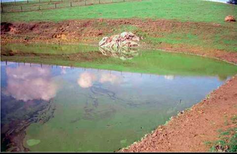
FIG. 54.1 Pond with Microcystis bloom in Petaluma, California. Pond water contained microcystin-LR and resulted in acute deaths and photosensitization of Holstein heifers.
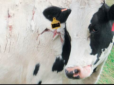
FIG. 54.2 Holstein heifer with hepatogenous photosensitization from microcystin-LR toxicosis; Central Valley of California.
warm months when water temperatures exceed 20° C and water is stagnant.1 High nutrient concentrations, especially phosphorus and nitrogen, increase the presence of cyanobacteria and cyanotoxins.2 Cyanobacteria are suspended in the water column and easily visible or are attached to the surfaces of stones and sediments along the bottom of lakes or rivers.3 Currently, of the 2000 species identified worldwide, more than 80 are categorized as toxigenic. Toxigenic and nontoxigenic species and strains coexist in water; thus the potential toxicity of a given water source must be carefully evaluated not only by identification of the actual cyanobacteria population but also by measuring toxin concentrations.4,5 George Francis first reported in Nature in 1878 a toxigenic bloom of a “poisonous Australian lake” with “a thick scum like green oil paint” and vividly described acute intoxications of sheep, horses, dogs, and pigs.6 Since Francis's publication in 1878, numerous case reports describing morbidity and mortality in animals exposed to cyanotoxins have been published.7-13
The most commonly reported cyanotoxin poisoning in large animals is acute hepatotoxicosis subsequent to microcystin exposure. Microcystins are produced by multiple cyanobacteria and detected in fresh and coastal waters worldwide.14 They present a realistic threat to pastured animals because of their presence in ponds and streams and because animals readily consume contaminated water (Fig. 54.1). Acute neurotoxicosis resulting from anatoxin-a or anatoxin-a(S) can occur in large animals, but reports are rare.
The frequency of cyanotoxin poisoning in animals is likely underreported due to absent diagnostic work-up of large animal mortalities and the current lack of well-established confirmatory methods in biological specimens. Diagnostic confirmation of suspect cyanotoxin poisoning cases of humans and animals requires extensive efforts from both toxicologists and clinicians, and resources are often not readily available. New cyanotoxins and expansion of existing cyanotoxins into new regions are discovered continuously, and oral bioavailability and toxicity data are often unavailable. It is probable that cyanotoxins poisonings are more common in animals than humans due to increased dependence and contact with surface waters.
Hepatotoxic Microcystins
Microcystin-poisoned animals typically die within a few hours after exposure. Severe destruction of the hepatocytes leads to hypovolemia and shock secondary to blood loss into the disintegrated liver lobules and embolism of hepatocytes into the lung. Clinical signs of nervousness, reluctance to move, recumbency, diarrhea, and pale mucous membranes are seen within 30 minutes to several hours of exposure.15,16 Many die within hours to days, but animals exposed to nonlethal doses may survive and develop hepatogenous photosensitization (Fig. 54.2).
Clinical chemistry changes of animals with microcystin toxicosis include elevated serum gamma-glutamyl transpeptidase (GGT), aspartate aminotransferase (AST), bile acids, alkaline phosphatase (ALP), hypoglycemia, and hyperkalemia. Acute hepatic damage results in a grossly enlarged and friable liver from intrahepatic hemorrhage and, histopathologically, in hepatocyte dissociation, degeneration, and necrosis. Pathologic findings should quickly lead to an environmental assessment for possible microcystin-contaminated water.17 Necrotic hepatocytes can also be seen in the pulmonary vasculature.
Microcystis, Anabaena, Planktothrix, Nostoc, Oscillatoria, and Anabaenopsis genera are among the known microcystin producers.18,19 Toxins are retained inside the cyanobacteria and released only upon cell damage, lysis, and death; of note, destruction of algal mats (either naturally or through the application of herbicides) may result in a pulse of microcystin release following destruction of the individual cell walls. Microcystins are cyclic heptapeptides that inhibit protein phosphatases; currently, more than 80 different structural variants have been identified. The most extensively studied congener, microcystin-LR, is found worldwide and causes acute, lethal hepatotoxicoses in farm animals.2,16,20 Most natural outbreaks are a result of oral exposure through drinking water; microcystin-containing algae release microcystins in the acidic environment of the stomach. Recently, the risk for microcystin exposure through blue-green algae- containing dietary supplements led to deaths in horses.21 Inhalation exposure to microcystins has been documented in humans recreationally using certain waters.22,23 All animal species, including fish and reptiles, and humans are considered susceptible to the toxic effects.
Animals with acute microcystin poisoning may have algal material in the GI tract (Fig. 54.3) or on the skin, especially the legs. Although identification of algae material in water and gastric contents is an important component of the diagnostic work-up, it does not confirm intoxication. As mentioned, the toxicity of cyanobacteria is strain specific, and morphologic observations alone cannot predict the hazard level. Detection of microcystins in gastric contents is confirmatory, but these tests are not routinely available at diagnostic laboratories and are limited to a few structural variants.24-26 Commercially available methods for microcystin analysis of liver and other tissues are not yet available. Consumption of milk, meat, or liver is unlikely to pose a significant health risk to humans.27,28 However, as a
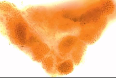
FIG. 54.3 Characteristic algal cell aggregates in rumen content of a heifer formed by Microcystis spp. Direct microscopic examination of rumen content in formalin (1: 1; v/v). 60?. Rumen content was confirmed to contain microcystin-LR.
tumor promoter, microcystins may pose a cumulative health risk as they become more ubiquitous.
Neurotoxic Anatoxins
Anatoxins are produced by many cyanobacteria genera, such as Anabaena, Aphanizomenon, Planktothrix, Oscillatoria, Microcystis, Cylindrospermum, and Phormidium. Reports of anatoxin-a poisoning in livestock are extremely rare; most cases are documented in dogs. However, all animals are considered susceptible. Anatoxin-a poisoning leads to muscle tremor, rigidity, respiratory failure, collapse, and death within minutes to a few hours.29-31 This is a result of anatoxin-a being a potent cholinergic agonist at nicotinic receptors.32 Animals poisoned with anatoxin-a(S) develop acute signs of excessive salivation, lacrimation, diarrhea, and urination (SLUD) because anatoxin-a(S) is a naturally occurring, irreversible acetylcholinesterase inhibitor.33 Animals often die of respiratory arrest within 30 minutes.
Because of the acute onset, CBC and chemistry profiles of animals with anatoxin poisoning are generally unremarkable. Furthermore, other than the possible presence of algae material in GI contents, there are no specific gross or microscopic lesions. Confirmation of anatoxin-a poisoning is by analytic detection of the toxin in urine, bile, or GI contents.31 Diagnosis of anatoxin-a(S) toxicosis is aided by the determination of blood acetylcholinesterase activity. In any suspect cases, environmental samples should undergo a toxicologic and phycologic analysis.
Other Cyanotoxins
Other freshwater cyanotoxins of concern to livestock include neurotoxic saxitoxins, hepatotoxic nodularins, and cylindro- spermopsin, which targets multiple organs (liver, kidneys, lung, heart, GI tract).
Treatment for cyanotoxin poisoning is supportive but often unsuccessful. Efficacy of decontamination with activated charcoal is limited.34,35 Symptomatic and supportive care to treat hypovolemia and electrolyte imbalances, as well as antioxidants such as vitamin E and selenium, may aid in the therapeutic regimen for large animals poisoned with microcystins. Animals that develop photosensitization must be protected from sun exposure. Because of the rapid onset of clinical signs in anatoxin-a poisonings, therapeutic options are even less pertinent; artificial respiration may be of benefit along with general supportive care. Administration of activated charcoal is suggested, but scientific evidence on its adsorptive capacity for anatoxin-a is lacking. Atropine can revert excessive muscarinic receptor activation in animals poisoned with anatoxin-a(S). It is important to note that pralidoxime (2-PAM) is not recommended.36
The frequency and extent of harmful cyanotoxins have increased with the addition of nutrients to surface freshwater ecosystems around the world. Steps must be taken to reduce fertilizer runoff and applications in fields surrounding ponds used for drinking water. Prevention of blooms is key to protecting animal and human health.