Mycotoxins
Timothy J. Evans • George E. Rottinghaus • Benjamin R. Landers
Importance of Mycotoxins
Mycotoxins are secondary fungal metabolites, which, if consumed in sufficient amounts, cause adverse health effects in animal species, including humans.1 They can be detected in various matrices, including cereal grains and forage, and are a major concern to international agricultural production and a threat to food safety worldwide.1-3 Individuals working within the United States swine and poultry industries are particularly cognizant of the potential for mycotoxin exposures, given the dietary dependency of those monogastric species on cultivated cereal grains.
However, the globalization of agricultural commodities, climate change-related weather conditions favoring fungal growth and/or mycotoxin production, subsequent reductions in the availability of foodstuffs not contaminated by mycotoxins, and economic pressures from higher costs of better-quality foodstuffs all underscore the need for individuals participating in other aspects of animal agriculture in the United States to have a greater awareness of the potential for mycotoxin contamination.In addition, the increasing risk to human and animal health posed by mycotoxins means that the veterinary profession must be vigilant in the surveillance of domesticated large animal populations as sentinels for mycotoxin contamination of agricultural products. Veterinary practitioners, regardless of whether their practices involve small companion animals or livestock, will need to be familiar with the “major” mycotoxins and the wide range of clinical signs, including subtle reductions in livestock production and food residues associated with these fungal toxins.4 It is only through an increased awareness of these toxins that mycotoxin-related illness in food animals can be minimized and companion animal and human exposure to mycotoxins limited or prevented.
General Principles of Mycotoxin Production and Detection
Many fungi are capable of producing toxins (i.e., are toxigenic), and a given mycotoxin could be synthesized by more than one fungal species.3 Some fungi produce more than one toxin or even multiple classes of toxins.1,3 Although a number of relatively common mycotoxins have already been identified, it is likely that many more potentially toxic, secondary fungal metabolites have yet to be isolated and characterized.2,3,5 Fungal colonization does not necessarily mean that a given mycotoxin is being produced.1,3 The substrate, moisture content, pH, ambient temperature, relative humidity, and other requirements for toxin synthesis are often more stringent than those for fungal growth.1,2 The “ideal” toxigenic conditions for the production of specific mycotoxins have been characterized, and some mycotoxins are more likely to be produced in a “field” or “storage” setting.1-3 The mere presence of fungal growth does not necessarily correlate with toxin production, and some mycotoxin-contaminated foodstuffs might not even be visibly moldy.3
From a clinical perspective, the inability to accurately predict whether mycotoxins will be present in a specific foodstuff means that fungal identification and spore counts are not adequate for identifying livestock diets contaminated with mycotoxins.1-3 High-performance liquid chromatography (HPLC), liquid chromatography-tandem mass spectrometry (LC-MS/MS),
and/or enzyme-linked immunosorbent assay (ELISA) are some of the more common analytic techniques used to directly measure mycotoxin concentrations in grains or processed foodstuffs. The use of an ultraviolet light to detect fluorescence associated with kojic acid, as an indirect indicator of aflatoxins, is outdated as the sole means of detecting these toxins.1,6 Likewise, use of semiquantitative methods, such as thin layer chromatography (TLC), as standalone evidence of fungal toxin contamination, is also less than optimal.
The consistent use of appropriate sampling techniques ensures the detection of suspected mycotoxins and guarantees that detected mycotoxin concentrations are representative of actual livestock exposure.3,,8 Grain “screenings” or “fines” are the grain by-products most likely to contain the highest concentrations of mycotoxins.1,3One of the biggest challenges pertaining to mycotoxin contamination in livestock rations is the accurate interpretation of analytic results, especially now, when low concentrations of mycotoxins are detected by increasingly sensitive laboratory methods.4 Table 54.8 shows “action levels” established by the U.S. Food and Drug Administration (FDA) for some mycotoxin-contaminated commodities fed to different species of livestock. These action levels (more specifically, “advisory levels” for deoxynivalenol and “guidance levels” for fumonisins) were originally intended as guidelines for “acceptable” concentrations of individual mycotoxins in common substrates being transported between states.6,9 The mycotoxin concentrations have often been interpreted as the highest dietary concentrations of these mycotoxins considered “safe” in the denoted species. However, the definition of “safe” is a relative term that depends on the end point being measured. Some concern has recently been expressed as to whether these action levels are low enough to prevent adverse health effects, especially with respect to carcinogenesis in humans.10
Mycotoxicoses
In discussing mycotoxin-related diseases, it is important to differentiate “mycosis” from “mycotoxicoses.” The term mycosis refers to a disease caused by fungal growth, rather than by the adverse effects of any toxins produced by that fungi.3 Mycotoxicoses, on the other hand, are diseases caused by the adverse health effects of mycotoxins, which in most cases have been ingested.1-3 For a specific mycotoxicosis to occur, growth conditions in substrates colonized by the appropriate fungal species must favor production of a specific toxin, and sufficient amounts of that toxin-contaminated substrate must be consumed by a susceptible animal species.
■ TABLE 54.8
Summary of Mycotoxins Important to Livestock
| Mycotoxins | Fungal Sources Substrates | Primary Toxic Effects | Dietary U.S. Food and Drug Administration Action Levels |
| Aflatoxins Bi, B2, G1, G2 | Aspergillus flavus and Aspergillus parasiticus Corn, nuts, cottonseed+ | Macromolecular binding enzymatic inhibition DNA adducts formed → hepatic insult/failure cancer (1° hepatic) All species | Total aflatoxinsa Immature 20 ppb Dairy/horse 20 ppb 100+ lb pigs 200 ppb Feedlot beef 300 ppb Breeding 100 ppb |
| Deoxynivalenol (DON/Vomitoxin) | Fusarium graminearum (formerly Fusarium roseum) Corn, wheat, barley+ | !Protein synthesis, GI irritation, Tvomiting → feed refusal, !nutrient absorption, !immunity 1° Swine Dairy cattle? | DONb Swine 1 ppm Horses/other 2 ppm Cattle 5 ppm |
| Zearalenone | F. graminearum (formerly F roseum) Corn, wheat, barley+ | Hyperestrogenism → reproductive tract Δs Pseudopregnancy Early embryonic death 1° Swine TSubfertility/ovary Δs Dairy cattle? | No action level determined Clinical signs Young gilts 1-3 ppm Mature sows >3 ppm Bred sows >15 ppm Heifers >10 ppm Cows >25 ppm |
| Fumonisins 1° B1 and B2 | Fusarium verticillioides (formerly Fusarium monilforme) 1° Corn | Sphingolipid Δs → leukoencephalomalacia Horses Pulmonary edema Swine Hepatic disease in both | Total fumonisinsb Horses 1 ppm Swine 10 ppm Ruminants 30 ppm or Repro/dairy 15 ppm |
| Ergot Alkaloids Ergopeptine alkaloids Ergovaline Lysergic acid+ ErgonovineLysergol Ergopeptine alkaloids Ergotamine+ | Epichloe coenophiala (genus formerly Neotyphodium and Acremonium) Tall fescue grass Neotyphodium Iolii Perennial ryegrass Claviceps purpurea Small grains and grasses | 1° Vasoconstriction → dry gangrene/Ttemp !Feed intake Ruminants/camelids? 1° Hypoprolactinemia → agalactia/long gestation Horses | Ergot bodies 0.1% by weight % Ergot bodies versus ergopeptine alkaloid concentrations |
aBased on corn action levels.
bActually “advisory” levels for DON and “guidance” levels for fumonisins, based on highest allowable grain level and percent of diet.
GI, Gastrointestinal.
From references 1, 3, 6, 8, 9, 25, 33, 37, 46, and 47.
The higher the dietary concentration of a given mycotoxin, the more likely and severe are the observed adverse effects.1-3 Similarly, the duration of exposure to a mycotoxin affects the likelihood and severity of the observed clinical signs, with longer exposures increasing the probability of more severe intoxications.2,3 Peracute and acute intoxications generally occur over a short period (usually days) and involve one or several “large” doses (orders of magnitude greater than “allowable” quantities) of a specific mycotoxin. In these instances, the diagnosis is often straightforward because contaminated feed is more likely available for testing. However, clinically recognized mycotoxicoses more often than not involve subacute (weeks) to chronic (months) dietary exposures to fungal toxins.1 Depending on the dietary concentration of a given mycotoxin, clinical signs in these types of exposures may be “classic” for a mycotoxin’s typical target organ (e.g., moderate to severe hepatocellular damage with sufficient amounts of aflatoxins) or more subtle in nature (e.g., immunosuppression, suboptimal growth, reproductive inefficiency, and/or carcinogenesis), reflecting impaired hepatic function.3,4 In subacute to chronic mycotoxin exposures, affected animals could still be ingesting specific toxins, with contaminated foodstuffs readily available for testing, or the consumption of specific mycotoxins may have already ceased, especially if the observed clinical signs are more subtle in nature and/or took some time to develop. Under these circumstances, analyses of representative samples of grains or forages harvested at the same time as the suspect foodstuffs or analyses of samples of prepared rations retained by the supplier or manufacturer might be the only way to establish the definitive cause of a suspected mycotoxicosis.
Another aspect of a potential mycotoxicosis, as illustrated in Fig. 54.4, using reproduction as an example of monitored animal performance, is the role that mycotoxins may play in diminished livestock performance, such as decreased feed consumption, growth rates, and fertility. In these instances, there may not be a single, definitive “cause” but rather a multifactorial etiology. It is probably overly convenient to hold feed manufacturers and distributors wholly responsible in these instances and to forget the important role of other factors, such as environmental stressors, infectious agents, and suboptimal management. In fact, the mention of this type of diagnostic conundrum can increase the overdiagnosis or misdiagnosis of a mycotoxicosis, especially when the detected concentrations are well below established levels of concern, and should not be misconstrued as an effort to indict trace amounts of some mycotoxins as the primary cause of clinical abnormalities in livestock.4,6 However, although it is not practical or even possible to eliminate all measurable fungal toxins from livestock diets, one should also recognize that there is no nutritional requirement or benefit related to dietary mycotoxins. It is critical, therefore, when no clear-cut, single cause of diminished performance is evident, to take an integrated approach and objectively evaluate the relative contributions of a wide variety of enzootic variables or “stressors” to the clinical problems being observed.4
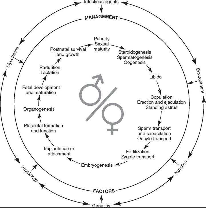
FIG. 54.4 The continuum of developmental stages and reproductive functions taking place in males and/or females, as well as the embryo and fetus, is shown schematically and illustrates the complexity of reproduction in ruminant species. The possible influences of genetic and physiologic predispositions, as well as nutritional, environmental, infectious, and especially mycotoxin stressors, on decreased reproductive performance are also represented, along with the role “management” can play in modulating those influences on reproductive function. (Modified with permission from Evans TJ: Diminished reproductive performance and selected toxicants in forages and grains. Vet Clin North Am Food Anim Tract 27:345, 2011. Modifications and artwork courtesy Don Connor and Howard Wilson.)
Major Mycotoxins of Veterinary Importance
AFLATOXINS. Aflatoxins arguably pose the most clinically relevant, mycotoxin-related threat to the worldwide health and well-being of humans and domestic animals. A number of fungal species, in particular Aspergillus flavus (A+fla+toxin) and Aspergillus parasiticus, can produce aflatoxins under appropriate growth conditions in a variety of nuts and grains, including cottonseed, peanuts, and especially corn.1-3 The major aflatoxins found in contaminated grains include aflatoxins B1, B2, G1, and G2 (B or G = fluorescence color), with aflatoxin B1 (AB1) generally being present in the highest concentrations.3 Although aflatoxins are sometimes described as “storage” mycotoxins, there are certainly instances when they are produced under “field” conditions.1,6 Drought and/or insect damage to corn or other susceptible plants, followed by warm (>25° C), humid (>70% humidity), ambient weather conditions, is the “classic” scenario that facilitates initial fungal invasion of compromised kernels and seeds and subsequent fungal growth with aflatoxin production.1-3,6 In fact, drought conditions are often associated with increased scrutiny on the part of grain elevator operators, livestock producers, and regulators, with respect to possible aflatoxin contamination, particularly in corn and corn by-products.
Ingested AB1 is metabolized by multiple cytochrome P450 subfamilies in the liver to produce several metabolites, including AB1-8,9-epoxide and AB1-dihydrodiol, which are toxic, and aflatoxin M1 (AM1), which can be found in meat as well as in milk.1 AB1-8,9-epoxide and other toxic metabolites bind to biologically important molecules, such as essential proteins, including enzymes involved in carbohydrate and lipid metabolism and protein synthesis.1,3 These AB1 metabolites also bind to macromolecular nucleic acids, inhibiting transcription and forming DNA adducts, which can be associated with carcinogenesis, especially hepatic tumors.1-3,8 In addition, toxic metabolites of AB1 can bind to subcellular organelles, such as mitochondria and ribosomes.1
Although excretion of violative AM1 residues (>0.5 ppb or 0.0005 ppm) in the milk is the first observed “abnormality” associated with AB1 exposure in lactating dairy animals, the liver is considered the major target organ of toxic AB1 metabo-
1 lites in all species.1-3,6 Clinical signs of aflatoxicosis range from prolonged prothrombin time (PT), hemorrhage, bloody diarrhea, and rapid death in peracute and acute intoxication, to anorexia, slow rumen motility, weakness, tremors, ataxia, and reproductive failure (abortion), to hepatic failure with icterus, coma, and death in subacute aflatoxicoses.1-3,6,8,11-16 Aflatoxin-induced hepatic insult and failure are usually accompanied by many of the typical clinical pathologic abnormalities expected with hepatocellular necrosis and/or hepatic failure, and elevations in the detected activities of liver-specific enzymes in serum often precede other clinical signs.6 Suppression of immune function, particularly those aspects related to cell- mediated immunity, have also been reported in animals following repeated exposure to moderately high dietary concentrations of AB1.1,6,8,15 Chronic exposure to dietary aflatoxins can be associated with decreased growth rates and feed efficiency, roughened hair coats and ill-thrift, impaired resistance to infectious disease, and hepatic fibrosis with regenerative nodules.1,3,6 Spermatogenesis can also be adversely affected by chronic exposures of male animals to AB1.17 Low-level subacute to chronic exposures to AB1 and its toxic metabolites also have the potential to be associated with teratogenesis, mutagenesis, and/or carcinogenesis, and experimentally, AB1 in grain dusts can be present in sufficient quantities to be converted to carcinogens by pulmonary cytochromes.3,18
Attempts to estimate the dosages or dietary concentrations of total aflatoxins likely to be “safe” or, conversely, the concentrations that will probably be associated with clinical disease can be challenging. Anecdotal evidence and reports of clinical cases of aflatoxicosis, which provide some dose-related data, may require additional scrutiny because some of the factors known to influence the toxicity of aflatoxins might be unaccounted for or unknown.6 However, such reports are still of value because they represent “real” clinical situations. An animal's plane of nutrition, rate of hepatic metabolism, age, and species, as well as the level and duration of aflatoxin exposure, are all important determinants as to whether and what signs of aflatoxicosis are likely to be observed.1,3,6,8 Animals fed a diet deficient in protein, selenium, and/or vitamin E and those with enhanced rates of hepatic metabolism are more likely to experience hepatic insult associated with dietary aflatoxin contamination.6,8 Immature animals, particularly young swine, are the livestock populations considered most susceptible to adverse health effects associated with aflatoxins.3,6 Weeks of exposure to total dietary aflatoxin concentrations of 200 ppb (0.2 ppm) have been associated with mild hepatic insult and suboptimal growth of piglets and decreased performance in calves.3,6,8 Horses and more mature cattle and swine have experienced toxic insult to the liver following weeks to months of exposure to aflatoxin concentrations exceeding 400 and 600 ppb (0.4 and 0.6 ppm), respectively, and hepatic failure and death are possible in animals consuming foodstuffs that contain a flatoxin concentration exceeding 1000 ppb or 1 ppm.3,6,8,11,12,14,15,19
The “threshold” AB1-induced “abnormality” usually first observed at the lowest dietary concentrations in livestock species is an actionable level of AM1 detected in milk obtained from exposed lactating dairy animals.3,6 The concentrations of AB1 causing short-term, minimally violative AM1 residues in milk are much lower than those likely to cause any liver-related adverse health effects, especially in ruminants.3,6,8,11-16,19 The ratio of the concentration of AB1 in the diet to the AM1 concentration detected in the milk varies widely from less than 50: 1 to almost 2000 : 1 (average of 300 : 1), and the proportion of dietary AB1 excreted as AM1 can range between 0% and 4% (average of 1%).6 Excretion of AM1 in the milk generally continues for 2 to 7 days after withdrawal from an AB1-contaminated diet.3,6
The accurate diagnosis of clinical aflatoxicosis usually involves the detection of high AB1 concentrations in the feed or, less optimally, the gastric contents of animals exhibiting appropriate clinical signs.3,6,8 The detection of AB1 and/or AM1 in the liver and kidney, in particular, can also be indicative of recent AB1 exposure (usually within 7 days) and may help establish the consumption of dietary AB1 by a deceased animal, when foodstuffs are unavailable for analyses.3,8
Treatment of aflatoxicosis in large animals is often cost prohibitive, especially in food-producing animals, and limited to therapy for hepatic insult and/or failure; provision of a balanced, AB1-free diet; possible supplementation with trace minerals and vitamins (selenium and vitamin E); and most importantly prevention of further exposure.3,6,8 Growth conditions are not conducive to AB1 production in Aspergillus-infected stored grains when the moisture content is reduced to less than 15%.8 Ammoniation has been reported to prevent fungal colonization and growth and to facilitate detoxification of aflatoxins.1,3,8 In “emergency” situations where it is difficult to find affordable, alternative foodstuffs free of AB1 contamination, producers may consider dilution of feeds to “safe” concentrations of AB1.3 However, this approach has some attendant risk, including the species of animal involved, exposure to enzootic stressors other than aflatoxins (e.g., infectious disease, poor nutrition), level of animal management and husbandry skills, willingness of the producer to thoroughly “blend” the feeds and collect frequent representative samples for AB1 analyses, and the targeted “safe” concentration of AB1.3,4 One possible approach to the dilemma of which targeted AB1 concentrations are likely to be “safe” is for producers to aim for total aflatoxin concentrations approximately one half of that shown in Table 54.8 for total dietary aflatoxin action levels, particularly in diets intended for lactating dairy cows, immature livestock, or animals used for breeding purposes.4,9 This approach can also be used for other mycotoxins for which there are action levels.4 Another consideration, especially for aflatoxins, may be the incorporation of clay binders in rations to prevent the absorption of fungal toxins.3,4,6,8 Hydrated sodium calcium aluminosilicates (HSCASs) have been shown to effectively prevent violative AM1 residues in milk and meat and to reduce livestock production indices, especially those parameters (e.g., growth and reproductive performance) likely to be affected by low-level chronic exposure to dietary aflatoxins.3,4,6,8,20-24 Before using a given binder, there should be in vivo evidence that the binder in question is effective at reducing AB1 absorption in live animals.1,3,4,6
DEOXYNIVALENOL (DON OR VOMITOXIN). Deoxynivalenol (DON or vomitoxin) is classified as a trichothecene mycotoxin and is produced in grains such as corn, wheat, and barley by toxigenic Fusarium species, especially Fusarium graminearum (formerly Fusarium roseum). This species is also the conidial stage of Gibberella zeae and associated with “head blight” and “scab” in wheat and “Gibberella ear rot” in corn.1-3,25 Although growth of this toxigenic fungal species occurs in the field when temperatures are warmer (20° to 25° C), DON production is favored under storage conditions when it is cooler (0° to 15° C).1,3 Rising and falling (undulating) temperatures, especially when associated with a cool, wet fall and delayed harvest, facilitate production of DON.1,3,26 Trichothecenes are stable compounds that persist much longer than the fungi producing them.1 Deoxynivalenol can also be found in forages (such as corn stover and wheat straw) associated with the grains most frequently contaminated by this mycotoxin.1,27 In addition, grains contaminated with vomitoxin are often contaminated with zearalenone (see below), which is also produced by F. graminearum (see Table 54.8) under similar fungal growth conditions.1,3,25
Deoxynivalenol is an extremely important trichothecene mycotoxin, from an agricultural perspective, because of its impact on indices of animal performance and the frequency with which it is detected in livestock diets, especially those intended for consumption by swine and dairy cattle.1-3,8,26 This particular fungal toxin inhibits protein synthesis and induces emesis by crossing the blood-brain barrier and directly stimulating the chemoreceptor trigger zone in the medulla oblongata, hence the alternative name of “vomitoxin.”3,25 Immunosuppression has been reported in some animal species, but feed refusal and gastroenteritis are the most common clinical signs, particularly in swine.1-3,25 Clinical pathologic abnormalities include those associated with decreased feed intake, impaired absorption of nutrients, vomiting, and diarrhea.8,25
Table 54.8 shows the guidance levels for DON in various species of livestock. Clinical signs of dietary exposure to DON, such as feed refusal, generally begin within 1 day of ingestion.3,25 Dietary concentrations of DON associated with clinical disease range from 1 ppm (feed refusal) to 5 to 10 ppm (vomiting and possible gastroenteritis) in swine; ruminants have been reported to refuse eating contaminated foodstuffs containing more than 10 to 20 ppm of DON.8,25 One dilemma often associated with DON occurs because of the low concentration frequently found in grains and their related forages. It is difficult to differentiate the direct adverse effects of the mycotoxin itself from clinical signs related primarily to reduced palatability and/or the decreased nutritional value of poorer quality foodstuffs, particularly when feed refusal and abnormalities arising from reduced feed intake are the only observed clinical signs.4 Similarly, any deviations from optimal dairy cattle performance should not necessarily be attributed solely to low-level mycotoxin contamination (10 ppm versus >25 ppm ZEA, respectively) to adverse reproductive effects associated with ZEA, there still remains doubt as to how often ZEA-related problems are observed in dairy cattle in real-world situations.4,8, There are reports that cattle are
insensitive to ZEA, while others report a 25% lower pregnancy rate in postpubertal heifers fed 12.5 ppm of ZEA.4,33,35,36 The disparity between these reports attests to the significance of the stage of sexual maturity, concurrent exposure to other enzootic stressors, and sensitivity of the criteria used to evaluate impaired reproductive performance in determining how ZEA affects dairy cattle fertility.4
FUMONISINS. Fumonisins, especially fumonisin B1 and B2 (FB1 and FB2), are produced primarily by the ubiquitous fungal species Fusarium Verticillioides (formerly Fusarium moniliforme).13 F. Verticillioides grows primarily on corn, and the highest concentrations of FB1, which is the most common and clinically important fumonisin, are most often found in association with corn “screenings” or “fines.”8,37,38 Although growth conditions facilitating FB1 production are not as well defined as those for AB1, DON, and ZEA, drought conditions during the growing season and subsequent cool, wet weather during ear development seem to correspond to higher FB1
837 1
concentrations.8,37
Fumonisins structurally resemble sphingolipids, which are found in highest concentrations in the brain and liver and are essential for cell growth and differentiation, as well as vascular integrity.1,3,8, One primary toxic mechanism of action proposed for fumonisins, in particular FB1, is inhibition of the conversion of sphinganine to sphingosine, resulting in a disruption of sphingolipid biosynthesis.3,8,, Horses and swine are the two livestock species most susceptible to fumonisin intoxication.1,3,37 The brain is generally considered the major target organ in horses, especially following more chronic, low-level exposures, but centrilobular hepatocellular necrosis, with clinical pathologic changes in the serum typical of toxic hepatic insult, can be observed in the absence of brain lesions when horses are acutely exposed to high FB1 concentrations.1,3,37 Subacute to chronic consumption of dietary concentrations of FB1 exceeding 8 to 10 ppm have been associated with the sudden onset of the clinical syndrome referred to as equine leukoencephalomalacia (ELEM) or “moldy corn poisoning.”1,3,8,37,40-42 Horses affected by ELEM (also see Chapter 35) exhibit altered mentation, ranging from depression to neuroexcitation, sometimes accompanied by head pressing; incoordination and apparent blindness; and finally recumbency and death, often within 5 to 7 days of the 13374042
onset of clinical signs.1’3’’ The pathognomonic lesion of
ELEM is leukoencephalomalacia, or degeneration of the white matter of the brain, which can be grossly visible involving one or both cerebral hemispheres or subtle and difficult to detect in 13374042
stained sections of brain.1’3’ ’ 42 Hepatosis of varying severity can also be observed in cases of ELEM, and transient increases in the activities of enzymes specific for hepatocellular damage might be observed under those circumstances.37,41
The primary clinical abnormalities associated with FB1 exposure in swine include porcine pulmonary edema (PPE), which is thought to be secondary to left-sided heart failure, and toxic insult to the liver.1,3,8,37 However, unlike ELEM, it is difficult to pinpoint any consistent “threshold” dietary concentrations of FB1 likely to cause heart failure and the resulting PPE in swine or, alternatively, histopathologic changes 837384344
restricted to only the liver.8,37,38,43,44 Some authors have suggested that FB1 concentrations in contaminated corn or especially in contaminated corn screenings, ranging from the dietary action level of 10 ppm of FB1 (see Table 54.8), should be considered actionable. Conversely, it is interesting to note that swine have been chronically exposed to dietary FB1 concentrations in excess of 100 ppm, without the development of signs consistent with PPE and with hepatic fibrosis being the only observed histopathologic finding.44 It appears that if PPE is going to develop, it will almost certainly do so within the first week of consumption of FB1-contaminated diets.37,43 This consistent observation suggests the role of various stressors, such as recent transport, excessive animal handling, and/or the presence of respiratory pathogens.44,45
The diagnosis of ELEM and PPE involves the observation of the appropriate, species-specific clinical syndromes, as well as the detection of sufficiently high enough concentrations of FB1 in corn-based rations, especially those diets prepared using screenings from homegrown corn.3,37 An elevated sphinganine- to-sphingosine ratio (SA/SO ratio) in the serum, urine, or tissues of animals suspected of being exposed to FB1 can also be an accurate indication of recent (1 ppm ergovaline in the seed heads), and LA+ are also present in this grass.50,51-53 The livestock species most likely to consume E+ forages are ruminants, especially cattle, and horses. The primary sources of the EP compounds—ergotamine, ergocristine, ergosine, ergocornine, and ergocryptine—are the dark-colored ergot bodies or sclerotia of C. purpurea. Ergot sclerotia, unlike endophytic mycelia, are externally visible and replace the individual seeds of many common pasture grasses, including E+ tall fescue and small cereal grains, such as oats, barley, wheat, and especially rye and triticale, but not corn.47,48,50 The cool, wet springs in the northwestern United States, the northern Great Plains, and parts of western Canada facilitate the germination and growth of C. purpurea.4'4''5 Those animal species likely to consume E+ tall fescue will also be exposed to the EAs produced by C. purpurea, if the E+ tall fescue or other nearby grasses are infected by this fungal species (“ergot- ized”). On the other hand, when small cereal grains are contaminated by ergot bodies, not only ruminants and horses but also swine, poultry, and even humans are likely to be exposed to EAs. The total concentration of EPs in C. purpurea sclerotia are extremely high, generally ranging from 2000 to 10,000 ppm, 4748505457
with LA+ also likely to be present.47,48,50,54-57
Although there is some overlap, the pathogenesis of clinical signs associated with dietary exposures to EAs can generally be explained in terms of vasoconstriction and/or hypoprolactinemia. Vasoconstriction involving interactions with dopaminergic, adrenergic, serotonergic, and glutamate receptors is responsible for many of the clinical signs observed in ruminants, especially cattle, and possibly in South American camelids.46,47,51 Hypoprolactinemia-induced agalactia in late gestational mares is the most sensitive indicator of dietary exposure to EAs.47,50,51 The stimulation of dopamine D2 receptors by EAs dramatically reduces prolactin secretion by lactotropes in the adenohypophysis.8,46-51,58 Abdominal fat necrosis in cattle is associated with chronic exposure to EAs in E+ tall fescue and arises from both vasoconstriction and the effects of hypoprolactinemia on lipid metabolism.46,51 The higher the dietary concentrations of EAs, the more likely horses are to exhibit vasoconstrictive effects and the more likely agalactia will be observed in ruminants, in which a placental lactogen helps prevent abnormal lactation from the amounts of EAs often present in E+ tall fescue.1,34,47,51 Swine exhibit vasoconstrictive effects and sub- optimal reproductive performance, including agalactia, when exposed to sufficient concentrations of EAs.1,47
When trying to define levels of exposure to EAs, the most readily available data pertain to concentrations of EPs measured using HPLC. If animals ingest total dietary concentrations of EPs between 0.1 and 1.0 ppm, regardless of the sources, this is equivalent to low-level exposures involving only E+ tall fescue without seed heads, plus or minus other grasses.50,51,53 Low to moderately high total dietary concentrations of EPs between 1 and 5 ppm correspond to what would be expected in pastures consisting entirely of E+ tall fescue in the seed head stage.51,53 Moderately high dietary concentrations of total EP ranging between 5 and 10 ppm and very high dietary EP concentrations, exceeding 10 ppm, would involve contamination of foodstuffs by ergotized grains.47,54-57,59
Due to vasoconstriction, the toxicity of EAs can be extremely dependent on ambient weather conditions (see Table 54.8), especially with lower dietary concentrations of EAs (0.1 to 1.0 ppm total concentration of EP), which are typical of E+ tall fescue.51 When it is hot, a syndrome referred to as “summer slump” or “summer syndrome” is observed in ruminants, in particular cattle, grazing E+ tall fescue.1,51 This condition is thought to arise from the limited peripheral dissipation of heat associated 13484951
with vasoconstriction.1,3,48,49,51 Regardless of the source of toxins, EA-related “heat stress” is characterized by hyperthermia, rapid breathing, lethargy, an unthrifty appearance, and reductions in feed intake and average daily gain, as well as suboptimal male and female reproductive performance.1,3,4,46-49,60-63 When it is colder, peripheral vasoconstriction in ruminants impairs the circulation to the distal extremities, resulting in cow discomfort, lameness, and eventually dry gangrene with sloughing of ear tips, switches, and hooves referred to as “fescue foot” or “gangrenous ergotism,” depending on the source of the EAs.1,3,8,46,47,51 Controlled experiments indicated that dietary concentrations of ergovaline between 0.4 and 0.8 ppm produced gangrenous effects in ruminants.64 However, the ambient temperatures in those specific experiments were not below freezing, so, considering the impact of ambient temperature on the vasoconstrictive effects of EAs, lower ergovaline concentrations could be expected to possibly cause “fescue foot” at temperatures below freezing.47,51,64 Given “average” ergovaline concentrations in E+ tall fescue and clinical signs in cattle eating other forages “free” of EAs, along with E+ tall fescue, it appears that adverse effects on production parameters can be observed in cattle, when total dietary concentrations of EP exceed 0.1 to 0.2 ppm, especially under extreme weather 47 51 53 54
conditions.4',51,53,54 In areas where cattle are commonly affected by fescue toxicosis, it stands to reason that suspected “fescue foot” or dry gangrene affecting the lower portions of the tail (“switch”) observed during the late spring or early summer tends to be more likely associated with the higher concentrations of EPs in ergotized grains or grasses (especially ergotized E+ tall fescue) rather than the lower EP concentrations in tall fescue infected only with endophyte.51,55-57,64
Exposure to EAs during the last month of pregnancy is the most common cause of agalactia in mares.47,50,51,58 Hypoprolactinemia, with minimal udder development, was consistently induced in pregnant ponies treated with a semisynthetic EP, and lactational abnormalities will invariably be the first signs observed in late gestational mares consuming EAs.50,58 Anecdotal reports suggest that dietary concentrations of ergovaline as low as 0.05 ppm can cause agalactia in some susceptible late gestational mares.50 Prolonged gestation, dystocia, and abortion, as well as fetal and placental abnormalities, are additional adverse effects observed in pregnant mares consuming higher dietary ergovaline concentrations (generally ≥0.1 ppm).47,50,51 Pellets containing screenings of ergotized grains or grasses can also be associated with EA intoxication in pregnant mares.50 The absence of normal changes in udder development (“waxing”), on which horse breeders and veterinarians are dependent to predict imminent parturition, results in unexpected and unsupervised parturition.58
The diagnosis of intoxications involving EAs requires the observation of appropriate clinical signs, identification of a source of EAs, and determination of EA concentrations in that source, as well as in the total diet.1,3,46 The accurate interpretation of those analytic results necessitates a thorough understanding of the analyses being performed and the pathogenesis of EA intoxications in different species.1,46 Using reproductive performance as an example of a monitored parameter, management factors and the stressors shown in Fig. 54.4, particularly those related to environment, should be considered when interpreting analytic results.3
The basic principles for the treatment and prevention of intoxications related to EA are no different than those outlined for other mycotoxicoses (i.e., removal from the source, stabilization, and supportive care). Incorporation of pasture management practices, including frequent mowing with removal of seed heads, careful use of nitrogen-containing fertilizers, dilution with legumes, production of hay, and pasture replacement with genetically modified varieties of continental tall fescue free from EAs, can provide added benefits to cattle producers and horse breeders living in enzootic areas.47,51 Drying and ammoniation of E+ tall fescue hay, manipulation of stocking rates, physical manipulation of ergotized grasses during the haying process, use of stockpiled E+ tall fescue during the fall, and copper supplementation can also help to further minimize EA-induced effects and maximize performance parameters in cattle.3,48,65,66 Veterinarians should also be aware of “fescue-associated edema syndrome” recently observed in horses consuming a genetically modified Mediterranean tall fescue in Australia.67
Provision of ample shade (also, possibly pond access) and plenty of potable water to cattle consuming low concentrations of EAs during the summer can help prevent “summer slump” or clinical signs resembling this condition, when the source of EA is not E+ tall fescue.47,49 It is also becoming increasingly apparent that genetic testing has the potential to be used as a management tool in the prevention of fescue toxicosis. A single nucleotide polymorphism (SNP) in the dopamine D2 receptor gene has recently shown promise as a biomarker for resistance to fescue toxicosis in Angus-based cattle, and it is likely to be commercially available in the very near future.68
Knowledge of mare breeding dates, confirmation of pregnancy, and withdrawal from any dietary sources of EAs at least 30 days before the anticipated foaling date are critical for the prevention of agalactia in pregnant mares.3,, Frequent evaluation of mammary development and, if deemed necessary, daily administration of the dopamine D2 antagonist domperidone (1.1 mg/kg body weight; watch for leaking colostrum) are additional ways to facilitate adequate lactation and prevent failure of passive transfer in foals.47,49-51 Domperidone can also be used to mitigate prolonged gestation, along with the 475051
attendant risk of dystocia.,,
Grain or grass screenings or “fines,” in general, and/or any grains containing visible ergot bodies should not be incorporated into feeds intended for consumption by pregnant mares or cattle maintained under extreme weather conditions.47,50,51 The only current FDA action level pertaining to EAs is expressed in terms of the percentage of ergot bodies (see Table 54.8) rather than concentrations of EPs.9 On the basis of the average EP content of ergot sclerotia in barley, 0.1% ergot bodies translates to approximately 2.5 ppm, which, depending on the proportion of the diet represented by this ergotized grain, may 475357
not be very protective in many clinical instances.,