Serum or plasma protein measurements constitute a vital component of laboratory diagnostic evaluations.
Globulin and total protein measurements will be higher in plasma than serum due to the presence of coagulation proteins, particularly fibrinogen. It is vital to use appropriate reference intervals for the sample type.
The following measurement techniques are widely used (units for the following serum or protein measurements are g/dL or the SI unit: g/L): Refractometry: The total protein concentration of serum, plasma, or body fluids can be determined via refractometry. The principle of measurement is that the degree of light refraction by a solution is proportionate to the concentration of solids. Refractometers are calibrated under the assumption that the solids are largely proteins and that other substances are present in constant concentrations.1 Increases in other substances can increase the refractive index of a solution and falsely increase the total protein reading; for example, marked hyperglycemia with glucose concentration of 700 mg/dL or azotemia with blood urea nitrogen (BUN) concentration of 300 mg/dL can falsely increase the protein reading by 0.6 g/dL.2 Hemolysis does not alter refractometer protein readings. The effect of marked icterus is unclear.3 Use of the urine specific gravity scale for direct measurement of serum, plasma, or body fluid protein concentration is inappropriate, as calibration differs for these different fluid types.1 Conversion tables are available for translation of specific gravity values into protein concentration values.4 Biuret reaction: Serum total protein is commonly measured via modified biuret testing on automated analyzers. The principle involves basic spectrophotometry measuring color change following copper binding to peptide bonds. Depending on the specific assay, hemolysis can cause a positive interference (e.g., hemoglobin of 400 mg/dL can cause a 12% positive bias).5 This method is not sensitive to very low protein concentrations (e.g., in cerebrospinal fluid).Bromcresol green (BCG) albumin binding: Albumin concentration in serum or plasma is usually measured via the BCG reaction. The principle involves basic spectrophotometry measuring color change when BCG binds to albumin. Nonspecific BCG binding to globulins can artifactually inflate the albumin value, particularly with a very low albumin concentration.6 Plasma albumin values can be increased over serum values because of nonspecific plasma globulin binding. Also, heparin interference can increase or decrease an albumin reading depending on the assay.7 Globulins are typically calculated by subtracting the albumin value from the total protein value.
Protein electrophoresis (PE): Serum or plasma proteins can be separated into albumin and globulin fractions electrophoreti- cally. The principle of measurement is that an electrical charge is applied to a cellulose acetate or agarose gel matrix, allowing serum and plasma proteins to migrate through the matrix and separate into bands based on their charge and size. The acetate or gel is stained to detect the protein bands and read via densitometry to generate a tracing. Albumin has a low molecular weight (roughly 66 kDa), is highly anionic, and migrates the furthest to form a band near the anode. The identical nature of all albumin molecules and therefore their migration to a single band generates a single sharp, narrow-based peak on the tracing. Globulins are divided into α-, β-, and γ-globulin regions in ruminants. In horses, globulins are further divided into α1, α2, β1, β2, and γ-globulin regions (Fig. 26.1). The heterogeneity of globulin fractions results in broad-based peaks for each, as the diverse proteins in each region migrate slightly differently. Rarely, a narrow-based peak will be seen in the γ-globulin region (see the Hyperglobulinemia section later). The presence of coagulation proteins will cause higher globulin peaks on plasma protein electrophoresis (PPE) than serum protein electrophoresis (SPE).
In particular, fibrinogen will migrate into either the “late-beta” peak or the β-γ junction and in rare cases might obscure the presence of a monoclonal peak.8,9 α- and β-globulins include many acute phase proteins (see the Acute Phase Response section later), whereas immunoglobulins (antibodies), primarily immunoglobulin G (IgG), comprise the γ-globulins. IgA and IgM are immunoglobulins that migrate in the β2 or β-γ junction regions but are not detected in health via PE testing.Immunoelectrophoresis: Immunoelectrophoresis is a specialized technique for quantifying each immunoglobulin subtype (e.g., IgG, IgM) via electrophoresis followed by immunoprecipitation using subtype-specific antibodies. Units are usually mg/dL. Other immunoglobulin measurements are designed specifically for detection of IgG in neonatal samples and are discussed under failure of passive transfer (Chapter 53).
Reference values for proteins vary by species (Table 26.1): When evaluating protein concentrations in a patient, some clinicians use the ratio of albumin to globulins (A/G ratio). However, it is often simpler to evaluate albumin and globulins separately, as the A/G ratio can change with alterations in one or both parameters.
Biological variation with life stage: Transfer of immunoglobulins from the dam to the neonate via colostral ingestion is a key process that determines the neonate's subsequent immunocompetence and is often reflected in serum globulin levels in the first few weeks of life. In addition, neonates generally have lower serum albumin levels; for example, calves had decreased albumin values in the first 8 days of life.10 Total protein levels in calves are decreased at birth, rise immediately following colostrum intake, then fall below the reference interval again at about 2 weeks of age and remain subnormal for some time.10 IgG measurement also reflects the combined presence of maternal and neonatal proteins (Fig.
26.2). Studies in cows found gradually increasing plasma globulin levels during pregnancy until 4 weeks preparturition, then a steady drop until parturition.11 IgG1 production is greatly increased at parturition, and the half-life is decreased, corresponding with transfer to colostrum.12 Serum total protein steadily increased during lactation in Danish Landrace goats.13 In pregnant and lactating mares, no significant differences in albumin, globulins, and total protein were found at any stage.14 Aging can be associated with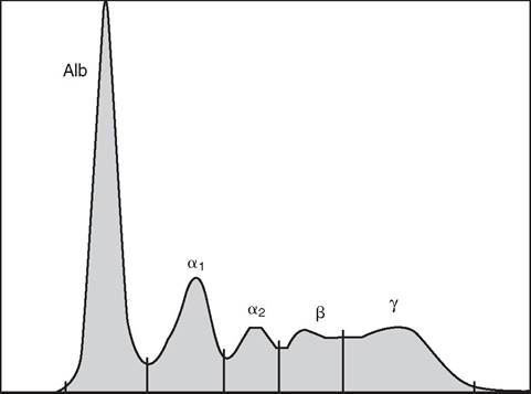
FIG. 26.1 Normal equine serum protein electrophoresis. Alb, Albumin.
alterations in some laboratory parameters. However, no significant differences in biochemical parameters or specific concentrations of IgG, IgA, or IgM were found in aged horses, despite alterations in lymphocyte populations.15,16