Spinal Tumors
Robert J. MacKay • Lisle W. George • Mary O. Smith
With the exception of lymphosarcoma in cattle, tumors involving the spinal cords of domestic animals are rare. In the horse, tumors within the spinal cord or dura (i.e., intramedullary or extramedullary-intradural) have included meningioma, epen- dymoblastoma, angioma, angioblastoma, ganglioglioma, schwannoma, and neurofibroma.1 Reported extradural tumors are lymphosarcoma, plasma cell myeloma, neurofibroma, fibrosarcoma, melanoma, hemangiosarcoma, and carcinoma.1-4 The tumor that most commonly affects the spinal cord of ruminants is lymphosarcoma, but others, such as histiocytic sarcoma, glioma, neuroblastoma, and embryonal neuroectodermal tumors, have been reported.5-9
■ Clinical Signs The clinical signs of tumorous invasion are usually referable to a single-site spinal cord lesion, although extradural tumors such as lymphoma can extend over multiple spinal cord segments.9 The rate of onset of the neurologic dysfunction varies.
Some neurofibromas, melanomas, and lymphosarcomas invade centripetally along the peripheral nerve rootlets. Affected patients develop slowly progressive limb ataxia and weakness caudal to the location of the lesion, which eventually leads to tetraplegia or paraplegia. In rare cases, the onset of tetraplegia may be peracute and unaccompanied by prodromal neurologic signs. Lymphosarcoma has a predilection for the lumbar segments of the spinal cord and the cauda equina in cattle older than 5 years (Fig. 35.19). A diagnosis of spinal cord tumor should be considered in cases of progressive neurologic disease characterized by flaccid tail and anus, dysuria, perineal scalding by urine, distended bladder, perineal analgesia or anesthesia, and paraparesis. Most animals with spinal tumors are mature to older adults; immature animals are affected in rare cases, mainly by embryonal tumors.8-11■ Clinical Pathology Examination of CSF may be useful when the tumor has infiltrated the cauda equina and is located in the lumbosacral cistern. In these cases, tumor cells may sampled for biopsy as the needle is inserted into the lumbosacral space.
In other cases the CSF may be normal, or albuminocytologic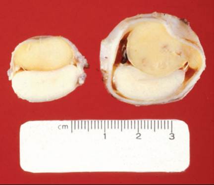
FIG. 35.19 Epidural lymphosarcoma in a 5-year-old Holstein dairy cow. The tumor had spread from neoplastic periaortic lymph nodes and had expanded to compress the caudal lumbar and cranial sacral spinal cord segments, which resulted in signs that resembled bilateral sciatic paresis. Tumorous masses are above the compressed spinal cord in the two transverse sections shown.
dissociation can be expected (elevated CSF protein levels with normal to mildly elevated nucleated cell count) with or without variable degrees of hemorrhage and xanthochromia. After necropsy, various immunohistochemical techniques can be used to identify the tumor type when routine histologic examination is insufficient.
■ Treatment There is currently no treatment for most spinal cord-associated tumors of large animals; however, protocols could be adapted from those used for other forms of lymphoma in horses or for other solid tumors in small animals. In one study, a cow survived for 57 days after three treatments with L-asparaginase at 10,000 IU∕m2 surface area. The nearly 2-month period of survival allowed the investigators to successfully superovulate the cow. When treating food animals with L-asparaginase, the benefits of the antimetabolite drug must be weighed against the potential for teratogenicity of the fetus, toxicity for humans, and the certainty of relapse in 12
the patient.12