Spondylitis and Diskospondylitis
Differential diagnoses for spinal pain include spondylosis, vertebral fracture, muscle strain, dorsal spinous process impingement, vertebral subluxation, vertebral infarcts, and aberrant parasite migration.13 Additional differentials for spinal cord ataxia in horses include cervical vertebral stenotic myelopathy, equine protozoal myeloencephalopathy, herpes myeloencepha- lopathy, and neoplasia.
■ Etiology and Pathogenesis Both spondylitis and diskospondylitis are thought to be septic conditions, although etiologic isolation is not often successful. Even in cases where bacteria are not isolated, response to antimicrobials may indicate a bacterial pathogenesis.7 Bacterial infection is more common than fungal infection, and the hematogenous route of infection is the most common. Spondylitis occurs most frequently in neonates, which may be due in part to failure of passive transfer leading to sepsis.1 In general, spondylitis is often secondary to a preexisting focus of infection elsewhere in the body.1 Tail docking wounds,14 umbilical infections,1,12 pneumonia,10 and lung abscesses1 are possible sources of infection. Septic thrombi embolize into the metaphyseal arteries of the vertebrae, where flow is sluggish and bacteria can colonize.14 In adults, direct injury to the intervertebral disc and/or the vertebral end plate may contribute to the formation of diskospondylitis. The injury disrupts the vasculature, increasing susceptibility to infection. Diskospondylitis is also reported to occur secondary to spread of local infection5 and traumatic injury.4 Infection results in destruction and remodeling of affected bone. Inflammation of the disc and vertebrae leads to spinal cord compression and the associated neurologic dysfunction.
The intervertebral disc actually may prolapse into the spinal canal.5 Lesions are usually confined to one intervertebral space. The neurologic signs may also be secondary to the infection eroding into the meninges and causing a suppurative meningitis.Pathogens that have been cultured from adult horses with vertebral osteomyelitis include Brucella abortus,15 Aspergillus spp., Streptococcus zooepidemicus,i Staphylococcus spp.,6,9 and Mycobacterium bovis.16 Vertebral osteomyelitis isolates in foals include R. equi,w,11,1'7 Streptococcus spp., Actinobacillus spp., Eikenella corrodens,18 E. coli, Salmonella typhimurium, Staphylococcus spp., and Corynebacterium pseudotuberculosis.13 Isolates from cattle with vertebral osteomyelitis include Aspergillus fumigatus, Bacteroides nodosus, Clostridium perfringens, Streptococcus spp., Staphylococcus spp.,2 E. coli,14 Actinomyces pyogenes, Fusobacterium necrophorum, and Pseudomonas spp.19 Staphylococcus spp. and Arcanobacterium pyogenes were cultured from the intervertebral disc of a goat.3
■ Diagnosis Diagnosis is based on clinical signs and imaging modalities. Clinicopathologic findings may include anemia, leukocytosis, neutrophilia, and hyperfibrinogenemia.1 One retrospective study, however, found that 80% of affected horses had a normal white blood cell count.20 Hyperproteinemia due to hyperglobulinemia may also be seen.7 Cerebrospinal fluid is usually normal unless the osteomyelitis has progressed to meningitis. Blood and urine cultures should be obtained but are often not diagnostic.
Radiography is often the basis of diagnosis. Radiographic signs of spondylitis include bony proliferation, lysis, sclerosis, and localized soft tissue swelling.1 Radiographic abnormalities in diskospondylitis include osteolysis of adjacent end plates with surrounding sclerosis and collapsed intervertebral disc space9 (Fig.
38.28). Contrast radiography may be used to delineate spinal cord compression.9 Radiographic changes may lag at least 2 to 8 weeks behind clinical signs, however, hampering the ability to make an early diagnosis.1 With vague clinical signs, the neuroanatomic location within the vertebral column may be difficult to identify, making targeted radiographs impossible. Nuclear scintigraphy may help localize the lesion to a specific vertebral section that can then be radiographed.1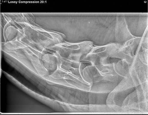
FIG. 38.28 An 11-year-old Arabian cross gelding with fever and neck pain. The cortical margins of the caudal end plate of C5 and the cranial vertebral body of C6 are markedly thin and ill-defined. In addition, there is osteolysis and sclerosis associated with the cranial vertebral body of C5. The C5-C6 intervertebral disc space is narrow.
Scintigraphy may also allow earlier detection due to the lag time in radiographic changes. Ciprofloxacin and labeled white blood cell scintigraphy may also prove useful in the diagnosis of vertebral osteomyelitis. Ultrasonography is becoming increasingly used as a diagnostic imaging modality. Abnormalities include irregular end plates or bone surfaces, wide or narrow disc spaces, vertebral step formation, and associated abscessation or muscle involvement.9 Transrectal ultrasonography of the lumbar vertebrae may be performed, while transcutaneous studies can image the rest of the vertebral column. Advantages to ultrasound are that it can be done in the field, it can be used for needle-guided aspirates of potentially infectious lesions, and it can be used as a screening tool to localize lesions.9 Depending on the size of the animal and the availability of equipment, MRI and CT may also be considered, as these modalities can be used to assess the vertebral end plates, the disc, and the spinal cord.3 CT has been used in a goat to diagnosis diskospondylitis.
Bone lysis, bone proliferation, mineral opacity within the intervertebral disc, and spinal cord displacement were seen.3■ Treatment Long-term antimicrobial therapy is indicated for cases of vertebral osteomyelitis. Blood, urine, or tissue cultures and sensitivities may be used to guide antimicrobial choices. When that is not possible, broad-spectrum, bacteriocidal antimicrobials should be used. Long-term treatment is necessary and may last for 3 to 6 months.10 NSAIDs can also be used for pain control. In nonresponsive cases, curettage of infected bone may be necessary.6,10 This also provides an opportunity to collect material for bacterial culture.10 In horses with instability and spinal cord compression, surgical decompression and stabilization may be indicated.
■ Prognosis The prognosis for horses with diskospondy- litis is guarded. If there are no neurologic deficits, and thus presumably no spinal cord compression, the prognosis is more favorable. Early diagnosis and long-term treatment is essential. In one retrospective, 13 of 19 adult horses survived to discharge, with 7 returning to their previous level of performance.20 Foals may have a worse prognosis, with only 7 of 15 surviving to
21
discharge.21