Temporohyoid Osteoarthropathy
Robert J. MacKay • Lisle W. George
THO is a progressive syndrome of horses involving periosteal reaction and enlargement and sclerosis of the stylohyoid bone, tympanic bulla, and petrous portion of the temporal bone (Fig.
35.14).1 Ultimately, there is fusion between the skull and hyoid apparatus, stricture of the external ear canal, and obliteration of the lumen of the tympanic bulla. These changes are thought to represent a primary degenerative process in the temporohyoid joint (THJ), although some cases may be secondary to chronic otitis media.1-3 The tympanic membranes of horses with this condition are usually not inflamed, and ruptures and associated discharges from the external ear are uncommon.1 Horses aged 6 months to older than 20 years have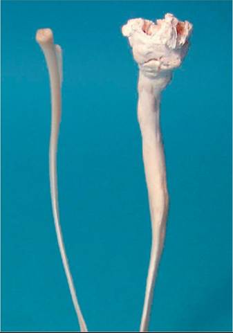
FIG. 35.14 Rostral view of left (right) and right (left) stylohyoid bones from a horse with left-sided temporohyoid osteoarthropathy.
Note fusion of the temporohyoid joint on the affected side and the extensive remodeling and enlargement of the stylohyoid.
been affected; the mean or median age reported in all studies is 10 to 12 years.1,4-8 Quarter Horse and Quarter Horse-type breeds accounted for more than 50% of cases in several but not all series.4-7 Both joints are affected, albeit asymmetrically, in most horses with clinical THO, and some cases are accompanied by associated bilateral clinical signs, including deafness.4-6,9 Fusion of the THJ causes interference with the interdependent coordinated movements of the tongue, hyoid apparatus, and larynx. Particular stress is placed on the fused joint or joints during chewing, swallowing, vocalizing, and combined head and neck movements. Veterinary procedures such as dental work and passage of a nasogastric tube also put unaccustomed pressure on the THJ.
Any of these mechanical forces applied to the immobilized THJ may result in fractures through the petrous temporal bone or proximal stylohyoid bone. THO is also probably a risk factor for fracture of the petrous temporal bone during head trauma, especially in horses that flip over backward and strike the poll. The facial nerve (cranial nerve VII) and vestibulocochlear nerve (VIII) and apparatus are within the petrous temporal bone and can be impinged on by bony proliferation or lacerated by fracture. After fracture of the petrous temporal bone, there may also be extension of middle/inner ear infection around the brainstem with signs of meningitis or extradural abscess.■ Clinical Signs Two sets of clinical signs are associated with the evolution of this disease process.1 Early in the syndrome, including the period before ankylosis of the temporohyoid joint, affected horses may show reluctance to chew, dysphagia, headshaking, ear rubbing, sensitivity to pressure at the base of the ear, facial hyperesthesia, wild tossing of the head and neck under tack, reluctance to take a bit, dropping of feed, or even weight loss; most THOs, however, are probably subclinical until development of neurologic signs. Fracture of the proximal stylohyoid bone increases the likelihood of pain- associated clinical signs. Because of bony proliferation around the ear canal, the external ear may be unusually narrow and difficult to examine by endoscope.
After fusion of the THJ, the disease may manifest as sudden onset of asymmetric signs of cranial nerve VII or VIII dysfunction, or both. Almost all affected horses have degrees of facial paresis, with drooping of the ear, upper eyelid, and lower lip on the affected side and deviation of the muzzle to the contralateral side.1,6,7 Buccinator weakness results in “quidding” (retention of feed material in the denervated cheek) and dropping of feed. Facial denervation may be evident as fine fasciculations of facial muscles and, after 2 weeks, obvious atrophy of the parotidoauricularis muscle behind the vertical ramus of the mandible.
Facial paralysis is occasionally bilateral.5 Schirmer tear tests commonly reveal reduced tear production (i.e., values of 5 to 19 mm; normal, ≥20 mm),5 indicative of injury to the parasympathetic lacrimal (great petrosal) branch of the facial nerve. By the time of presentation, most such horses already have a deep horizontal corneal ulcer with corneal edema and miosis. Because of facial weakness, affected horses cannot mount the typical response to corneal ulceration—namely, blepharospasm and epiphora—and so ulcers often go undetected by horse owners. Signs of vestibular dysfunction accompany facial paresis in the majority of cases. Of 29 horses with THO that had involvement of VII, 23 (almost 80%) had vestibular signs.6 Typically, affected horses exhibit sudden onset of head tilt, neck turn, body lean, and tight circling, all toward the affected side, and a staggering ataxic gait with preservation of limb strength. These signs may be revealed or exacerbated by blindfolding of the horse. In the acute stages, horizontal nystagmus may be present, with the fast phase away from the side of the lesion. In a study of 24 horses with THO and neurologic signs, all had electrodiagnostic evidence of partial to complete hearing loss on the affected side; 12 also had partial hearing loss on the contralateral side.10 Vestibular signs can occur without concurrent involvement of the facial nerve, although this is unusual.10 Of interest is that 12/19 (61%) of horses with THO had concurrent left laryngeal hemiparesis.10 Horses may also have difficulty eating and swallowing because of either pain (e.g., from acute stylohyoid fracture) or involvement of cranial nerves IX and X. A few horses have seizure-like convulsions at the onset of neurologic signs. Bacterial abscess adjacent to the brainstem or meningitis may be sequels to fracture of the petrous temporal bone and result in the death of the horse.■ Diagnosis Endoscopic examination of the inside of the guttural pouches is the diagnostic imaging test of choice because of its convenience in standing horses and its high sensitivity for detection of THO.6 Evaluation of the external contour of the THJ from within the guttural pouch can reveal asymmetric enlargement and discoloration of the stylohyoid bone and THJ and, in some cases, hyperemia, edema, or hemorrhage in the guttural pouch lining surrounding the joint.
Previously healed fractures of the stylohyoid bone may be evident as angulation and distortion of the bone close to the THJ. CT of the THJ allows appreciation of changes not always evident on endoscopy (or plain radiographs), including involvement of both sides, enlargement of the ceratohyoid bone, fractures of the petrous temporal and stylohyoid bones, and the presence of fluid in the tympanic bullae.4,11 A system has been developed to quantify the remodeling changes found in horses with THO.4 The main disadvantage of CT is the expense and requirement for general anesthesia. Where standing CT units are available, this is less of a concern, although adequate positioning can be difficult in horses with severe vestibular dysfunction.8 Radiographic examination of the skull with ventrodorsal, lateral, and lateral oblique views reveals cases in which the bony proliferation has progressed to cause osteitis and fusion of the temporohyoid joint. Optimal images are obtained with the horse under general anesthesia and in dorsal recumbency; however, adequate views can be obtained with the use of a dorsoventral projection while the horse is standing but heavily sedated. Sensitivity for detection of THO by plain radiography in one series was 20/24 (83%).6 MRI and nuclear scintigraphy have also been used successfully for THO diagnosis but have not been widely applied.6,8,12BAER testing of vestibulocochlear nerve function has been described for horses with THO and is the only way of evaluating auditory function in these horses.5 It is also a sensitive method for detecting involvement of the contralateral side.
In horses with neurologic signs other than dysfunction of CNN VII and VIII, CSF tap, analysis, and possible culture are recommended. Two of four horses tested in one report had increased nucleated cell counts or protein concentrations suggestive of septic or nonseptic meningitis.6 Xanthochromia and increased RBC counts are also expected in acute cases.3
■ Treatment and Prognosis Medical and surgical approaches to THO have been described.1,3,13 Conservative medical therapy involves prolonged rest and treatment with an NSAID (e.g., 2 mg/kg phenylbutazone PO bid, or 1.1 mg/kg flunixin IV or PO bid for 3 days, then once daily for 1 to 2 weeks more) and broad-spectrum antibiotics (e.g., trimethoprim-sulfamethoxazole, 30 mg/kg PO twice daily for 30 days).
The lever action of the hyoid apparatus on the petrous temporal bone can be resected surgically by complete removal (ostectomy) of the ipsilateral ceratohyoid bone or removal of a 1- to 2-cm section from the middle of the stylohyoid bone.3,13,14 Because midbody partial stylohyoid ostectomies tend to heal,14 the ceratohyoid procedure is now favored. If necessary, this surgery can be performed bilaterally when the horse is standing and sedated. With facial nerve paralysis and keratitis sicca, corneal ulcers are a medical emergency and must be treated or prevented with topical antibiotics, artificial tears, and, preferably, a multifocal partial split-thickness tarsorrhaphy.3 For this purpose, shallow 3- to 5-mm-long incisions are made in the tensed lid margins of each eyelid immediately posterior to the line of tarsal gland openings. With 5-0 absorbable suture, a bite is taken in each eyelid deep in the incision, in the plane of the eyelid, and then the suture is carefully tied so that margins evert away from the corneal surface.3 One or two shallow horizontal mattress sutures are then placed in the everted lid margin. The eyelids are joined in this manner at two or more sites, leaving open the nasal third of the palpebral fissure. The tarsorrhaphy is maintained permanently or until resumption of lacrimal and eyelid function, which may take up to 18 months.Both medical and surgical approaches reportedly have returned most treated horses to athletic activity after a period of weeks to months, and so evidence to support the need for surgery in all cases is not yet clear.1,6,7 On theoretical grounds, however, it is clear that a horse with THO will remain at risk for catastrophic relapse of neurologic signs as long as the hyoid apparatus and skull remain connected through a fused THJ. Relapse is particularly likely to occur if the temporal bone is fractured. In a series of horses with THO, 13 of 20 (65%), 1 of 25 (4%) and 1 of 8 (12.5%) horses treated by medical therapy, ceratohyoid ostectomy, and partial stylohyoid ostectomy, respectively, died as a consequence of THO.9 In comparison with surgical therapy, medical therapy was significantly associated with nonsurvival.
If possible, the management of convalescent horses should be adjusted to minimize stress on any remaining THJ. Cribbing collars can be tried for crib biting, bitless bridles can be used for riding, and dental work and other professional procedures can be minimized and performed cautiously.