The Lethargic Crab Disease (LCD)
LCD is known solely from the crab species U. cordatus. The great majority of the mortality outbreaks associated with LCD occurred during the summer of the southern hemisphere, but some anecdotal accounts reported mortalities during the winter
W σ>
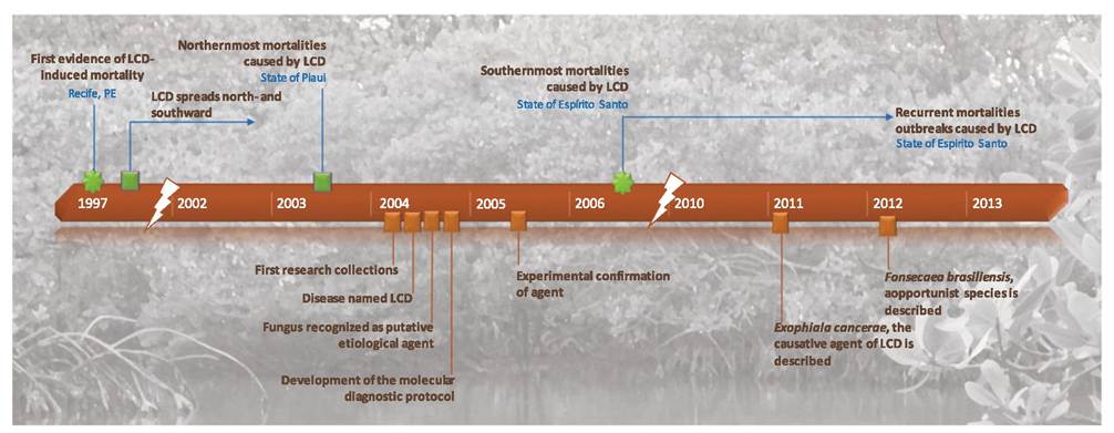
Fig.
11.2 Chronological order of the main facts and pioneering events that contributed to the epidemiological studies on lethargic crab diseaseV. A. Vicente
months as well. The infected crabs become increasingly lethargic and irresponsive as the disease progresses, resulting in the inability to feed or escape from predators and fishermen. In the mangrove, crabs apparently crawl out of their burrows and die outside. When captured, sick crabs die quickly, usually during the transfer from the mangrove and the point of commercialization. Tetany was observed in many moribund animals. These signs strongly suggest injury to the nervous and respiratory system, as revealed in subsequent studies (Boeger et al. 2007; Orelis-Ribeiro et al. 2011). The rapid death of sick crabs, associated with capture and transport, appears to be correlated with their inability to respond adequately to corresponding stress.
These clinical signs are explained by the histopathology of the disease (Boeger et al. 2007). Free or encapsulated (hemocytic encapsulation) yeast-like cells of a fungal agent are abundant in epidermal and connective tissues, gills, intestinal wall, thoracic ganglion, hepatopancreas, hemolymph, and heart of moribund animals showing clinical signs of LCD (Fig. 11.3). Gonads, somatic muscles, and digestive system are less affected by the fungus. In advanced stages of the disease, hyphae may also be present. Necrosis, tissue degeneration, and congestion of hemal sinuses and vessels are present in heavily infected organs (Fig. 11.3). Nerve fibers, especially those associated with the ventral thoracic ganglion, may be compressed by accumulation of yeast-like cells. Cardiac musculature is greatly compromised. In heavy infections, the tissue of gill lamellae, including epithelium and pillar cells, is destroyed with subsequent dilation or compression of the lacunae. Cellular immune responses include hemocytic infiltration, agglutination and encapsulation, and phagocytosis. Phagocytosis of yeast-like cells is abundant in the connective tissue associated with the exoskeleton.
11.3