The traditional approach to parasite control programs has focused on using the appropriate anthelmintic at appropriate intervals.
Parasitic disease in domestic animals was assumed to be the result of not dosing the animals often enough with anthelmintics. Scant consideration has been paid to the interaction of the parasite with the host and the environment because of the reliance on anthelmintics.
These drugs have been placed directly into the hands of the livestock owner because the expertise of a veterinarian did not seem necessary in the control of parasites. However, reports of resistance to anthelmintics and emergence of new manifestations of parasitism are surfacing throughout the world. It has become increasingly apparent over the past 25 years that this approach to parasite control is no longer sustainable.Secondary to the notion that parasitism is under control is the decrease in research to develop new anthelmintics. There has been little pharmacologic development other than in variations on the current anthelmintics. Recently two new anthelmintic classes have been developed but are not commercially available internationally. Research programs in parasitology of domestic animals are facing funding reductions as research priorities shift to other diseases. As producers and owners struggle to deal with the realities of anthelmintic resistance, veterinary medicine must reassess traditional approaches to parasite control programs. Veterinarians need to reeducate themselves away from the traditional tools of deworming, anthelmintic rotation, and pasture rotation. Integrated and holistic management strategies incorporating selective use of anthelmintic agents, enhancement of host immunity to parasitic infection, and grazing and environmental management have become increasingly important in the design of sustainable parasite control programs.
The impact of parasitic infection varies widely with geographic area and management system. General guidelines may be suggested for parasitic control, but it is inadvisable to adhere to any rigid anthelmintic schedules or even management recommendations.
The best parasite control programs are those designed with the goals of the producer in mind, as well as the costs and returns of treatment. Other factors that must be considered include the animal's environment, climatic variations, and geographic location. Although many producers and owners would like a “cookbook” approach to parasite control, these are rarely effective across the various management conditions. It is unfortunate that an epidemiologically and economically sound parasite control program designed for animals in one geographic area may be neither efficient nor effective in another location.The most important concept in the design of sound parasite control programs is the interaction of the parasite with the host and the environment. An understanding of the life cycle and epidemiology will suggest the most effective methods for parasite control. In this chapter, parasite factors, host factors, and environmental factors affecting transmission and disease expression are discussed for each major class of parasites in each host species (horses, cattle, small ruminants). The methods of monitoring parasite infections and anthelmintic resistance are presented in detail. Coccidiosis in cattle and small ruminants is summarized and, finally, the classes of anthelmintics and their modes of action are discussed at the end of the chapter.
Equine Parasitic Disease
Martin K. Nielsen
The changed approaches being promoted for control of equine gastrointestinal helminths are based on the scientific concepts outlined in the following section. The recommendations given today are based on our best knowledge of parasite and host biology as well as mechanisms of anthelmintic resistance. Some of these recommendations are likely to change as new information is generated. In essence, the traditional strategies used for equine parasite control were based on the best scientific evidence available at the time, and the recommendations now given are merely a response to the experiences gathered over the past decades.
The concepts underlying the modern approaches for equine parasite control are presented here.Anthelmintic Resistance
Anthelmintic resistance can be defined as “the loss of treatment efficacy of a given anthelmintic formulation that used to have efficacy against the same parasite species and stage in the same host animal, at the same dosage, and by the same route of administration.” From this sentence, we can learn several things: (1) If a drug formulation never had efficacy against the parasite and stage in question, we are not facing resistance. (2) In cases of nonlabeled use of anthelmintics, we cannot make any conclusions regarding resistance because we have no information about the expected level of efficacy. (3) Horses are always infected with several different species of parasites at the same time, and resistance often occurs in some but not all of these species. (4) The expected level of efficacy needs to be defined for each parasite species for each drug in order to evaluate the possible presence of resistant parasites.
■ TABLE 49.1
Current Levels of Resistance by Major Nematode Parasites to the Three Anthelmintic Classes in Managed Horse Herds
| Drug Class | Cyathostomins | Large Strongyles | Parascaris spp. |
| Benzimidazoles | Widespread | None | Early indications |
| Pyrimidines | Common | None | Early indications |
| Macrocyclic lactones | Early indications | None | Widespread |
The current levels of anthelmintic resistance in equine parasites are presented in Table 49.1. Resistance has been widely documented in cyathostomins (small strongyles) and Parascaris spp. It is worth noticing that as a general pattern, the resistance profiles are almost completely complementary between the two parasite categories.
The drugs that seem to still work well against Parascaris spp. often have no or little efficacy against cyathostomins, and vice versa. However, levels of anthelmintic resistance are constantly developing, and this pattern therefore may very well be shifting. As described in the following section on egg reappearance periods, signs of emerging ivermectin and moxidectin resistance have been reported in cyathos- tomins,1-4 and total drug failure is a realistic possibility in this parasite group. No new anthelmintic formulations with new modes of action are expected to be introduced to the equine market in the foreseeable future.The only available method for evaluating anthelmintic efficacy in horses is the fecal egg count reduction test (FECRT). This method is outlined in Box 49.1 along with some suggested cutoff values for determining anthelmintic resistance.
Egg Reappearance Period
Egg reappearance periods (ERPs) are defined as the period of time from anthelmintic treatment until parasite eggs are found in the feces again. Although ERPs could potentially be established for a number of different parasite categories, they are most often referred to for strongyle (i.e., cyathostomin) parasites. The ERPs were initially defined for each anthelmintic class to help identify the optimum interval between anthelmintic treatments, and this was the basis for launching the so-called interval dose program in the 1960s.5
Nowadays, ERPs are used for an entirely different purpose. It has been observed that the first sign of developing anthelmintic resistance to a given drug is often a gradual shortening of the ERP until reduced efficacies can finally be found with the FECRT. This concept is highly relevant for monitoring the efficacy of ivermectin and moxidectin against cyathostomin parasites. Initially ERPs were reported to be at least 9 and 16 weeks for ivermectin and moxidectin, respectively,6-8 but recent publications reported egg reappearance at around week 4 to 5 for both drugs.1,4 Necropsy-based studies have suggested that these observations were due to survival of luminal yet premature stages of cyathostomin larvae.2,3
■ BOX 49.1
Fecal Egg Count Reduction Test (FECRT)
The FECRT evaluates the efficacy of an anthelmintic drug based on its ability to reduce the fecal egg output after treatment.
Fecal egg counts (FECs) are performed just before (or at the time of) and 14 days after treatment. The fecal egg count reduction (FECR) is calculated for each individual horse according to the following formula:
It is recommended to use an egg counting technique with a detection limit of less than 25 eggs per gram (EPG). Always use the same technique consistently. Include horses with the highest possible pretreatment egg counts, and never use horses with counts below 200 EPG.
The FECRT should be established on the farm level by calculating the FECR for a number of individual horses and then calculating the average FECR for the treated group. It is recommended to include at least 5 to 10 horses on each farm if possible.
Suggested cutoff values for resistance depend on the drug tested and the number of horses investigated, but the following cutoff values are recommended as general guidelines for strongyle and ascarid nematodes:

If the farm average FECR falls below these values, anthelmintic resistance should be suspected. However, it is important to rule out other causes of decreased efficacy, such as misdosing, inadequate storage, etc. One must also consider how many horses were tested and how high the starting FECs were. Due to inherent variability in the measurement of FEC when performing FECRT, interpretation of the data can sometimes be difficult when results fall into the borderline zones. In such cases, it is recommended to repeat the FECRT.
There are currently no available methods for diagnosis of anthelmintic resistance in equine tapeworms.
Prepatent Period
Prepatent periods (PPPs) should not be confused with the egg reappearance periods defined above. Although the latter defines a response to anthelmintic treatment, PPPs are defined in the absence of anthelmintic intervention.
The PPP for any given parasite is defined as the time elapsed from the uptake of the infective stage of the parasite until it reaches patency and starts shedding eggs to be detected in the feces. Thus the PPP represents the duration of the parasitic part of the life cycle. Table 49.2 presents PPPs for some of the most important equine helminth parasites. It should be noted that although the PPPs for cyathostomin parasites generally are relatively short, they can undergo arrested development for up to several years, which thereby dramatically lengthens the PPP.9Information about PPPs can be considered when identifying optimal times for routine collection of fecal samples and anthelmintic intervention.
Parasite Distributions
Regardless of parasite and host species, distributions of parasites among their hosts always seem to follow the same pattern. Hosts of similar age and breed and kept under identical management grazing in the same pasture can have widely different parasite
■ TABLE 49.2
Prepatent Periods of Some Important Equine Parasites
| Species | Prepatent Period | Reference |
| 2-3 monthsa | 113 | |
| Parascaris equorum | iy -3 months | 114 |
| Anoplocephala perfoliata | ∖yι -4 months | 115 |
| Strongylus edentatus | 11-12 months | 116 |
| Strongylus equinus | 8 months | 117 |
| Strongylus vulgaris | 6-7 months | 116 |
aEncysted larval stages can remain dormant for years.
burdens. Parasites never follow a symmetric normal distribution but are rather said to be overdispersed.10 Technically speaking, this means that the variance is larger than the mean, but in practical terms this has been translated to the so-called 20/80 rule.11 This refers to the observation that about 20% of the host animals harbor approximately 80% of the parasites. Similarly, about 20% of horses are shedding 80% of the total strongyle egg output. This phenomenon is most pronounced in adult horses, where a large majority is shedding low numbers of strongyle eggs and very few individuals can be observed with very high counts. This pattern has been found to be consistent in individual horses over time, and the tendency is particularly strong among horses with zero or low strongyle egg counts.1214 Taken together, these observations have become the foundation for the selective therapy principle discussed later in this chapter.
Parasite Refugia
The term parasite refugia has become generally accepted as an important factor affecting the rate of development of anthelmintic resistance in large animal parasites.15 Parasites in refugia are the parts of any given parasite population (including all stages, both internal and external) that are not exposed to the anthelmintic drug at the time of treatment. It can be said that these parasite stages in a way “escaped” the drug and can be viewed as refugees, hence the terminology. Logically, all stages on pasture, such as eggs and preinfective larvae, are always in refugia. In addition, animals left untreated are contributing significantly to the refugia, and some parasitologists therefore distinguish between “pasture refugia” and “animal refugia.” Finally, some anthelmintic formulations have no efficacy against some parasite stages within the horse, and these can be regarded as part of the refugia as well. Examples of this include pyrantel formulations that have no efficacy against parasite stages present outside the gastrointestinal lumen and ivermectin, which has no documented efficacy against encysted cyathostomin larvae.
The parasites in refugia are hypothesized to play a role in diluting out resistant parasites whenever the animals are dewormed. A simplistic explanation of this concept is the larger the refugia, the more the dilution of resistant parasites, and the slower the development of resistance. In a theoretical example, let us consider a horse herd that is dewormed with a drug that has efficacy against all parasitic stages of the parasites, and the treatments are carried out at time of the year when no eggs or larvae are present on pasture. This would represent a scenario with no parasite refugia. Because no anthelmintic treatment is 100% efficacious, there will always be a few parasites surviving and passing their genes to the next generation. In a situation with low or even no refugia, all eggs being passed in the feces after deworming will be progeny of resistant parasites mating in the intestinal tract. However, if some of the horses in the herd were left untreated or treatments were carried out under circumstances with eggs and larvae present on pasture, then the few resistant worms surviving treatment would be diluted out by the many nonresistant worms, eggs, and larvae, and the progression of resistance in the parasite population would be much slower.
The role of parasite refugia in the development of anthelmintic resistance was initially shown in a sheep study,16 and recent work with a combination of computer simulations and field studies with ruminants has provided solid evidence behind the concept.17-19 Most recently, computer simulation models have been established for Parascaris spp. and cyathostomin parasites,20-21 and studies confirm the value of parasite refugia in these parasites.
Important Parasites Infecting Horses
Over the past five decades the emphasis has shifted several times in terms of which parasites are considered the major targets of the control programs. When the first modern paste dewormers were introduced in the 1960s, the large strongyle Strongylus vulgaris was considered the most important threat to equine health and was therefore identified as the main target for the interval dose program.5 As time went on and new anthelmintic drug classes were introduced, this parasite went from being present in virtually all horses to becoming very rare.22-23 At the same time, cyathostomins were recognized as major pathogens and identified as the new primary target of parasite control programs.24 More recently, the tapeworm Anoplocepbala perfoliata has been associated with an increased risk of specific colic types.25 In addition, Parascaris spp. are identified as the major parasitic pathogens in foals.26-27 These are the four major parasite pathogens, but others can play a role as well. Less important but widely occurring parasites include Strongyloides westeri, Oxyuris equi, and the bot larvae of Gasterophilus spp. Finally, the insect-borne nematodes Thelazia spp., Habronema spp., and Setaria spp. can occur at significant levels in certain habitats and are included in this chapter.
Parascaris Species
Recent work has recognized the presence of two Parascaris spp. infecting horses: Parascaris equorum and Parascaris univalens. These two species are morphologically identical but can be differentiated by karyotyping and counting the number of chromosomes. Available data suggest that P univalens is the predominant species, while P. equorum may be extremely rare.28 The equine roundworm has gained status as perhaps the most significant parasitic threat to equine health in managed horse populations. The major reason for this shift of emphasis is the high level of ivermectin and moxidectin resistance observed worldwide (see Table 49.1). Parascaris spp. are the primary parasites in foals until about the time of weaning. Between 6 and 12 months of age, most horses clear infection with this parasite, and the strongyles become the predominating parasite group instead. However, a smaller second wave of ascarid infections can be observed around 8 to 10 months of age.29 Equine ascarids are ubiquitous in foaling operations, although prevalence differences can be observed between individual farms.30-31 Patent infection is not uncommon in adult horses, particularly brood mares that are highly exposed to infection.
The life cycle is classic ascarid. Eggs are often claimed to be highly resilient to environmental influences, but very little evidence exists to substantiate these claims. Work with eggs of the pig roundworm, Ascaris suum, has substantiated this, but very little work has been done with P equorum eggs. The infective stage is the embryonated egg, which hatches in the stomach and small intestine of the horse. Larvae then penetrate the mucosal lining and migrate the classical hepatotracheal route. Passing through the lungs has been associated with airway symptoms in young foals,32 but the major pathogenic role is played in the small intestine. Here, the parasites compete with their hosts for nutrients, which can result in ill-thrift, weight loss, rough hair coat, and pot-bellied appearance.26,32 The major clinical impact is associated with verminous small intestinal impactions.26 Surgery is often required to relieve the condition, and prognosis for long-term survival is guarded with only 11 of 37 published cases surviving beyond 1 year after the colic incident.26 Rather ironically, anthelmintic treatment has been shown to be a significant risk factor for this condition.26 The instant paralysis elicited by most dewormers can cause the worms to aggregate in the lower parts of the jejunum and ileum. Benzimidazole-type drugs are not acting through paralysis of the worms and appear to be less associated with this condition.26 Given that benzimidazoles have maintained efficacy against roundworms, they appear to be a good first choice for this parasite in most instances.
Cyathostomins
Historically, this group is also referred to as small strongyles. It consists of 50 different species belonging to 14 different genera.33 However, some of these species and genera have been described as infecting equids other than horses, such as donkeys and zebras. Horses are always infected by multiple species, and there currently is limited knowledge about the pathogenic role of individual cyathostomin species. Cyathostomins are truly ubiquitous, and all grazing horses are exposed to infection.
The cyathostomins all follow the same basic strongyle life cycle. Strongyle eggs are passed in the feces, where they will hatch, and larvae develop through the first (L1) and second (L2) stages until they finally reach the third, infective stage (L3). Hatching and subsequent larval development is highly temperature-dependent. At temperatures below 6° C, eggs do not hatch but remain viable. Hatching and larval development occurs at increasing rates up to about 40° C. At temperatures above this level, eggs and larvae quickly die out. Optimal conditions for development of eggs and larvae are in the temperature range of 25° to 33° C, where L3 is reached within 3 to 4 days. In comparison, this takes several weeks at around 10° C. When molting from L2 to L3, the larvae retain their L2 cuticle on the outside of the new sheath. This makes L3 particularly resistant to environmental influences such as desiccation and freezing. Thus, for this stage, the widespread notion of the “killing frost” is a myth. In addition, intact fecal balls provide excellent protection of larvae, enabling them to further withstand desiccation and freezing temperatures well.34 Due to the double-layered cuticle, L3 cannot uptake feed and thus live on carbohydrates and lipids stored in their cells. The more active these larvae are, the shorter they live. As larval activity is highly temperature dependent, it can be deduced that the higher the temperature, the shorter the survival. Dry conditions, as achieved with freezing or desiccation, limit larval movement and therefore facilitate their survival. However, repeated cycles of frost and thaw have a deleterious effect on most free-living stages of strongyle, although this can be mitigated by snow cover, which tends to stabilize temperatures right at the freezing point, which is optimal for survival of eggs and L3.34
Inside the horse, the L3 exsheath in the stomach and subsequently reach the colon and cecum. Here, they enter the glands of Lieberkuhn and penetrate cells at the base. From here, some species appear to penetrate deeper into the submucosa, while others remain in the mucosa. A fibrous capsule is formed around each larva, and then a fluid-filled cyst is formed. Larvae can remain encysted for several years,9 but encystment appears to be a strategy for the parasites to make it through the winter, where conditions on pasture are unfavorable for parasite transmission. Interestingly, in warmer climates, encystment appears to occur over the hot summers. Horses can harbor several hundred thousand encysted cyathostomin larvae without showing any sign of discomfort. The major clinical consequences are associated with the process of excystment, where the L4 penetrate their cysts and migrate the short distance back to the intestinal lumen. This causes a small local inflammatory reaction around the vacated cyst. In rare cases, synchronous emergence of large numbers of larvae from their cysts can cause a severe generalized acute typhlocolitis, called larval cyathostominosis.24 This condition is characterized by profuse watery diarrhea and pronounced dehydration as well as protein loss and ventral edema. The case fatality rate is reported to be 50%.24 Identified risk factors for larval cyathostominosis include horses aged 1 to 4 years, anthelmintic treatment within 14 days prior, and late fall, winter, or early spring in temperate climates.35 The luminal worm burden appears to elicit some sort of a repressive signal, which prevents the encysted stages from resuming development. If the luminal burden is removed by anthelmintic treatment, this repressive signal disappears abruptly, and a synchronous emergence is triggered. Treatment of acute larval cyathostominosis involves intensive fluid therapy, which can be combined anti-inflammatory drugs and antibiotics. The anthelmintic drug of choice is a single dose of moxidectin (400 μg∕kg PO) because of its efficacy against encysted larvae. The 5-day elevated dose of fenbendazole (10 mg∕kg PO once daily) is also labeled for treatment of encysted cyathostomins, but larvicidal resistance has recently been reported to this regimen, making it unlikely to have maintained full efficacy once adulticide resistance has been documented to the single dose.36
Chronic cases of larval cyathostominosis also occur. These are typically characterized by weight loss and loose feces or intermittent diarrhea, often over an extended period. Plasma protein and albumin levels can be lowered, and ventral abdominal edema can be observed. Horses usually recover, although it may take time for them to regain full body condition.
Tapeworms
Three species of tapeworm infect horses: A. perfoliata, Anop- Iocephala magna, and Anoplocephaloides (Paranoplocephala) mamil- lana. Of these, A. perfoliata is by far the most common, and it is the only species that has been associated with clinical disease. Being a cestode, the life cycle involves intermediate hosts, which are oribatid mites that feed on the organic material present in the feces. During this process, the mites can ingest tapeworm eggs present in the feces. Inside the mite, eggs develop to infective cysticercoids over a period of approximately 2 to 4 months. Horses inadvertently ingest these mites during grazing. In the gastrointestinal tract, the cysticercoids are liberated from the mites, and the scolexes attach to the mucosal lining around the ileocecal valve in the cecum.
Prevalence of A. perfoliata has been reported to vary widely between farms and regions, often ranging between 20% and 80% of horses.25 Prevalence appears to be habitat-dependent, and it is likely associated with conditions favoring survival of the oribatid mites. However, the most significant factor appears to be access to pasture. The longer the time spent on pasture, the higher the tapeworm exposure seems to become.37 Horses younger than 2 years and older than 15 years of age have been reported to have significantly larger Anoplocephala burdens than horses between these ages.38-39 While this could be explained by the assumed incomplete levels of immunity in these age groups, it is equally likely that these age groups simply spend more time on pasture. Tapeworm burdens appear to be accumulating through the grazing season to reach their highest point in the fall and winter.38-43
An increasing number of case-control studies has associated A. perfoliata infection with colics originating from the ileal region.25 These include ileal impactions and ileocecal intussusceptions, which are sometimes complicated by intestinal rupture.44-45 Taken together, these studies indicate that tapeworms can play a role in the development of colic, but this is often largely confounded by the multifactorial nature of colics and regional differences in climate and management.
Two anthelmintics are labeled for treatment of equine tapeworms: praziquantel and pyrantel pamoate. Praziquantel is a sole tapeworm drug that has no efficacy against nematodes. For this reason, it is most often formulated in combination with ivermectin or moxidectin, although it exists in a standalone formulation for horses in Europe. Pyrantel, on the other hand, is widely acting against luminal stages of nematodes as well as cestodes. At the dosage labeled for nematode treatment (6.6 mg/ kg), the efficacy of pyrantel pamoate is greater than 80% against A. perfoliata.4 A double dosage of pyrantel pamoate (13.2 mg/ kg) is a substitute for tapeworm treatment, and the efficacy of this has been found to be greater than 95%.47
Strongylins (Large Strongyles)
The strongylin group comprises the three Strongylus species—S. vulgaris, Strongylus edentatus, and Strongylus equinus—as well as Triodontophorus spp., Craterostomum acuticaudatum, and Oesophagodontus robustus. As mentioned, large strongyles have generally become quite rare in managed equine establishments. The most common of these nowadays is probably Triodontophorus spp., but the Strongylus species, particularly S. vulgaris, are considered the most pathogenic of helminths infecting the horse.
The development of the external stages of the strongylins is virtually identical to what is already described for the cya- thostomins. Inside the horse, larval migrations are widely different. The three Strongylus species all have PPPs of 6 months or more, and they spend several months migrating in various tissues and organs of the horse. Major pathologic lesions have been ascribed to these migrations, whereas the adult worms are found to do little harm to their hosts. The lesions caused by migrating stages of S. vulgaris are classic in equine parasitology and very well described.48 L3 invade the mucosa of the small intestine, cecum, and colon. They then molt to L4 before they enter local arterioles. Inside these, they migrate beneath the endothelium toward the root of the cranial mesenteric artery, leaving characteristic fibrous tracts on the intimal surface. Upon reaching their destination about 14 days post infection, they enter the lumen of the vessel and remain there, embedded in thrombus masses. Here, they grow in size and molt to L5, while causing a pronounced verminous endarteritis with roughened intima, fibrosis of arterial walls, and increased diameter of the vessels. After about 4 months, the L5 are transported by the bloodstream to the walls of the ventral colon and cecum, where pus-filled nodules are formed around them in the submucosa. These nodules eventually open into the intestinal lumen, and the young adult worms emerge. After another 6 weeks, the worms become sexually mature and start shedding eggs. Although the lesions are pronounced, they are relatively rarely accompanied by disease. A classical syndrome of thromboembolic colic has been described,49 where thrombi detach from the arteritis lesions and are carried distally until they reach a terminal branch sufficiently small to become occluded. This causes ischemia and infarction of intestinal segments, which is very painful to the horse. However, the primary manifestation in many cases appears to be peritonitis due to bacterial overgrowth of the devitalized intestinal segment, and the prognosis for survival is reserved.50
Larvae of S. edentatus migrate via the portal system to the liver, where they molt to the fourth stage within the parenchyma. They migrate within the liver, and then migrate sub- and retroperitoneally via the hepatorenal ligament to the adipose tissue in the abdominal walls. Larvae can also be found in the perirenal fat tissue. Here, larvae eventually molt to L5 and migrate retroperitoneally back to the intestinal walls. Described lesions include a filamentous peritonitis, especially around the liver and diaphragm, as well as hemorrhagic and inflammatory lesions in the abdominal wall.51 The third Strongylus species, S. equinus, is probably the rarest, at least in managed horses on the northern hemisphere, where it is hardly encountered at all. As opposed to S. edentatus, it migrates within the peritoneal cavity and passes through the pancreas, where it can cause significant damage.52 Despite their obvious pathologic impact, neither S. edentatus nor S. equinus have been associated with defined clinical syndromes.
None of the strongylins have been reported as resistant to any of the anthelmintic drug formulations available. Treatment efficacies against S. edentatus appear to be more variable than in the other species,53 but this seems to always have been the case and thus cannot be concluded to be resistance.
Other Parasites
Other parasites frequently infect equines but are generally considered of lower importance. For completion, they are briefly covered here.
S. westeri, or the equine threadworm, parasitizes the small intestine of suckling foals. This parasite is remarkable because it is able to maintain a full life cycle outside its host, and only female worms seem to infect the foals. Infection occurs via three possible routes: skin penetration by L3, ingestion of these larvae from the environment, or lactogenic transmission from the mare.54 Infection usually occurs within the first weeks of life, and infected foals are usually asymptomatic. However, one study reported an association between diarrhea and high Strongyloides egg counts (>2000 eggs per gram [EPG]),55 and another study described a “frenzy” syndrome in foals exposed to apparent percutaneous penetration of L3 from the environment.56 Because of S. westeri, it has become a widely used practice to deworm mares at or just before foaling. However, it remains unclear how effective this measure is, as S. westeri prevalence has been observed to increase in managed equine 57
populations in recent years.
O. equi, the equine pinworm, is widely prevalent, but eggs are rarely encountered in fecal samples, as they are laid outside the digestive tract. Adult worms live in the dorsal colon. The female worm migrates through the descending colon and rectum to protrude from the anus and deposit her eggs in patches on the perianal skin. When these egg patches dry up, they become very itchy to the horse, and horses begin rubbing their tails against various objects. This serves as an excellent means to spread eggs in the environment. The infective stage is the embryonated egg, which is inadvertently ingested from the contaminated environment. Besides the tail-rubbing, O. equi does not cause disease or discomfort to the horse, and there is no justification for routinely targeting this parasite in the parasite control program. Recent studies have reported 5859 apparent resistance to ivermectin in isolates of O. equi.58,59
Gasterophilus spp. are the widely occurring botflies, whose larval stages overwinter in the intestinal tract and then pass in the feces during the spring. They then pupate in the loose soil for a couple of months until the adult flies emerge. The two most common species are Gasterophilus intestinalis and Gasterophilus nasalis. Eggs are glued onto the hair coat, and larvae make their way into the oral cavity either by crawling on the skin (G. nasalis) or when the horse grooms itself or another herd member (G. intestinalis). In the mouth, the larvae spend several weeks burrowing into the tongue and subsequently spend time in the interdental spaces before reaching the stomach.60 In the stomach, G. intestinalis attaches to the mucosal lining by the margo plicatus, whereas G. nasalis can be found in the pyloric region. Characteristic lesions have been described at the attachment site,61 but these have not been consistently associated with disease. With the emergence of equine dentistry over the past decades, the lesions in the oral cavity are gaining more attention, as Gasterophilus spp. can cause oral discomfort.62
The equine eye worm, Thelazia lacrymalis, is transmitted by muscid flies and can be found within the conjunctival sac of horses, where they generally appear to do very little or no harm to the eye. However, abscess formation and inflammation of the lacrimal glands have been described.63 Prevalence rates in the range of 20% to 42% have been reported in managed horses.64,65
The group of stomach worms comprises Habronema spp., Draschia spp., and Trichostrongylus axei. Habronema and Draschia spp. have very similar life cycles. Like Thelazia spp., they are both transmitted by muscid flies. The flies acquire the infective larvae while feeding on the feces. Horses get infected by accidentally ingesting infected flies. Here, the larvae eventually reach the stomach, where Habronema larvae apparently cause no gross lesions, but adult specimens of Draschia spp. can be found embedded in large tumor-like fibrous masses around the margo plicatus.66 Common to both of these species is that the larvae can be deposited by the flies in or near wounds or mucocutaneous junctions. For the parasite, this represents a dead-end pathway, from which the life cycle cannot be com- pleted.67,68 When larvae are deposited in this manner, they cause a condition usually referred to as cutaneous habronemiasis or draschiasis, or “summer sores.” This is characterized by persistent, eosinophilic granulomatous lesions with eosinophilia and fibrosis. A rare, pulmonary form of habronemiasis has been described as well.69 Anthelmintic resistance has not yet been reported in Habronema or Draschia spp., but several veterinary practitioners from subtropical and tropical regions report that skin lesions consistent with cutaneous habronemiasis do not resolve upon treatment with a macrocyclic lactone (ivermectin or moxidectin). The prevalence of Draschia spp. seems to have decreased dramatically after the introduction of ivermectin.70
T. axei possesses the rare capacity of infecting both ruminants and monogastrics such as pigs and horses. The external part of the life cycle is similar to that of the strongyle parasites described in this chapter. Inside the horse, it develops within the gastric glands, where it can cause hypertrophy of the mucosa.71 In horses, T. axei has not been found to affect pH or levels of plasma pepsinogen.72 The parasite is a rare finding in horses, but prevalence has been found to increase when horses are co-grazed with sheep.'3,'4
Setaria and Onchocerca spp. are filarial nematodes infecting horses. Infective L3 are transmitted by blood-sucking arthropods feeding on the horses. The larvae then migrate to their predilection site, which is the connective tissues (Onchocerca spp.) or the abdominal cavity (Setaria equina). Here, they develop into adult worms, which then release microfilariae intro the bloodstream. These microfilariae in turn will enter the blood-feeding arthropods, and the cycle is complete. The intermediate hosts for Onchocerca spp. are biting midges (Culicoides spp.) and black flies (Simulium spp.), whereas the hosts for S. equina are mosquitoes. The latter parasite is regarded as nonpathogenic, whereas Onchocerca spp. have been associated with pronounced skin reactions. Microfilariae of Onchocerca cervicalis tend to congregate in certain regions of the body, including the ventral midline and face, where they are ingested when Culicoides feed in these regions.75 This results in a verminous dermatitis characterized by intense pruritus.76 Lesions can be effectively treated with macrocyclic lactones.77 However, there is no known therapy to eliminate adult O. cervicalis from the connective tissue around the ligamentum nuchae of horses. Therefore infected horses will need periodic retreatment with ivermectin or moxidectin to prevent or control recurrence of clinical signs.78,79 Treatment of the skin-dwelling microfilariae has been associated with an inflammatory reaction to the dying parasites,80 which can be confused with the widely occurring Culicoides hypersensitivity (summer eczema, sweet itch).
Adult Parafilaria multipapillosa is another filarial nematode that occurs in subcutaneous and intermuscular connective tissue of horses outside North America. Nodules form in the overlying skin and may rupture and bleed or leak tissue fluids. This condition is often referred to as “summer bleeding,”81 not to be confused with the “summer sores” described with cutaneous habronemiasis or draschiasis. L1 are present in the exudate from bleeding lesions and ingested by feeding horn flies (Haematobia irritans). Larvae develop to the infective third stage within the fly and are transferred to horses when flies feed on lachrymal secretions or skin wounds. The larvae then migrate in the subcutaneous tissues and develop to the adult stage within a year. Eggs and microfilariae can readily be identified in smears taken from lesion exudates.82 Little is known about anthelmintic efficacy, but lesions have been observed to recur after treatment with macrocyclic lactones.
The equine lungworm, Dictyocaulus arnfieldi, is covered in the Lungworm Infection in Large Animals section later in this chapter.
Halicephalobus gingivalis (formerly Micronema deletrix) is a free-living nematode belonging to the order Rhabditida. It is widely present in the soil and humus but possesses a capacity to also invade living tissues. Infection appears to occur through soil-contaminated wounds or nasal mucosal membranes with apparent predilection in the head region,83-86 but penile infection has also been described.87 Inside the host, the nematode reproduces tremendously, and the larvae invade deeper into the tissues. Larvae appear to have a special affinity for the central nervous system (CNS) and the kidneys.85 Lesions are multifocal, eosinophilic, and pyogranulomatous. Once the CNS is affected, symptoms often slowly progress into a very grave state. Symptoms include blindness, ataxia, loss of 858889 proprioception, head pressing, coma, recumbency, and death.85,88,89 Treatment options are very limited. Surgical debulking can be combined with repeated high doses of fenbendazole (50 mg/kg) or ivermectin (0.55 mg/kg),90 but the overwhelming majority of published cases had a fatal outcome. Similarly, Halicephalobus spp. infection has been reported to be extremely rare but fatal in humans.91-93
Diagnostics
The cornerstone of diagnostics in equine parasitology remains the fecal egg counting methods. Before these are discussed, it should be emphasized that egg counts can be performed for at least three purposes: (1) evaluation of anthelmintic efficacy with the FECRT (see Box 49.1); (2) identification of low, medium, and high strongyle egg shedders for a targeted treatment approach; and (3) diagnosis of parasite infection in clinical cases. When discussing the usefulness of fecal egg counts, it is important to note that this largely depends on the purpose. In other words, fecal egg counts are not equally useful for all three purposes.
Numerous techniques exist with various names such as Stoll,94 Wisconsin,95 or McMaster,96 but they are all based on the same simple principle. A flotation medium is usually constructed with some sugar or chemical salt solution to have a specific density higher than most parasite eggs but lower than the fecal matter. Examples of components used in flotation media include glucose, sucrose, sodium nitrate (NaNO3), sodium chloride (NaCl), magnesium sulfate (MgSO4), and mercury chloride (HgCl). Very frequently used media are saturated glucose-NaCl and Sheather’s sugar solution, which are relatively cheap and easy to compose. The numerous different egg counting techniques and the even more modifications of these can appear confusing, but in reality there are only three factors to consider when the choice of method has to be made: (1) the level of accuracy and precision associated with the technique, (2) the detection limit of the technique, and (3) the equipment requirements and time consumption in the laboratory.
The accuracy of a given technique is a measure of how close the determined egg counts are to the true egg count in a given sample. All of the different available techniques are associated with a certain degree of egg loss in the process of mixing, filtering, and flotation, so the eggs counted are only a subset of the true number of eggs per gram of feces. Similarly, precision is a measure of repeatability between repeated counts from the same sample. As a general rule of thumb, the simpler and perhaps more user-friendly methods come with lower accuracy and precision, whereas the more refined techniques that use centrifugation-enhanced flotation and larger counting chambers usually have higher precision and accuracy. The McMaster technique, for example, comes with low to moderate accuracy and precision,97 whereas the FLOTAC system is very accurate and precise.98 However, the FLOTAC is much more time consuming and includes a centrifugation step. A recently developed modification of the FLOTAC principle, the Mini- FLOTAC, is relatively user friendly as it does not involve centrifugation and has been found to have better precision and accuracy than the McMaster method.97 These features are important when interpreting the FECRT. Low precision allows for high levels of variability in the FECRT. This is the reason why the FECRT always must be performed by averaging values across several horses on each farm.
The choice of detection limit will depend on the purpose of the egg count. If FECRTs are performed, it is crucial to be able to detect low egg counts in the posttreatment samples to possibly discover early signs of reduced anthelmintic efficacy. A crude McMaster, which is performed by many practice laboratories, usually comes with a detection limit of 25 or 50 EPG, which makes it very unsuitable for the FECRT, unless the pretreatment levels are very high. The FLOTAC technique mentioned above comes with a detection limit of 1 EPG. While this can also be achieved by a more traditional technique such as the Wisconsin, FLOTAC has been shown to have much higher precision.98 For identification of high strongyle shedders for a targeted treatment approach, the detection limit is not important, and McMaster methods can be used.
Until recently, there were no published studies evaluating egg counts as a diagnostic test for horses. From the database generated over 50 years of equine parasitology research at the University of Kentucky, fecal egg counts were related to total intestinal worm counts from close to 700 horses.99 Results indicated a high positive predictive value, indicating that an egg count-positive horse is very likely to also harbor worms in the intestine. However, the negative predictive value was low, emphasizing that 0 EPG does not necessarily indicate zero worms. Further, there were no direct linear correlations between strongyle and ascarid egg counts and the corresponding intestinal worm burdens. Thus higher egg counts do not necessarily equate to larger worm burdens. However, strongyle egg counts in the range of 100 to 500 EPG did correspond to significantly smaller strongyle burdens than egg counts above this level.99 Taken together, this information illustrates that fecal egg counts can be used to qualitatively detect presence of strongyle or ascarid burdens, but that there are considerable limitations to these techniques as quantitative diagnostic tools. Furthermore, it should be borne in mind that much of the parasitic disease observed in horses is caused by migrating or encysted larvae that are not shedding any eggs. Detecting eggs in the feces cannot be used to estimate the probable presence of larval burdens. Also, strongyle eggs are practically ubiquitous in equine fecal samples, and their mere presence cannot be used to suggest parasitic disease. In foals and weanlings, egg counts can be very useful for determining whether parasite burdens are dominated by strongyles or ascarids, which is crucial information for selecting the right anthelmintic (see Table 49.1).
Another weakness of strongyle fecal egg counts is that up to 50 strongyle species have been described infecting horses,33 and it is not possible to morphologically distinguish between their eggs. Larval cultures can be performed to differentiate large and small strongyles and for species-specific detection of the three Strongylus species.100,101 In a retrospective validation, larval cultures were found to have high positive and moderate negative predictive values with no direct linear correlation between larval counts and worm counts.99 Thus larval cultures should be interpreted with the same caution as egg counts. A pragmatic approach has been to pool feces from several horses into one pool, but this cannot be recommended, as diagnostic sensitivity is greatly lost with this approach and the negative predictive values are likely to be much lower.
Diagnosing tapeworm infection is a particular challenge despite the availability of several different techniques. Anop- locephalid eggs may show up on routine egg counts from time to time, but validation studies have shown that the diagnostic sensitivity of the simple McMaster is less than 10%.25 The reason for this is believed to be the uneven distribution of tapeworm eggs in the feces. Eggs are released in clumps often retained within a tapeworm segment, which then disintegrates on its way through the cecum and colon. The eggs remain in their clumps, and egg counting methods examining just a few grams of feces can easily miss detecting these eggs. A modified egg counting technique that analyzes 40 g of feces has been found to have a diagnostic sensitivity and specificity of 0.61 and 0.98, respectively.102 However, this was based on detection of cestode burdens comprising only a single worm. If the threshold were adjusted to detect 20 or more tapeworms, the sensitivity was improved to about 90%.25 Thus the tapeworm infections most likely to not be detected with this method are small burdens unlikely to cause clinical disease. Several enzyme- linked immunosorbent assays (ELISAs) have been made commercially available for detecting anti-Anoplocepbala antibodies.25 These assays have been found useful for evaluating the level of tapeworm exposure on the herd level. Optical density (OD) values correlate positively with worm burdens, and the assays can be helpful for generating information to help balance the level of tapeworm-directed treatments on each farm. It should be emphasized that serology is generally less useful for diagnosis on the individual level. Antibody levels are too variable, horses remain titer-positive for months after tapeworm treatment,103 and one recent study suggests cross-reactivity with the less common A. magna.m In the United States, the diagnostic laboratory at the University of Tennessee, College of Veterinary Medicine, offers a modified version of the Anoplocepbala ELISA, but no information is available on the performance of this test. There are currently no available methods for diagnosis of anthelmintic resistance in equine tapeworms.
Integrated Parasite Control
It is often said that a “one size fits all” parasite control program does not exist. There is no simple chart to hang on the wall in the horse barn, and follow the same guidelines for all of the horses. To the frustration of many horse owners, farm managers, and veterinary practitioners, it is much less straightforward to identify what to do. This section outlines the elements to incorporate in a modern evidence-based equine parasite control program.
The most important evidence to incorporate on each farm is the efficacy of the anthelmintic drugs used. Although Table 49.1 can be used to predict the drugs more likely to still have efficacy on a particular farm, the only way to really know is to evaluate the efficacy with the FECRT, as described in Box 49.1. Ideally, all drugs used should be tested on every farm each year. Regardless of the deworming program incorporated, it is very important to know the efficacy of the drugs used. Veterinary practitioners who provide recommendations on the choice of anthelmintic should realize that these recommendations cannot be given without knowing the efficacy on the given farm. For ivermectin and moxidectin, the efficacies measured on day 14 post treatment should still be 100% in the large majority of instances, but the ERP may be shortened considerably on some farms. Thus it may be a pragmatic solution to perform the posttreatment egg counts in week 4 or 5 after treatment to evaluate whether there is evidence of a shortened ERP. A shorter ERP should not necessarily discourage people from using the drug, as there may not be valid alternatives, but it provides useful information about the level of parasite control achieved by using these drugs.
Egg counts can also be used for two other purposes. In foals, they can readily tell whether parasite burdens are predominantly Parascaris spp. or strongyles. This is crucial information, as the drugs that are likely to work against Parascaris spp. would not have efficacy against strongyles on many farms, and vice versa. In adult horses, a few egg counts repeated over time can be used to identify horses that are consistently shedding low, moderate, and high egg numbers.11,105,106 This can then be used in a targeted selective approach, where the consistent high shedders can receive more anthelmintic treatment and horses consistently shedding low numbers can receive less treatment. Simulations have shown that treating all horses shedding 200 strongyle EPG or more with a drug with a 99.9% efficacy yields an overall reduction of the strongyle egg shedding of 94%.11 However, when using 200 EPG as a treatment threshold, only about 50% of adult horses will need treatment, so with just half as many treatments, close to the same overall egg count reduction can be reached. This selective approach increases parasite refugia, slows down the development of anthelmintic resistance, and is widely implemented in some countries.
Recent studies have illustrated that in a system basing all anthelmintic treatments on fecal egg counts, some horses will not be dewormed for years. This gives room for the large strongyle species, particularly S. vulgaris, to reemerge, which has been documented in Denmark.107,108 Here, about 80% of farms using selective therapy had at least one horse infected with S. vulgaris, which was twice as much as in farms not using selective therapy. Although none of the horses showed clinical signs of disease, the pathogenic potential of S. vulgaris does suggest that horse populations would benefit from a basic foundation of anthelmintic treatments applied to all horses. Given the long life cycles and no apparent development of resistance in large strongyle species, just one or two yearly treatments with ivermectin or moxidectin would effectively reduce the occurrence. Further treatments beyond this basic foundation should be directed at high strongyle shedders in a selective approach as described above.
Tapeworms need to be considered in every parasite control program. They should be assumed to be present under most circumstances, although large regional differences in prevalence can occur.25 As indicated in the previous section, the serum ELISA could be useful for generating information about the level of tapeworm exposure in a herd. If OD values are consistently high (i.e., many horses above the level of 1.0 to 2.0) or if confirmed clinical cases have occurred, a more aggressive treatment approach can be taken. In most cases, however, annual treatments in the fall appear to be of greatest benefit to minimize burdens and prevent the development of gravid forms that will otherwise contaminate pastures in the coming season. With an ivermectin-praziquantel combination formulation, this could easily be incorporated into the basic treatment foundation described above.
It has been a widely used strategy to regularly rotate between anthelmintic drugs when deworming horses. The theoretical basis for this strategy all makes good intuitive sense; parasites carrying genes that enable them to survive treatment with one drug can be killed by applying another drug with a different mode of action. Unfortunately, there is no evidence to support this hypothesis. In fact, one equine study clearly suggested that rotating drugs with each treatment did not appear to slow development of resistance. Computer modeling studies with both ruminant and equine parasitology data have convincingly shown that rotating drugs does not prevent accumulation of resistant genetic alleles and therefore does not slow down the development of resistance.111,112 Besides, rotation would only make sense if several anthelmintic drugs to which no resistance had already developed were available. As shown in Table 49.1, there are only three drug classes to choose from. Thus it will be difficult to find many horse farms without resistance already developed to one or maybe even two of them. Further, when rotation is carried out blindly without testing the efficacy of all drugs used, alternating between drugs may very well mask resistance so that it takes longer for it to be discovered. In summary, drug rotation does not appear to prevent or reduce the development of resistance, and it may create a false sense of security for the horse owner, who will believe that a solid strategy is in place.
Foals and young horses require specific consideration with regard to parasite control, as they are the most susceptible to infection, often shed the highest egg numbers, and are more at risk for parasitic disease than their older counterparts. As a rough guideline, most foals should require approximately five anthelmintic treatments during their first 15 months of life. Higher treatment intensity can be justified in cases of on high infection pressure or documented clinical problems in the herd. Less than four treatments would be considered inadequate unless low parasite transmission levels or absence of Parascaris spp. can be consistently documented.
The major helminth pathogen in foals younger than 6 months of age is Parascaris spp. Treatments targeting this parasite should be timed around the age of 2.5 to 3 months of age, when the first worms will have reached the intestine and started shedding eggs. Considering the increased risk of impactions from paralytic anthelmintics, benzimidazoles may be the best choice at this time. If this treatment is delayed just a couple of months, the worms will have grown much larger and increased in numbers, posing an increased threat of verminous impaction. A second deworming treatment should be targeted around or preferably just before the time of weaning. This is a stressful period for the foal, and large parasite burdens are undesired. At weaning, the main parasitic threat is still likely to be Parascaris spp., but strongyle parasites may start to play a role as well. Therefore fecal egg counts yield useful information about the presence of ascarids and strongyles and help guide the veterinarian to select the right anthelmintic.
Benzimidazoles are unlikely to be effective against cyathos- tomins on many farms, so other drug classes should be considered. Similarly, ivermectin has lost efficacy against Parascaris spp. across the world. Again, it is crucial to routinely evaluate the efficacy of the anthelmintics chosen with the FECRT. A third treatment should be considered for the weanlings or yearlings at about 8 to 10 months of age, which would be in the late autumn or early winter in most cases. This should target strongyles and tapeworms, while ascarids should be considered only in the case of positive egg counts. The next treatment could coincide with the fecal testing and treatments that are carried out in the spring for adult horses on many farms. In areas with defined grazing seasons, a fifth treatment should be considered about midway through the grazing season. Yearlings are considerably more susceptible to parasitic infection than older horses, and leaving them untreated for an entire grazing season of 5 to 9 months' duration puts them at risk of acquiring large parasite burdens.
Gastrointestinal Nematode Infections in Cattle ostertagi and species of Cooperia have been the most prevalent parasites across the United States, followed by species of Haemonchus, Trichostrongylus, and Oesophagostomum. Other species within the genera Nematodirus and Trichuris complete the GIN population in the United States. The various GINs do differ somewhat with respect to their site of infection and pathologic effects, but their general life cycle patterns are quite similar. The hookworm Bunostomum phlebotomum, the threadworm Strongyloides papillosus, and the roundworm Toxocara vιtulorum are not widely distributed within the United States and not considered a part of the “normal” GIN population.
■ Life Cycle Adult female nematodes produce eggs that pass out of the host with the feces. Under optimal conditions in the external environment, first-stage larvae (L1) can develop and hatch from eggs within 24 hours. L1 grow and develop to second-stage larvae (L2), which in turn grow and develop into third-stage larvae (L3). In general, the third stage is the infective larval stage. After ingestion, L3 develop into fourth-stage larvae (L4), which then develop into immature adults. Sexually mature adult nematodes develop within 2 to 4 weeks after ingestion of the L3 unless arrested development occurs. The life cycle of Nematodirus is the same except that development to infective L3 occurs within the egg before hatching. For Trichuris, development to the infective L1 occurs within the egg, which does not hatch until ingested by the animal. Approximately 8 weeks are required before sexually mature Trichuris are present.1,3
Climate and management of pastures and animals are among the numerous factors that influence the level and extent of parasitism. Although temperature is considered the driving force behind larval development, larval development can proceed only in the presence of adequate moisture. Larvae of all stages can be killed by extremely low or high temperatures, desiccation, and/or direct sunlight. Larval development and transmission tend to occur in predictable seasonal patterns based in part on regional climatic differences.1-4 In the southern United States, infective L3 persist longest when conditions are cool and wet (October to May), but die off quickly during the summer after rain-induced liberation from the fecal pat.5,6 Nematodes acquired by grazing cattle during the summer months come from eggs recently deposited on pasture. In the northern United States, infective L3 may be on pasture year-round. Significant numbers of Cooperia and Nematodirus may be acquired for up to 12 months after deposition of eggs on pasture, with acquisition of fewer numbers for up to 24 months. Acquisition of low levels of Ostertagia can occur for at least 14 months after deposition of eggs. In subtropical climates, seasonality may be much less marked and pasture infectivity may follow the rainfall pattern. In arid climates, large numbers of larvae may be present on the pasture whenever local conditions permit lush grass growth.2,7-9 Sufficient moisture, usually in the form of rainfall, is also of critical importance to transmission, to release larvae from the fecal pat and provide a film of moisture for migration onto vegetation.1,4
Not only can larvae survive on pasture, but some species can arrest development within the host. This usually occurs when adverse environmental conditions would decrease larval survival in the external environment. Best known for this phenomenon is O. ostertagi. In northern temperate climates, pasture larval populations peak in the summer and early fall, and L4 tend to overwinter in the host, resuming development in the spring. In warmer climates with hot, dry summers, the highest numbers of infective larvae may be found in the late spring to early summer, and L4 tend to oversummer in the host, resuming development in the fall.2,10,11
■ Pathophysiology Of all the cattle nematodes, O. ostertagi
111 ’111 1 ’ 1’
has long been considered the most pathogenic nematode in temperate regions. The pathophysiology of ostertagiasis centers on the development of larvae within the gastric glands of the abomasum. As the larvae develop within the lumen of the glands, hyperplasia and intense eosinophilic infiltration occur. Mucosal glandular cells lose their differentiation, and cell junctions are weakened. Albumin is lost into the lumen. Parietal cells cease to function, causing a decrease in hydrogen chloride (HCl) production. The change in pH stimulates overproduction of gastrin, which initiates cell proliferation and hyperplasia. Alkalinity also decreases the bacteriostatic activity of the abomasum. When the pH exceeds 5, the conversion of pepsinogen to pepsin is inhibited. As a result, pepsinogen is released into the blood through permeable cell junctions. Initially, cellular changes occur within the parasitized glands, but as the glands distend due to nematode growth, the changes encompass the surrounding, nonparasitized glands as well. Widespread hyperplasia creates the typical “Moroccan leather” appearance of the abomasum. In experimental models, Ostertagia infection in calves is also associated with elevated peripheral eosinophil counts and decreased lymphocyte counts.1,3,12
■ Populations at Risk Although infections with some GINs readily induce immune responses that limit future populations of nematodes within the gut, cattle remain susceptible to O. ostertagi for many months. Protective immunity is usually not evident without prolonged exposure and may not occur until the animals are 2 years of age or older.1 Consequently, clinical type I ostertagiasis occurs primarily in young cattle (up to ≈18 months of age) during their first grazing season (primary exposure), and type II disease is present in older animals (2 to 4 years of age). Adult cattle, after the second grazing season, rarely show signs of nematode infection or require anthelmintic treatment. Although mature cattle ingest infective larvae, fewer larvae establish infections, so parasite numbers and the magnitude of fecal egg shedding are generally decreased. Most preventive and treatment strategies therefore are directed at young grazing stock, primarily beef calves and dairy replacement heifers.
■ Clinical Manifestations In young animals, GINs may simply cause poor growth and ill thrift, or they may cause serious clinical illness and even death. Inappetence, a common feature of PGE, contributes to reductions in weight gain, growth, and possibly onset of puberty.
The synchronous development and maturation of inhibited larval O. ostertagi can result in severe clinical disease, called type II ostertagiasis. Usually seen in cattle months after their first grazing season on contaminated pastures, it is characterized by intermittent profuse watery diarrhea accompanied by thirst, anorexia, ill thrift, and hypoproteinemia. Fever, anemia, and submandibular edema may also be present. Conversely, type I ostertagiasis results from the rapid acquisition of large numbers of larvae that complete development to the adult stage within the usual 3-week time frame. The primary physiologic change is appetite suppression. Although the underlying mechanism for the two types is the same, the seasonal occurrence of each type varies in accordance with the epidemiologic patterns of the area.1,3
For both types I and II ostertagiasis, reduced feed intake and diarrhea have a negative effect on animal weight; however, leakage of endogenous protein into the gastrointestinal tract has greater impact on the loss of production. The need to replace these proteins (e.g., albumin, immunoglobulins) occurs at the expense of muscle proteins and fat deposition.1,3
Compounding the effects of O. ostertagi are the other GINs. Larval and adult Haemonchus parasites inhabit the abomasum and are avid bloodsuckers, capable of producing severe anemia. T axei inhabits the abomasum, producing local and systemic changes like those produced by O. ostertagi with similar clinical signs. Cooperia species live in the small intestine, inducing tissue changes resulting in fluid, electrolyte, and protein losses and reduced feed intake. Infection with Oesopbagostomum radiatum produces structural and functional changes, including anemia, hypoproteinemia, diarrhea, anorexia, and weight loss.1,3,13,14
■ Control of Gastrointestinal Nematodes GINs affect ruminants in one of two ways—economically or clinically. Clinical disease is the manifestation of abnormal signs resulting in severe morbidity or mortality. In contrast, economic disease is the level of parasitism preventing an animal from reaching its genetic potential, which translates into loss of milk or meat production. Most parasite losses are economic rather than clinical.1 The goal of any control program should be to decrease the chances of acquiring large numbers of GINs. Control programs must consider the expected species present, the environment, host nutrition, husbandry, and time of year. Given the higher stocking rates today versus 30 years ago, the concomitant reduction in alternative control practices, and the widespread availability of inexpensive, safe, and effective dewormers, maintaining acceptable levels of animal production in the face of GINs has relied heavily on the use of anthelmintics. Management, however, provides a much cheaper and more effective means for controlling GINs than anthelmintics alone.1,15,16
ANTHELMINTICS. Anthelmintics and their doses used in cattle are given in Table 49.3. Animals benefit from the removal of the parasites, as well as from preventing immediate reestablishment of infections by those compounds with persistent activity. Macrocyclic lactones and benzimidazoles are currently the most popular compounds in use. Levamisole and morantel tartrate are less commonly used, and availability in some areas is limited. Drug withdrawal times must always be considered, and the manufacturers’ recommendations must be followed.
Historically, anthelmintic-resistant GINs have been far less of a problem in cattle than in sheep or goats. Unfortunately, this situation has changed. Anthelmintic-resistant nematodes of cattle have been confirmed in the United States and elsewhere, with resistance reported in all classes of compounds currently available.1,16-20 Species of Cooperia, which are the dose-limiting parasites for the macrocyclic lactones, are most commonly associated with these reports; however, resistant species of Ostertagia, Haemoncbus, and Tricbostrongylus have also been documented.15-18 Regrettably, producer concerns regarding anthelmintic resistance remains secondary to the production benefits gained by their use.21 Even where anthelmintic resistance is identified as a problem for the beef cattle industry, the preventive steps taken actually contribute to the selection for anthelmintic resistance alleles.22 Such strict reliance on anthelmintics without regard for good pasture parasite management will lead to more rapid selection of nematodes with resistance alleles.
Producer concerns may also be influenced by the fact that Cooperia species are most commonly associated with anthelmintic resistance, and these species are considered of minor pathogenic importance.1 Thus resistance in Cooperia would be a less severe problem than resistance in the highly pathogenic O. ostertagi. Recently, though, negative production effects associated with Cooperia punctata have been demonstrated,23 as has an increased pathogenicity of an isolate of macrocyclic lactone-resistant Cooperia oncophora.1 In addition, single-species infections are uncommon in cattle entering feedlots, and production losses where anthelmintic-resistant Cooperia was a component of the GIN population have been documented.24
Simultaneous administration of anthelmintics with a similar spectrum of activity but different mechanisms of action has been suggested as a means of providing GIN control in the face of single- or multiple-drug resistance while prolonging the effective life of available compounds. The expectation is
■ TABLE 49.3
Efficacy of Various Anthelmintics Against Gastrointestinal Nematodes in Ruminants Without Resistance Issues
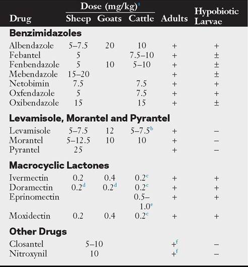
aAll doses are for oral administration unless otherwise indicated.
b10 mg/kg for topical administration.
c0.2 mg/kg for oral, subcutaneous, or intramuscular administration; 0.5 mg/ kg topically as a pour-on.
dInjectable not recommended for use in sheep and goats by injection or pour-on. e0.5 mg/kg for topical administration; 1.0 mg/kg in extended-release formula for injectable administration; not recommended for sheep.
fEffective against Haemonchus contortus only.
+, Highly effective; ±, moderately effective or effective according to some authors; —, ineffective.
Note: Administration of some of these products may constitute extralabel use in sheep and goats; follow manufacturers’ guidelines for meat and milk withdrawal in cattle; not all compounds are available in all countries.
that GINs surviving one anthlemintic will likely be killed by the second.25,26 Computer simulation models also indicate that reversion toward susceptibility is more likely to occur when drugs were used in combination rather than in rotation.27 Although combination anthelmintic products are available elsewhere in the world, licensure in the United States has not yet occurred.
Treatment Intervals. The choice of drug and treatment interval should be formulated for an individual herd or farm, as part of an overall GIN control program. Factors to consider include the geographic location, time of year, and grazing management.1 There are several options for preventing clinical disease and maximizing gains in first-season grazing calves using strategic anthelmintic treatments. It should be noted, however, that these recommendations reflect the practices currently in use and assume anthelmintic resistance is not an issue on the farm in question. Recommendations are based on the calendar in the northern hemisphere, and not all products are available in every country.
• Prophylactic anthelmintic use. (1) Two or three treatments between turnout and midsummer to minimize the number of eggs deposited on pasture. For calves turned out in early May, two treatments 3 and 6 weeks later are used. For calves turned out in April, three treatments at 3-week intervals are recommended. If using parenteral or pour-on macrocyclic lactones, the interval is extended to 5 (ivermectin) or 8 (doramectin, eprinomectin, moxidectin) weeks due to the residual activity against ingested larvae.3,20 (2) Use of an intraruminal bolus at turnout or weaning. This provides either the sustained release of anthelmintic drugs or pulse release of therapeutic doses of anthelmintic at 3-week intervals. This strategy may be most cost-effective on farms where pasture infectivity is high. However, evidence suggests that young cattle protected with these products are more susceptible to infection in their second year at grass.3 Likewise, the use of extended-release injectibles at turnout provides the ability to treat existing infections while preventing new infections with anthelmintic-susceptible GINs for approximately 120 days.28
• “Dose-and-move” strategy. This strategy consists of treating calves with a single dose of anthelmintic, then moving them to a “safe” pasture (risk of infection is low) just before the anticipated peak in pasture infectivity (e.g., early to midsummer in temperate climates). This strategy minimizes the number of anthelmintic treatments during the grazing season. However, it is effective only on farms where such pastures are available. Furthermore, any residual nematodes left behind will likely possess resistant genes; therefore contamination of the “safe” pasture will be with resistant nematodes. This must be considered when planning for the future use of the pasture (discussed later). Also, sufficient numbers of overwintering L3 O. ostertagi may be present in the spring in some years, resulting in heavy infections and clinical disease.
• Targeted strategic treatment. This strategy relies on the selective use of anthelmintics by targeting individual animals within a herd rather than using whole-herd treatments. Promoted for sheep and horses, this strategy has not been embraced by the beef cattle industry. However, a recent investigation of dairy cows indicates that selective treatment strategies can compete economically with whole-herd treatment strategies.29
Integrating effective pasture management can reduce the number of anthelmintic treatments necessary1,30; however, there currently is no realistic alternative to the continued use of available compounds in intensive production systems. Therefore it is imperative that the efficacy of these compounds be maintained for as long as possible. Recommendations designed to promote this in beef cattle include the following: (1) do not treat second-year (unless not previously pastured) or adult cattle to maintain a population of unexposed nematodes (refugia) on the farm; (2) do not graze first-year calves on the same pasture each year (avoids exposure to larvae produced from resistant nematodes); and (3) do not use the same family of anthelmintic year after year in calves.31
Adult Cattle. In general, the cattle most at risk for clinical disease and production losses are beef calves and dairy replacement heifers in their first season at pasture.2 Development of immunity should protect the animals during their subsequent grazing seasons. Treatment of adult beef cattle is generally unnecessary, unless immunity is inadequate or pasture infectivity is exceptionally high. Anthelmintic treatment may be warranted in first-calf heifers and newly acquired cows that may not have been pastured as heifers. In some situations it may be beneficial to treat beef cows after spring calving. Despite these recommendations, a recent survey showed that approximately 86% of U.S. beef cow and calf producers routinely treat their cows one or more times per year.22 Benefits of treating adult dairy cattle depend on a variety of factors, including grazing management options and levels of GINs.1,27
OTHER STRATEGIES. Alternate grazing of cattle and sheep is promoted as an effective control measure. Ideally, a 3-year rotation of cattle, sheep, and crops is used. Few GINs crossinfect cattle and sheep. Adding this to the infective L3 life span on pasture of less than 1 year, this scheme could provide good control of bovine ostertagiasis. Annual rotation of beef cattle and sheep in marginal areas has been reported to provide adequate parasite control.3 However, in areas where Haemonchus is prevalent or where Haemonchus contortus cycles in cattle,15,17,24 co-grazing these two species may be dangerous. Rotational grazing of calves with adult cattle has been shown to be an 23
effective control practice.2,3
Vaccination for the control of O. ostertagi has been an ongoing area of research for many years with mixed results.32,33 Given that it has taken more than 20 years for development of a commercially available vaccine against H. contortus, it is unlikely that a viable O. ostertagi vaccine will come to market in the near future.
EVALUATION OF CONTROL PROGRAMS. Success of control programs is based on the producer’s expectations. Because most control programs are based on anthelmintic use, the response to treatment is the criterion by which control programs are currently evaluated.15,22 As long as animal production meets producer expectations, the control program is considered to be a success. Fecal egg counts are most useful as a tool to evaluate pasture contamination and assess treatment efficacy; however, most U.S. producers do not have this done. In fact, most cow and calf producers do not even involve a veterinarian in the diagnosis or treatment of PGE.22
Lack of expected response to anthelmintic treatment may be the first indication of program failure in the form of a resistance problem.15,17 However, inappropriate administration or underdosing of the anthelmintic may also be responsible, and determining the root cause of the treatment failure is vital for future control efforts. Detection of anthelmintic resistance currently depends on the FECRT. This test only detects clinical resistance or when less than an expected response to treatment occurs. This usually occurs only when the frequency of resistant alleles in the population reaches 25%.34 The FECRT estimates anthelmintic efficacy by comparing pretreatment and posttreatment fecal egg counts. Fecal samples are collected immediately before or at the time of treatment and then 10 to 14 days later. Although guidelines for using FECRT in cattle were proposed in 1992, they lack detail, particularly in interpretation of data.19,35 Comparing FECRT with controlled efficacy studies showed FECRT results to be reliable for detecting ivermectin-resistant C. oncophora and O. ostertagi and moxidectin-resistant O. ostertagi. However, due to reduced fecundity of moxidectin-resistant female C. oncophora, FECRT results were equivocal, with day 7 results showing efficacy but day 14 results indicating resistance.36 Thus, for cattle, waiting the full 14 days before collection of the second sample is vital. A method for using composite fecal samples in the FECRT has been described with the aim of reducing costs to the producer; however, it still requires the collection of individual fecal samples.37 Because cattle tend to have lower fecal egg counts than sheep, use of appropriate flotation techniques capable of handling low fecal egg counts is important.1
■ Clinical Management
DIAGNOSIS. Antemortem diagnosis of ostertagiasis in young animals is primarily based on clinical signs of inappetance, weight loss, and diarrhea at a time of the year that is consistent with the parasite’s epidemiology.3 In temperate areas, type I disease usually occurs in fall, and type II occurs in the late spring. Grazing history is important because clinical disease can develop in older animals that have not previously grazed pastures. Affected farms usually also have a history of previous outbreaks. Plasma pepsinogen levels are useful in animals up to 2 years old; however, the test is less reliable in older cattle.1 The most reliable method for diagnosis of ostertagiasis or PGE is necropsy. The “Moroccan leather” abomasal lesion is pathognomonic for Ostertagia. For PGE, casual examination of the lumen of the gastrointestinal tract is unlikely to find any of the strongyle-type nematodes because the worms are small and easily overlooked. Adult Nematodirus are larger and may be more readily seen, but alone they are unlikely to be the cause of PGE. Nodules associated with larval Oesophagos- tomum can be found in the wall of the lower small intestine, as well as the upper large intestine, and adults can be found in the contents of the large intestine. Trichuris will be partially embedded within the cecum; removing the contents is necessary in order to see them. Abomasal wall digestion techniques can be used to identify the presence of larval OstertagiaN
TREATMENT. Animals with type I ostertagiasis can be expected to respond well to treatment with the standard dose of any of the current anthelmintics, as long as resistance is not an issue. These animals should also be moved to a less contaminated pasture. Because any parasites not removed by proper administration of the drug will have resistance alleles, targeted strategic treatment, rather than whole-herd treatment, may be beneficial in keeping the population of resistant nematodes low.
Successful treatment of animals with type II ostertagiasis requires compounds that are effective against arrested larvae, as well as the developing larvae and adults. Only the modern benzimidazoles and macrocyclic lactones are effective at standard doses. Even though the larvae may be killed, the damage to the abomasal mucosa may limit complete recovery. Animals with profound hypoproteinemia and dehydration respond poorly compared with those showing only mild diarrhea and slight hypoalbuminemia. Severely affected animals may need supportive treatment to survive. Recovered animals often fail to thrive.
Gastrointestinal Nematode Infections in Sheep and Goats
SherriU A. Fleming
Gastrointestinal nematode (GIN) infections in sheep and goats are responsible for severe clinical syndromes and profound production and death losses. Young animals, periparturient ewes and does, and animals on substandard planes of nutrition are most susceptible to outbreaks of parasitic disease. The GINs of small ruminants include H. contortus, Teladorsagia circumcincta (formerly Ostertagia), T. axei, Nematodirus spp., and Cooperia spp.1 The proportions of each of these nematodes in small ruminant populations vary according to geographic location. H. contortus is usually the most significant pathogen in wet, temperate climates such as the southeastern United States. T. circumcincta may be the predominant infection in northern or arid climates such as England. H. contortus and T. circumcincta represent the majority of parasite burdens seen in small ruminants.2 Anthelmintic resistance is present in all these parasites, but the prevalence is highest for H. contortus, making it the most economically important GIN of sheep and goats.3
The problem of anthelmintic resistance in GINs of small ruminants has been reported in South Africa, Australia, New Zealand, Malaysia, Spain, France, Denmark, the United Kingdom, Brazil, and the United States.4-7 In the United States, resistance to all classes of anthelmintics has been documented.5,7 Resistance to two and three classes of anthelmintics was found on 14 of 15 farms and 6 of 18 farms, respectively, in a survey of 18 goat flocks in Georgia and South Carolina.5 The first report of total anthelmintic failure was made on a meat goat farm in Arkansas in 2005.7 It is no longer acceptable to plan parasite control programs solely on the basis of the use of anthelmintics. Veterinarians and producers must customize programs to control exposure to infection and reduce the use of anthelmintics. A thorough knowledge of the biology of GINs is necessary to plan effective control programs.
■ Life Cycle and Epizootiology3'9 The life cycle of a GIN is direct and consists of a host phase and a free-living phase. Worms mate in the host, and females lay eggs that pass in the feces. Eggs hatch and develop to infective larvae while remaining in the fecal mass. Infective larvae then move from the fecal mass onto the surrounding forage, where they can be consumed during grazing, thus completing the cycle. The time from ingestion of infective larvae to egg-laying adults, the prepatent period, is about 3 weeks. The time for development from egg to infective larvae can be as short as 4 to 10 days (especially during the summer months); therefore transmission (reinfection) and continual pasture contamination can be rapid. During the colder months, development is delayed and may take 1 to 2 months to reach the infective larvae stage, so pasture contamination and reinfection are minimized. The infective larvae have a protective sheath, making them relatively resistant to adverse environmental conditions, and they can survive for months, thus extending transmission potential. As long as the temperature and moisture conditions remain favorable, development and survival continue; conditions that are too hot, too cold, and/or too dry threaten parasite survival.
The life cycle of a GIN has four phases.
PHASE 1—SYMBIOTIC/PARASITIC PHASE. Phase 1 is the interaction between host and parasite. During this phase, the parasite must develop and survive in the host. After ingestion, infective larvae lose their protective sheath and invade the mucosa of the abomasum, small intestine, or large intestine depending on the GIN species involved. While in the mucosa, larvae develop to the next larval stage and then return to the surface of the gut mucosa, where they become adult worms. The host defense mechanism is immunity. When the infectious agent enters the body, the immune system reacts to mobilize various components (antibodies, killer cells, etc.) to inhibit or kill the invaders. These components act on larval stages in the mucosa and the adults in the lumen. The strength of the immune response depends on the age of the host, nutritional status, and concurrent stressors. The immune system matures with age with young naive animals susceptible to infection and older animals more resistant. As a result, young animals usually harbor the highest GIN populations and suffer the most severe consequences, whereas adult animals have lower infection levels. Under poor nutrition and/or stressful conditions, the immune system is compromised and cannot respond adequately, regardless of the age of the animal.
The prepatent period of most worms is about 3 weeks, but this period can be extended for worms that can enter a period of delayed development called hypobiosis. This occurs during the season of the year when the environmental conditions are unfavorable for development and survival of the free-living larval stages. Depending on the worm and the environment, this happens during either summer or winter.
PHASE 2—CONTAMINATION PHASE. Phase 2 involves eggs being passed in the feces. The magnitude of this phase is affected by stocking rate, age of the animals, season of the year, and hypobiosis. The higher the stocking rate, the more feces are deposited on the grazing area, thus the more eggs are passed, and vice versa. More eggs are also passed from young versus older animals. Most worms have a definite seasonality, resulting in higher egg production and deposition during different times of the year. Of particular note is the periparturient rise (PPR) in fecal egg count (FEC). This occurs at or around parturition and extends through most of the lactation period because these are stressful conditions when the dam's immune system is compromised. The existing female worms increase the number of eggs deposited in the feces. If a worm species undergoes hypobiosis, the development time to the adult stage is extended. This will result in fewer adult worms over time and fewer eggs deposited in feces. However, when these hypobiotic larvae resume development, massive numbers become mature adults over a short period of time, and the resultant egg production and deposition in the feces can be high.
PHASE 3—FREE-LIVING PHASE. Phase 3 involves larval development and survival, which depend on prevailing environmental and nutritional conditions. The development and survival from egg to L1, then to L2, and finally to L3 or infective larvae occur within the fecal mass. L1 and L2 are unprotected and require oxygen and energy (feeding on nutrients and microorganisms) to grow. L3 are enclosed in a protective sheath and do not feed. Temperatures conducive for normal development and survival are between 65° and 85° F. With lower or higher temperatures, development and survival are reduced. Moisture is also crucial for development and survival. Because the initial development and survival occur within the fecal mass, moisture is usually adequate to allow development to the infective larvae; however, if the fecal mass dries out quickly due to high temperatures and/or physical disruption of the fecal mass, the L1 and L2 are susceptible to desiccation and will die. Generally, L3 can survive very low temperatures, but sustained temperatures above 95° F are usually lethal. When infective larvae migrate out of the fecal mass, they are relatively resistant to environmental conditions encountered due to their protective sheath. Temperature is usually the only factor that may adversely affect L3. The moisture conditions at ground level under forage cover are usually adequate for L3 to move around and survive. Because they do not feed, length of survival depends on how fast they use energy reserves. The hotter it is, the faster they move and use up energy stores, which decreases survival. Infective larvae move up and down the forage when there are moist condiitons (i.e., advancing and receding dew). Rain also provides a moisture medium for larval movement on forage. For the most part, infective larvae do not move much past 30 to 50 cm from the fecal mass or 5 to 6 cm up the forage. Therefore the lower the animals graze and the closer they are to the fecal mass, the greater the consumption of L3.
PHASE 4—INFECTION PHASE. Phase 4 occurs when available L3 are consumed during grazing. This phase is affected by stocking rate in two ways. The stocking rate determines how many eggs initially contaminated the pasture and, consequently, how many L3 will be available to consume. If the initial contaminating animals are removed and replaced by new animals, the new stocking rate will determine the level of exposure each animal has to infective larvae during grazing (i.e., the higher the stocking rate, the more chance of exposure, and vice versa). Grazing animals usually do not graze close to fecal masses, so the greater the distance between masses, the less the exposure. Eventually fecal masses disintegrate, forage grows well with the fertilization, and animals will graze over the area where exposure can be high. Natural sources of water, such as streams, ponds, or lakes, provide moisture along the banks, where forage can grow readily. When animals congregate to drink and consume the attractive forage, defecation in these areas usually leads to increased contamination and eventually to more L3. Essentially, a high stocking rate has been artificially created in a relatively small area. The same can be said for areas where supplements, especially hay, are fed if conditions are favorable. Once L3 are consumed, phase 1 is repeated.
■ Pathophysiology of Gastrointestinal Nematode Parasites The damage done by H. contortus is the result of the blood sucking by L4 and adult parasites. The pathogenesis of Teladorsagia in sheep is similar to that described for Ostertagia in cattle and is a result of destruction of abomasal mucosa. T. axei causes abomasitis, whereas Tricbostrongylus Colubriformis and other Inchostrongylus spp. penetrate beneath the duodenal mucosa and result in generalized enteritis, including hemorrhage and plasma protein loss into the intestinal lumen.
■ Clinical Signs of Gastrointestinal Nematode Parasites Most animals acquire mixed infections of nematodes. Clinical signs may therefore reflect the effects of more than one species of parasite. There appears to be some synergism between T circumcincta and T. colubriformis, which makes the effect of the combined infection more severe than that of either alone. Although mixed infections with GINs are assumed, the actual population of parasites in small ruminants varies depending on the geographic location. In general, most clinical illnesses results from H. contortus infections. Most parasitic infections in small ruminants are associated with altered gut function, anorexia, ill-thrift, weight loss, and hypoproteinemia. Diarrhea is a variable sign (black scours). Animals may die suddenly without overt clinical signs or may exhibit chronic wasting. Clinical signs of H. contortus infection can vary from peracute to chronic and result from decreased nutrient use and anemia/hypoproteinemia. The most common signs are failure to thrive, weight loss, and decreased appetite. Weakness, bottle jaw, pale mucous membranes, poor capillary refill time, and possibly diarrhea develop with more severe or long-term infections. Following sudden exposure to large numbers of L3, animals can die acutely, even before the infection is patent. Other diseases such as pneumonia and heat stress may result secondarily.
■ Anemic Crisis and Patient Management The anemia created by H. contortus is due to blood consumed by the parasite and is usually chronic. Parasitized patients present with lethargy and weight loss and often have hematocrits of less than 10%, yet many are still capable of rising and walking. Addressing the anemia by blood transfusion must be weighed carefully against stress to the patient caused by restraint, jugular catheterization, and blood administration, as well as by the hemolysis of these same cells several days later. Obviously, in life-threatening circumstances (a patient unable to stand), administration of whole blood can be critical. In a chronic situation (patient rising and/or eating), treatment with an effective anthelmintic should eliminate the parasite and halt blood loss. Because H. contortus consumes blood, the affected animal actually loses substrates essential to erythrocyte production (iron, cobalt, copper). Providing supplemental iron and B-complex vitamins should speed reversal of the anemia. Assuming blood loss has been halted, a 1- to 1.5-point rise in the hematocrit per day is expected when the bone marrow is maximally stimulated. In most parasitized patients, a rise of one half of this value is a more realistic expectation.10
If blood is to be given, it is essential to be certain that the donor has a normal packed cell volume (PCV). Blood collected from animals in the same environment may be as anemic as the patient, but the individual is more capable of dealing with the anemia. In lambs weighing less than 45 kg, 1 unit of whole blood (≈450 mL) is often sufficient to survive a crisis, provided H. contortus has been controlled.
■ Populations at Risk Young animals are most susceptible to infection and clinical manifestations of disease. Lambs and kids may become heavily infected with parasites and shed large numbers of nematode eggs. In sheep, some degree of immunity develops as the animal approaches 1 year of age. Adult animals typically have complete immunity against Nematodirus and variable resistance to Tricbostrongylus spp. Some immunity to Haemonchus and Teladorsagia spp. develops with age; compared with lambs or kids, adults are more resistant to infection with these species. However, even mature animals may succumb to parasitic infection when malnourished or challenged with heavily contaminated pasture. A periparturient rise in fecal egg production is seen in ewes and does.
Goats are more susceptible to GIN infections than are sheep. The difference lies in part in the host’s immunologic responses to nematode antigens. Goats prefer to browse on brush and trees and only graze grass when forced to by management. Under natural conditions, goats would have a low level of exposure to infective larvae, and this may explain their lower level of natural immunity compared with sheep.11,12
■ RefugiG Most parasitologists now consider levels of refugia (see Parasite Refugia section earlier) as the single most important factor in selection for anthelmintic-resistant parasites.13,14 Worms in refugia provide a pool of genes susceptible to anthelmintics, thus diluting the frequency of resistant genes. For many years parasitologists and veterinarians have recommended that all animals on a premise be treated with an anthelmintic at the same time. However, this strategy has proven problematic, and a selective approach is now recommended. Only those animals in danger of severe parasitism should receive medication. This selective approach is highly compatible with host-parasite dynamics because 20% to 30% of animals harbor about 80% of the parasites.15 Animals with low worm burdens are an important source of refugia, are not in danger of the negative effects of parasitism, and should not be treated.
■ Management of GGstrointestinGl Nematodes in Small Ruminants The traditional approaches of deworming often, rotating class of anthelmintics, and moving to clean pastures has ensured development of anthelmintic resistance to all classes of drugs and disseminating these resistant parasites over a wide area. It is obvious that drastic changes in the management of GIN in sheep and goats are long overdue. Several countries have developed websites and/or companies that make recommendations for specific geographic areas.16 WormBoss in Australia and SCOPS in Great Britain are two good examples that attempt to give specific management advice.16,17 A New Zealand company sells a system of record keeping and testing complete with a computer system for keeping farm records.18
FAMACHA OR SMART DRENCHING METHOD.19 To maintain adequate levels of refugia, it is necessary to leave a portion of the herd or flock untreated. FAMACHA is a selective approach for H. contortus that targets the portion of the herd or flock with high worm burdens, including those animals that are poorly resilient to worm infections.20 This approach will successfully control parasites in the entire group and delay the development of anthelmintic resistance. Failure to observe animals closely for clinical signs may result in the death of some individuals. It is necessary to know which anthelmintics are effective before beginning this system. The differences among farms in overall quality of management, stocking rates, breeds of animals, preexisting levels and spectrum of anthelmintic resistance, presence of nematode species other than H. contortus, and production targets need to be considered before implementing FAMACHA on individual farms. This system was developed in South Africa for identifying sheep that are anemic, and it has been extended to use in goats.21 In this method the ocular mucous membranes of sheep and goats are categorized by comparison with a laminated color chart bearing pictures of sheep conjunctivae classified into five categories ranging from red (A/1; normal) to practically white (E/5; severe anemia). Because anemia is the primary pathologic effect from infection with H. contortus, this system can be an effective tool for identifying animals that require treatment (but only for H. contortus). FAMACHA has been tested extensively and validated in both sheep and goats in South Africa and in the southern United States.22,23 In all studies, the numbers of false negatives were low, suggesting that when using FAMACHA according to recommended guidelines, death from anemia would be rare.
Based on the results of recent studies in the United States and numerous studies performed in South Africa, guidelines for using FAMACHA have been developed, and it is suggested that these guidelines be read in their entirety before FAMACHA is practiced.20 Individual animals are scored 1 to 5 by comparing their mucous membrane color with a laminated chart. It is recommended that treatment be withheld until animals score a 4 or 5 as long as animals are in good body condition and good overall general health, animals are examined frequently (e.g., every 2 weeks), and good husbandry is used to identify animals in need of treatment (e.g., unthrifty, anorexic, lagging behind, bottle jaw) between FAMACHA examinations. Using this approach, the number of anthelmintic treatments administered will be reduced greatly, resulting in significantly diminished selection pressure for resistance and therefore a reduction in drug costs. Due to the increased handling of animals, labor costs will be increased. Only adult animals should be managed with this system. Lambs and kids have comparatively small blood volumes, poor immunity, and poor resilience and can progress rapidly from moderate to severe anemia. This precaution should also be extended to ewes and does during the periparturient and early lactation period because these animals have decreased immunity to GINs.19,24-26 These and other animals that may be stressed by disease or in poor body condition should always be treated if scored as 3. Alternatively, in the northern parts of the country where H. contortus is an important problem but resistance prevalence is much lower, it may be reasonable to be more liberal when making treatment decisions (i.e., treat all 3, 4, and 5). Many more treatments are given when all animals with scores of 3 and higher are dewormed, but a significant number of animals will remain untreated to supply refugia. These refugia combined with a relatively short transmission and treatment period are likely to produce a slow evolution of resistance, but the more intensive treatment protocols will improve animal productivity.
On farms where low to moderate levels of resistance have been diagnosed to one or more drugs (60% to 90% reduction in FEC), a useful strategy to help gain the full benefits of both treatment and resistance prevention could be to use these “less effective” drugs either singly or in combination on all animals scored as 3. There is evidence that using dewormers with different modes of action in combination has greater efficacy and does not cause a more rapid development of resistance.27 Using drugs that are less effective in this group is unlikely to lead to clinical problems because the few 3 scores that are moderately anemic and in need of treatment should receive a sufficient reprieve from infection until the next FAMACHA examination, and the majority of the 3 scores that are not anemic do not need to be treated. This strategy helps to preserve the efficacy of the drugs that are still fully effective by saving them only for the 4 and 5 categories and also helps to decrease egg contamination of pastures.
Training of producers is critical when using this method. It is the responsibility of veterinarians and other animal health professionals to ensure that standards of training are maintained by using the “Train the Trainer” seminars through the American Consortium for Small Ruminant Parasite Control (ACSRPC, formerly Southern Consortium or SCSRPC) at www.acsrpc.org or www.wormx.info. When using FAMACHA, it is extremely important that efficacy of anthelmintics is known because animals are not treated until they become anemic. Treating anemic animals with a drug that has moderate to poor efficacy due to worm resistance may result in animal deaths. Other important precautions when using FAMACHA include but are not limited to (1) the card is an aid in the control of Haemonchus spp. only; (2) the system should be used by producers only where technical assistance is available from a veterinarian or other animal health professional; (3) other management-based worm control practices must be maintained; (4) smart drenching principles should be used; (5) paleness or reddening of the conjunctivae may have other causes; (6) animals should always be scored with the help of the chart, not from memory; (7) animals are examined at least every 2 to 3 weeks at the beginning of the expected period of Haemonchus spp. challenge in climates where a seasonal incidence of infection occurs, and during critical periods weekly examinations may be necessary; (8) the card is protected from light when not in use and replaced after 1 year of use.4,28
Maintaining records of treatments that are included with the FAMACHA kits gives the owner the ability to rate the genetic merits of individuals on the premises. Host resistance to infection with H. contortus measured on the basis of FEC and PCV is a moderately heritable trait, and it has been demonstrated that the same animals tend to exhibit the highest FEC and lowest PCV on each occasion they are measured.29-31 Importantly, data from recent investigations examining the heritability of resistance and resilience of Merino sheep to infection with H. contortus indicate a high heritability for the clinical estimates of FAMACHA scores.22 Removing the most susceptible animals from the breeding pool each year will have the long-term effect of improving the overall innate genetic resistance and/ or resilience of the herd or flock to H. contortus.
FIVE-POINT CHECK.32 Because FAMACHA is based on anemia and focuses on parasitism by H. contortus, a combined system that could identify problems with a variety of other potentially pathologic parasites (Oestrus ovis, Teladorsagia spp., Trichostrongylus spp., Nematodirus spp., and Oesophagostomum columbianum) has been proposed. This is the Five-Point Check, which includes checking for nasal discharge, submandibular edema, back body condition scoring, and fecal staining of perineum in addition to ocular membrane color. Anthelmintic recommendations were made based on the points that identified poor scores. Pilot studies showed good acceptance and usefulness of this system, but further studies need to be completed to determine the efficacy and economy of this approach.
GRAZING STRATEGIES. The goal of pasture management is to provide safe pastures for grazing by reducing the exposure of susceptible hosts to infective larvae.33,34 A safe pasture is one that has had no sheep or goats grazed on it for 6 months during cool or cold weather or for 3 months during hot, dry weather. Weaning sheep and goats at 2 months of age and rotating them through pastures ahead of the adults while forage is longer will minimize the exposure of susceptible animals to infective larvae. Pastures should be subdivided into smaller lots to allow longer rest periods between grazings. Pastures that are overgrown provide a good environment for larval survival because ultraviolet light and dry conditions are effective in killing larvae. Keeping pastures clipped will also assist in weed and parasite control and improve forage quality. Short-duration grazing carries pasture rotation to a level that maximizes forage production and harvesting by controlled animal grazing. It is management intensive but can be effective in controlling parasite burdens. Tilling and reseeding heavily contaminated pastures will convert them to safe pastures and provides an opportunity to improve the forage quality. Taking a cutting of hay from a pasture assists in reduction of infective larvae, but one report indicates GINs and tapeworms developing in “worm-free” lambs after feeding hay from heavily infected pastures. During the most dangerous part of the grazing season, it may be necessary to dry lot the flock and feed hay and grain from elevated feeders.
Stocking rate is an important consideration in parasite control because it affects the exposure to infective larvae and the contamination of the pasture. It is impossible to make a general recommendation on stocking rate because this will vary according to the type of pasture, time of year, current weather conditions, and type of animal being grazed. Thumb rules include five to seven goats or five sheep being the equivalent of one cow and suggest five to seven goats per acre. Goats prefer to browse brush and trees, whereas sheep prefer to graze near the ground. Pasture management must include monitoring the condition of herbage to ensure that overgrazing does not occur and to maintain a productive pasture.
In the early spring or at the onset of the rainy season, reduced pasture contamination is the most important aspect of control. The ewe or doe in the periparturient relaxation of resistance, even if she has the genetic capacity for resistance, will be a source of eggs for the environment. Strategic deworming to remove arrested or recently emerged larvae before they contaminate the pasture will have a great impact on pasture contamination. Treatment 2 weeks after a rain that removes recently acquired worms before they can begin passing eggs will also decrease pasture contamination. Providing sufficient dietary protein is vital during the periparturient period and during rapid growth so that animals will tolerate the worm burden better, as well as increase resistance to infection.35-37 A strong link between nutrition and parasitism has been illustrated between protein intake and resistance to GIN infection. The most dramatic has been the abolishment of the periparturient egg rise in lambing ewes by providing protein at 130% of requirements. Immunity is closely related to protein repletion. GINs increase the demand for amino acids by the sheep. Lambs will voluntarily select a higher protein diet when infected with GINs compared with uninfected lambs. There is conflicting documentation that sheep will decrease feed intake when initially infected with GIN. Some authors hypothesize that the decrease in intake may be due to stimulation of the immune system or that the host is becoming selective in its diet.
Pastures may be used for hay cropping and grazed during the last half of the grazing season to effectively reduce GIN challenge. When plants high in condensed tannins are grazed, incoming larvae are adversely affected, and these plants provide bypass protein for the host.38-40 There is growing evidence in work from New Zealand and Europe that grazing or feeding of plants containing condensed tannins (CTs) can reduce FEC, larval development in feces, and adult worm numbers in the abomasum and small intestine. Preliminary tests with Sericea lespedeza (also known as Lespedeza cuneata), a perennial warmseason legume, have shown positive effects of reduced FEC in grazing goats, and in sheep and goats in confinement when the forage was fed as hay. In addition, an effect on reducing worm burden has also been reported. S. lespedeza is unfortunately considered to be an invasive exotic species in the United States. Similar results have been observed using quebracho extract for small intestinal worms, but not abomasal worms. In addition to its potential use in controlling worms, S. lespedeza is a useful crop for limited resource producers in the southern United States. It is adapted to hot, dry climatic conditions and acid, infertile soils unsuitable for crop production or growth of high-input forages, such as alfalfa. It can be overseeded on existing pasture or grown in pure stands for grazing or hay. In addition to hay, S. lespedeza is being evaluated in the form of meal, pellets, and cubes to be fed as a supplement to grazing animals, or as a deworming method under temporary short-term confinement. The physical structure of some plants may challenge larvae to ascend vegetation or may provide protection from adverse pasture conditions. If animals are allowed to browse, their chances of acquiring larvae diminish as the distance from the ground increases. Most infective larvae are found within 2 inches (50 mm) of the soil surface.
Alternate grazing or co-grazing with other species of livestock may harvest Haemonchus larvae from the pasture. Small ruminants can graze after cattle, and this is considered a safe pasture, assuming adequate parasite control in the cattle. For the most part, each livestock species harbors its own parasite fauna except for overlap between sheep and goats. Only T. axei, a minor abomasal worm, is found in all livestock species. In general, the Haemonchus in sheep and goats do not do well in cattle, and vice versa. However, some populations of
H. contortus may thrive in calves. If practical, cattle and small ruminants can be grazed together where each consumes the parasites of the other, which reduces available infective larvae for the preferred host species.
Anthelmintic administration should be coordinated with the weather. During hot, dry weather, there will be little or no exposure to L3. As soon as there is significant rainfall (0.5 to 1 inch), larvae exposure goes up exponentially as previously inactive larvae become active and new larvae are hatched. Producers should be trained to plan deworming within 3 weeks of significant rain after a dry spell. Similar strategies can be used during cool weather. Once ambient temperatures drop below 50° F, the flock can be dewormed and no further treatments are necessary until temperatures become favorable to larval development and activity.
ALTERNATIVE THERAPIES
I. Copper oxide wire particles (COWPs) have been marketed for years as a supplement for livestock being managed in copper-deficient areas.41 COWPs come in adult cattle, calf, and ewe boluses (25, 12.5, and 4 g, respectively). Only the cattle boluses are available in the United States. Due to potential toxicity in sheep, only one dose per year is recommended. It is also well known that copper has some anthelmintic activity against abomasal worms but not other gastrointestinal worms. That makes COWP a narrow-spectrum product. However, in view of anthelmintic resistance by H. contortus, recent work has revisited the possibility of using COWP to specifically target H. contortus. Such work has shown that as little as a gram or less and 2 g may remove substantial numbers of H. contortus in lambs and ewes, respectively. Similar work in goats has not been tested adequately to establish what is needed, but similar doses may be appropriate. As mentioned, copper must be used cautiously in sheep because toxicity can develop due to liver accumulation. Toxicity may not be an issue in goats because they have been reported as not being as sensitive to excess copper intake. Thus higher doses and/or more treatments during haemonchosis season may be useful in goats.
2. Other nutritional considerations include phosphorus, cobalt, and molybdenum.42 Supplementation with phosphorus has been shown to prevent worm establishment. Cobalt deficiency has also been associated with reduced immunity to GINs. The addition of molybdenum at 6 to 10 mg/day decreased worm burdens in lambs. This effect was not attributable to the expected copper deficiency. Molybdenum may have a role in increasing jejunal mast cells and blood eosinophil numbers.
3. Nematode-trapping fungi included in feed or supplements have demonstrated potential for biological control of the free-living stages of GIN parasites under experimental and natural conditions.43-45 These fungi naturally inhabit soil throughout the world where they feed on a variety of free- living soil nematodes. These fungi capture nematodes by producing sticky traps on their growing hyphae. Of the various fungi tested, Duddingtonia flagrans possesses the greatest potential for survival in the gastrointestinal tract of ruminants. After passing through the gastrointestinal tract, spores germinate and looped hyphae trap the developing larval stages in the fecal environment. This technology has been applied successfully under field conditions and is an environmentally safe biological approach for control of worms under sustainable, forage-based feeding systems. The major drawback is that the fungal spores must be fed daily. Daily feeding, which ensures that all animals consume an equivalent amount of feed, is necessary. For adequate control of larvae during the transmission season, spores must be fed for a minimum of 60 days. This can be expensive and time consuming. A bolus prototype that would allow a single administration where spores would then be slowly released over a 60-day period is being developed. This product is not available in the United States at this time.
4. Vaccines have been explored for management of the negative consequences of parasitism.46-48 Successful vaccines have been developed for lungworms in cattle and tapeworms in sheep. The most promising vaccine for small ruminant worms has been what is called a “hidden gut” antigen and specifically targets H. contortus. This antigen is derived from the gut of the worm, and when administered to the animal, antibodies are produced. When the worm ingests blood during feeding, it also ingests these antibodies. The antibodies then attack the target gut cells of the worm and disrupt the worm's ability to process the nutrients necessary to maintain proper growth and egg production. This vaccine is currently available in Australia and has been shown to alleviate losses from H. contortus in sheep when used according to directions (Barbervax®). Multiple vaccinations are required prior to and during the active parasite season.49 There are also reports of successful use in goats.50,51 Vaccines for other worms that do not feed on blood have focused on using antigens found in worm secretory and excretory products. Protection has been variable, and marketing of such products has not been pursued.
5. Genetic improvement in resistance to nematode infection is most likely based on inheritance of genes that play a primary role in expression of host immunity.52-54 Based on survival of the fittest management conditions, several breeds around the globe are known to be relatively resistant to infection. Such breeds include Scottish Blackface, Red Maasai, Romanov, St. Croix, Barbados Blackbelly, and the Gulf Coast Native. Katahdin sheep have been considered more parasite resistant, but studies to document this are few and not conclusive. Using such breeds exclusively or in crossbreeding programs would certainly lead to improved resistance to worm infection, but some level of production might be sacrificed. Selection of more resistant stock can be accelerated by identifying sires that produce relatively resistant offspring. Computer programs have been used in New Zealand and Australia to rank sire genetics based on resistance to GIN infections, but change takes up to 8 to 10 years. Heritabilities for FEC, a common measurement for assessing parasite burden, range from 0.22 to 0.40, which is relatively high. Thus selection for resistance using a measurement such as FEC has been moderately successful.
6. Replacement of resistant nematodes with susceptible parasites. Genomic studies and identification of resistance genes are working on inserting susceptible genes to create susceptible populations that can be introduced to contaminated pastures
5558
to compete with resistant parasites.
ANTHELMINTIC USE. Anthelmintics have deliberately been left to the end of this section because drugs cannot be considered the most important aspect of a parasite control program. Guidelines for drug dosages are contained in Table 49.3. At this time, only oral administration of anthelmintics is recommended in small ruminants. In general, goats require 1.5 to 2 times the dosages of sheep, with the exception of Ievamisole, due to more rapid gastrointestinal transit times. It is critical that producers accurately assess weights of individual animals and dose appropriately. If an approximate dose is going to be used in all animals, that dose should be for the heaviest animal in the group.
The challenge of anthelmintic resistance is life threatening in small ruminants. There is no point in repeatedly deworming with expensive drugs if that drug is no longer effective. See the following section, Evaluation of Parasite Control Programs, for information on detecting anthelmintic resistance. On farms where low to moderate levels of resistance have been diagnosed to one or more drugs (60% to 95% reduction in FEC), a useful strategy to help gain the full benefits of both treatment and resistance prevention could be to use these “less effective” drugs either singly or in combination. Drugs in the benzimidazole family may be effective if multiple-day dosing regimens are used.
Evaluation of Parasite Control Programs as they come through the chute, blocked by FAMACHA score, and then randomly assigned to treatment within blocks. This approach is a bit more complicated but will result in groups that are balanced, which will result in a more accurate test. Calculations are performed using the following formula:

where Xc and Xt are the geometric mean or arithmetic mean EPG for control (c) and treated (t) groups before (1) and after (2) treatments, respectively. Free software that performs all calculations and gives data interpretation is available. If the RESO calculator is used, the assignment of resistance status is based on both percent reduction and the 95% confidence intervals. If the RESO calculator is not used, the following guidelines can be applied: reductions of greater than 98% indicate high efficacy, reductions of 90% to 98% indicate efficacy, reductions of 80% to 89% indicate moderate efficacy, and reductions of less than 80% indicate insufficient efficacy. FECRTs yield reliable data only if FECs are sufficiently high to properly measure a reduction from treatment. If the control group's mean FECs are below 150 EPG, objective assessment of resistance will not be reliable. Group mean FECs of less than 150 EPG can be common in adult sheep; therefore when FECRT is performed on a sheep farm, it is preferable to use weaned lambs if available. With goats, low FECs are usually not a problem.
Larval Identification
Ideally, if there is less than a 90% egg count reduction, the eggs should be hatched and the larvae species identified. In the majority of cases these will be H. contortus, but other species, especially Tricbostrongylus species, readily develop resistance as well. One can sometimes be fooled into an improper interpretation of egg count reduction results if mixed species are present and only one is resistant.
Egg Hatch Assays and Larval
Development Tests
Eggs from feces are incubated with concentrations of the anthelmintic to be tested and the eggs hatched. A dose-response curve is generated (DrenchRite test from Horizon Technology, Sydney, Australia). * The advantage of this test is that a single fecal sample can be tested for all available classes of anthelmintics simultaneously. The cost of this test has recently increased and may discourage owners from pursuing this diagnostic. However, when compared with the cost of using anthelmintics that are ineffective, it is easy to justify the use of this test. It is important to read instructions for submitting this test and scheduling a testing date before the samples are collected.
Larval Culture
Larval cultures can be used to distinguish between large and small strongyles in horses and to identify the various nematode species in ruminants. Most parasitology laboratories can perform this examination. It requires submission of 200 to 400 g of fresh feces. Pooled samples from several herd members are often used.
Pasture Larval Counts
Counts of parasitic larvae on herbage are useful to indicate the level of exposure experienced by the grazing animal. This examination is somewhat tedious but can be performed by many university laboratories. A 2-kg sample of forage is gathered for submission to the laboratory. The grass is sampled by walking a V pattern across the acreage and stopping every three paces to sample grass. The samples are subsequently washed and passed through screening to isolate and identify larvae.
■ BOX 49.2
This is one method for performing a McMaster fecal egg count. Similar protocols are used routinely in many laboratories, so you may see a slightly different procedure recommended elsewhere. The important point is to use the same procedure each time.
The first step is to collect freshly passed feces uncontaminated by soil or bedding. The best way to accomplish this is to wear a rubber glove and extract feces directly from the rectum. Alternatively, feces can be picked up off the ground if done soon after deposition. The collection container should be labeled with the identification of the animal and the date of collection. Fresh samples work best, but accurate results can be obtained if the sample is refrigerated during the interim. If samples are not refrigerated, the eggs will hatch within 12 to 24 hours. Once hatched, eggs cannot be counted.
Materials
Compound microscope
Scale Saturated sodium chloride (table salt) 50-mL centrifuge tube, with screw cap and marked with milliliter increments
Pipette (1-mL syringe or eye dropper works well)
McMaster egg counting slide (Chalex Corporation, Issaquah, Wash.) Paper towels
A fresh fecal sample, refrigerated until tested
Procedure
1. Weigh out 2 g of feces into a 50-mL centrifuge tube and fill to 30-mL line with salt solution. It is recommended to weigh feces on a small scale, but if you do not have a scale you can still get a close estimation by putting 28 mL of salt solution into a 50-mL centrifuge tube first and then adding feces until a volume of 30 mL is achieved.
2. Pour off approximately 25 mL of the salt solution into another small container, keeping feces in the tube (use tongue depressor).
3. Soak feces for a few minutes and mix (soft feces) or break up (fecal pellets) with tongue depressor.
4. Add back approximately one half of the salt solution and mix well, breaking up any remaining feces as well as possible.
5. Add back the remaining salt solution and screw the cap back onto the tube.
6. Shake tube vigorously for approximately 1 minute to disrupt any remaining feces as much as possible.
7. Set tube aside for a few minutes to let bubbles dissipate.
8. Wet McMaster chamber with water and dry top and bottom on paper towels.
9. Rock (do not shake) tube several times to thoroughly mix solution without causing large air bubbles to form.
10. Immediately pipette (using 1-mL syringe or eye dropper) a sample of the suspension and fill both sides of counting chamber Work quickly. If it takes more than a few seconds to load the first chamber, mix the fecal solution again and refill pipette before loading the second chamber
11. Let the sample stand for 1 to 2 minutes to allow eggs to float.
12. Count all eggs inside of grid areas (more than one half of eggs should be inside grid) using low-power (10?) objective. Focus on the top layer, which contains small air bubbles (small black circles; if numerous large air bubbles are visible, remove the fluid and refill).
13. Count only trichostrongyle and strongyle eggs (oval shaped, approximately 80 to 90 microns long). Do not count Stron- gyloides species (oval, ≈50 microns long), tapeworm eggs (triangular or D shaped), or coccidia (various sizes). Note the presence of other species but count only the trichostrongyle and strongyle eggs.
14. Once filled, the chambers can sit for no longer than 60 minutes before counting without causing problems. After 60 minutes, drying and crystal formation may begin.
15. Total egg count (both chambers) ? 50 = EPG (eggs per gram).
Notes
This is a dilution technique, and theoretically this ratio of feces to flotation solution will not detect infections with fewer than 50 EPG of feces (one egg seen on slide), so it is not accurate for samples with low numbers of eggs. On a practical level this is not important because from a clinical standpoint, slight differences in results when egg counts are low do not matter
Fairly soon after counting is complete, thoroughly rinse out the McMaster chamber with warm running water. Doing so will keep the chamber clean and ready to be used again. If fecal solution dries in the chamber, do not soak in soapy water for long periods because this will cause the chamber to become cloudy. If the chamber gets dirty, soak for only a few minutes in water containing dish soap and then rinse completely with tap water
Necropsy Evaluation
The nature and magnitude of parasitic infections can be established by necropsy examination. Gross examination and an estimate of adult worm population in the gut lumen are often sufficient. Many worms detach from the mucosa as the carcass cools; however, the damage done by the parasites can be seen on gross or histologic examination. Occasionally it is necessary to use digestion techniques or histologic examination to document the presence of hypobiotic larvae.
Future Evaluation Techniques
Research is developing to make identification procedures more rapid. Validation and refinement of automated smatrphone apps for performing egg counts and identifying egg species has been reported and is expected to improve with continued use.1 The use of deep amplicon sequencing (rDNA ITS-2) has great potential for improving the identification of populations of nematodes, the “nemabiome” that will identify the species present and their population percentages.9 These approaches have been validated for cattle and small ruminants. These techniques have great potential for being able to identify the populations present, the anthelmintic resistance level, and response to treatments more quickly.9,10
Lungworm Infection in Large Animals respiratory disease is obscured by a parasite-induced hypersensitivity response or by superimposed secondary bacterial infections.
■ Life Cycle The life cycle of D. viviparus is direct, with no intermediate host involved. Adults produce embryonated eggs that hatch shortly after oviposition. L1 migrate up the trachea, are swallowed and passed in the feces, where they develop to L2 and infective L3 within a week under optimal environmental conditions. After the L3 are ingested, they exsheath within the abomasum and migrate through the wall of the intestine to the mesenteric lymph nodes, where they molt to the L4. The L4 then migrate through the lymphatics and bloodstream to the lungs, arriving as early as 7 days after infection. L4 may then develop to sexually mature adult worms, or they may arrest development as late L4 or molt to immature adults and then arrest. Arrested development can prolong the normal prepatent period from 3 to 4 weeks to approximately 5 months.1,2
Most problems with cattle lungworms in North America occur in areas of moist climates or areas of intense irrigation where larvae on pasture are protected from desiccation and larval dispersal away from the fecal pat onto herbage is facilitated. Under favorable conditions, infective L3 can survive on pasture for as long as 11 months.1 These larvae, plus the larvae produced from carrier animals harboring small numbers of adults or adults that developed from arrested larvae, can result in high levels of contamination on spring pastures. In subtropical climates, larvae are virtually absent during the hot summers; larval contamination of pastures peaks in the autumn because of carrier animals.2
The life cycle of D. filaria is similar to that of D. viviparus, including the ability of the larvae to arrest development. The prepatent period is approximately 26 days, with peak larval output occurring 39 to 57 days post infection. As with D. viviparus, D. filaria is more prevalent in moist climates, and larvae can survive on pasture throughout the winter. Spring pastures contaminated with overwintering larvae and larvae produced from adults developing from arrested larvae in carrier animals are sources of infection for susceptible lambs or kids.1,2
The life cycle of D. arnfieldi is similar to that of other species of Dictyocaulus, except that larvated eggs are passed in the feces with the L1 hatching almost immediately thereafter. The prepatent period is 2 to 3 months.2,4
Unlike species of Dictyocaulus, M. capillaris and P. rufescens have indirect life cycles that involve molluscan intermediate hosts. Adults, occurring in the parenchyma (M. capillaris) or small bronchioles (P. rufescens), produce eggs that develop and hatch. L1 are passed with the feces; once on pasture, larvae invade the foot of a susceptible species of snail or slug and develop to infective L3. Sheep and goats become infected when ingesting the mollusk while grazing. The larvae penetrate the intestine and migrate to the lung in much the same manner as Dictyocaulus. Early L4, developing L4, and adult M. capillaris become embedded in fibrous nodules within the parenchyma. Protostrongylus larvae mature in the alveoli and enter the bronchioles as adults. The prepatent period for both species is 5 to 6 weeks, although arrested development may prolong the prepatent period of M. capillaris. Patency for both species is very long (>2 years).2-4
■ Pathophysiology Clinical bovine dictyocaulosis is characterized by bronchitis and pneumonia. The disease process is divided into four phases: penetration, prepatent, patent, and postpatent. In the penetration phase (days 1 to ≈7), no clinical sign or significant respiratory pathology occurs as the larvae migrate to the lungs. The prepatent phase (days 7-8 to 25) starts with the arrival of larvae in the alveoli and ends with immature adults in the bronchi. An eosinophilic infiltrate blocks bronchioles; excess mucus production and alveolar collapse occur. Respiratory failure resulting from severe interstitial emphysema and pulmonary edema can lead to the death of heavily infected animals beginning about day 15 onward. The patent phase (days 25-26 to 55-60) is associated with egg-laying adults in the bronchi and trachea. Bronchial and tracheal epithelial damage occurs, air passages are blocked by exudate, and consolidation of lobules results from aspiration of eggs and L1 into bronchioles and alveoli. Bronchitis, tracheitis, and pneumonia follow. The postpatent phase (days 61 to 90) is normally the recovery phase and is associated with the expulsion of adult nematodes. Clinical signs begin to diminish; however, peribronchial fibrosis and epithelialization of alveoli may remain. A frequently fatal syndrome, called postpatent parasitic bronchitis, can occur in about 25% of heavily infected animals during the postpatent phase. This syndrome is characterized by proliferation of type 2 pneumocytes in the alveoli, impaired gas exchange at the alveolar surface, interstitial emphysema, and pulmonary edema and is thought to result from the aspiration of dead and dying worm material into the alveoli. Fatal secondary bacterial infections, resulting from weakened pulmonary defenses, may also occur.1,2
Goats tend to be more susceptible to infection with D. filaria and more severely affected than sheep, although individual susceptibility to infections does occur. Heavy infections in both species produce bronchitis, pulmonary edema, atelectasis, and emphysema, although death is uncommon. As with D. viviparus, secondary bacterial infections can occur.1
The characteristic lesions of dictyocaulosis in horses and donkeys are similar. Pathologic changes include discrete circular areas of overinflation (not emphysema) surrounding affected bronchi primarily in the caudal lung lobes. Histologically these areas contain a small bronchus packed with nematodes. The small airways are often occluded with exudate. Adult worms may also be found in the main bronchi surrounded by small amounts of mucus with little cellular reaction; however, if L1 are present, they are surrounded by an intense mucopurulent reaction. In horses, in addition to these types of lesions, many immature nematodes are present in smaller bronchi, accompanied by bronchial epithelial hyperplasia with large amounts of mucus and pus. Many areas of alveolar hemorrhage and 24
edema are also present.2,4
Pathologic changes caused by P. rufescens result from occlusion of small bronchioles by mature worms, resulting in clogging of lesser branches with eggs, larvae, and cellular debris. Other changes include desquamation of alveolar and bronchial epithelium and cellular infiltration and proliferation of connective tissue. The results are small areas of lobular pneumonia that appear as gray to yellowish conical lesions whose base is on the surface of the lung. In sheep and goats infected with M. capillaris, distinct subpleural nodules containing adults and L1 are common. However, infections in both hosts may be more diffuse with interstitial pneumonia, bronchopneumonia, or fibrinous pleuritis. Secondary bacterial infections can also occur.1,2,5
■ Populations at Risk Clinical dictyocaulosis occurs most frequently in pastured calves, lambs, and kids in the first grazing season. Clinical disease is also reported in adult cattle as a result of primary exposure as adults, exposure of a previously immune animal whose immunity has waned due to lack of reinfection, or exposure of an immune animal to a massive challenge of infective larvae (reinfection syndrome))'2'6'9 Although dairy or dairy-cross calves are most frequently affected, beef calves are also susceptible to D. viviparus)'9,10 Many infections with either M. capillaris or P rufescens are subclinical; however, goats do appear to be affected clinically more often than sheep with M. capillaris),2
Immunity to D. viviparus occurs after first exposure and can develop before the end of the first grazing season. However,
it is variable in both degree and duration. Cattle may be immune for 7 to 12 months after infection. In subsequent infections most larvae are either killed before reaching the lung or are inhibited from maturing into adults. Usually the immunity developed during the first grazing season is boosted by reinfection in subsequent years and prevents manifestations of disease. However, without reexposure to infective larvae or vaccination, immunity wanes and disease can occur in adult cattle.1,2,8
Occasionally, outbreaks of acute lungworm disease are seen in adult cattle on pasture whose immunity is overwhelmed by ingestion of large numbers of larvae. This condition is called the reinfection syndrome. Although most ingested larvae are killed or fail to mature, some reach the lung and incite an acute, immune-mediated reaction.2
Both sheep and goats develop a strong immunity to D. filaria. Thus disease is more common in young animals. As with cattle, the strength and duration of immunity depend on the level of and number of reinfections, as well as the length of time between events.1 Although not common, cattle can be infected with D. filaria. Patent infections do not establish, but clinical disease may occur.1,11
Individual horse or pony susceptibility to infection with D. arnfieldi is evident, with infections occurring during any time in the life of the animal. Patent infections can establish in foals (in an effort to limit disease severity and decrease pasture contamination.1,2,,
Control of lungworms in horses and ponies depends on control in donkeys. Co-grazing with donkeys should not occur. If it does, donkeys should be monitored for L1 and treated to reduce larval output and pasture contamination.2
ANTHELMINTICS. Several anthelmintics are effective against lungworms (Table 49.4).1,3,4,16-20 The macrocyclic lactones are particularly effective against both adult and developing L4 stages and have prolonged residual activity that prevents appearance of larvae in the feces for at least 60 days.21,22 Concerns that such highly efficacious anthelmintics would prevent induction of immunity in first-season grazing animals by killing ingested larvae, before they can penetrate the intestine and thus stimulate an immune response, appear to be largely unfounded. The immune response may be attenuated, but exposed animals still develop some degree of immunity to lungworm infection despite prophylactic treatment with these drugs.22-26 The exception to this may be sustained-release intraruminal devices containing ivermectin, levamisole, or a benzimidazole or long-acting injectables. Although their use can prevent clinical disease for several weeks or months during the grazing season,26-31 some studies have shown that use of these delivery systems impaired the development of immunity in treated calves.26,30 Other studies, however, showed that treated calves can develop immunity during the grazing season.32 Regardless, vaccination of treated animals before their second grazing season may be advisable.2,8
VACCINES. An effective vaccine has been developed against D. viviparus in cattle and is available in the United Kingdom and parts of western Europe. Two doses of live, irradiated larvae are administered by mouth 4 weeks apart. The larvae migrate to mesenteric lymph nodes and provoke an immune response but die before they reach the lung. Management considerations associated with use of the vaccine include the
■ TABLE 49.4
Drugs Used to Control Lungworm
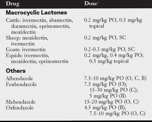
aDose depends on the product and species; follow manufacturers’ recommendations; multiple treatments may be warranted depending on host species; sheep and goats should be dewormed by mouth; be aware that some recommendations constitute off-label use.
B, Bovine; C, caprine; O, ovine; PO, by mouth; SC, subcutaneously.
following: (1) calves must be at least 2 months old; (2) vaccinated calves should not be placed on pasture for at least 2 weeks after the second dose; (3) vaccinated calves must not be placed onto heavily infected pastures, nor should they be mixed with animals showing signs of lungworm disease or with unvaccinated calves; (4) animals should not be treated with an anthelmintic during vaccination or the larvae in the vaccine will be killed;
(5) immunity is not long-lasting, so animals must continue to ingest low levels of infective larvae to maintain immunity; and
(6) the vaccine has a short shelf life and is relatively expensive. Farms with contaminated grazing areas and a history of lungworm disease benefit most from vaccination.8,14,33
■ Evaluation of Preventive Programs The efficacy of preventive programs is best assessed by evaluating the number of susceptible animals that demonstrate signs of infection. Because total eradication of the lungworms is difficult and low numbers of parasites produce minimal problems and maintain immunity, a useful goal is the control of clinical signs and achievement of production goals.
■ Clinical Management
DIAGNOSIS. Presumptive diagnosis of clinical lungworm infection is based on clinical presentation, farm history, seasonal prevalence, and response to treatment.1-4 Verminous pneumonia may mimic respiratory diseases caused by other agents that require specific treatment. Although definitive diagnosis is difficult, it is important.
To document the presence of lungworm infection, it is necessary to demonstrate larvae or adult worms. In cattle and small ruminants, the presence of specific L1 in fresh feces indicates lungworm infection.1,14,34 The Baermann technique is the technique of choice for detection of L1 in feces and is considered to be very sensitive for the diagnosis of patent primary bovine lungworm infections beginning about 3.5 weeks after infection, provided a sufficient number of animals are sampled.34 Antibody detection tests have also been developed and are commercially available for use in cattle. Evaluation of their application for testing bulk milk tank samples indicates these tests appear to be most useful in diagnosing suspected dictyocaulosis in moderate to severe outbreaks. In mild outbreaks where just a few animals are coughing or on farms where prevalence is low (are ingested by the host with contaminated food or water. Digestive enzymes activate the sporozoites within the oocysts, which excyst in the intestine and enter intestinal cells. The asexual phase of development, in which two or more cycles of merogony occurs, is then initiated. Asexual fission results in the production of merozoites. Mature meronts rupture (along with the host cell), releasing merozoites, which enter other cells and repeat the cycle or progress to the sexual phase of development (gamogony). In this phase, merozoites enter new cells and produce macrogametes and microgametes. Microgametes are released by cellular rupture and fertilize a macrogamete to form a zygote. A cyst wall then forms around the zygote, resulting in the next generation of oocysts. Once again the host cell ruptures, releasing oocysts into the lumen of the intestine, where they are then passed with the feces. The prepatent period is generally 1 to 4 weeks, depending on the particular species of Eimeria.lA
■ Pathophysiology Most intestinal Eimeria develop exclusively in the intestinal epithelial cells, although a few exceptions (e.g., Eimeria bovis) do exist. As a result of the developmental cycle, infected host cells are destroyed. However, the degree of damage depends on the species of Eimeria involved; the site of infection (discussed later); the number of oocysts ingested; and various host factors, including age, physical condition, and premunition.1-4 If the number of oocysts ingested is low, nonimmune healthy animals may tolerate infection and show no signs of disease. However, if nonimmune animals ingest many oocysts, widespread rupture and exfoliation of intestinal cells alter gut function, allowing loss of blood, fluid, albumin, and electrolytes into the gut. Secondary bacterial invasion can also occur. Sections of sloughed intestinal mucosa and fibrin casts may be seen in the feces, which may be blood tinged.
The most pathogenic species of ruminant coccidia are those that destroy the cells of the large intestinal mucosa (Eimeria alabamensis, E. bovis, and Eimeria zuernii in cattle; Eimeria crandallis and Eimeria ovinoidalis in sheep; Eimeria ninakohlya- kimovae and Eimeria caprina in goats) or involve deeper layers of the lower small intestine (Eimeria christenseni in goats).3 Because the ruminant small intestine is very long, providing many host epithelial cells, enormous parasite replication can occur with minimal damage. If nutrient absorption is compromised, the large intestine can compensate to a certain extent. Conversely, the rate of cell turnover in the large intestine is much lower than that in the small, it is much shorter in length, and other regions of the gut cannot compensate for its impaired function.3
A condition called nervous coccidiosis has been described, primarily in weaned beef cattle.4-8 The pathophysiology of this manifestation has never been definitively established, and conflicting theories have been proposed, including copper imbalance, plasma electrolyte imbalances, a neurotoxin, and a combination of stressors in which coccidia are only one factor.4,8
■ Populations at Risk Infections with mixtures of pathogenic and nonpathogenic species of Eimeria occur throughout the life of most ruminants and usually do not cause clinical signs because species-specific immunity is acquired quickly and maintained by continuous reinfection. The net effect of the immune response is a reduction in clinical signs and a decrease in oocyst production.3 Therefore adults help maintain the parasite but are not usually the source of heavy environmental contamination; rather, nonimmune animals, whose initial infections result in excretion of massive numbers of oocysts, are primarily responsible.3,4 Clinical coccidiosis is therefore a disease of young, nonimmune animals that ingest large numbers of sporulated oocysts from heavily contaminated environments.1-4 Calves 6 to 12 months of age are most susceptible to infection, as are lambs and kids 1 to 6 months of age; however, most clinical disease in lambs and kids occurs at 4 to 8 weeks of age. Conditions favoring coccidiosis include overcrowding, housing multiple age groups together, and pens that are not cleaned regularly.1-4,9 Thus coccidiosis is a disease of management. Although stress, such as from shipping, weaning, dietary changes, and/or adverse weather, appears to facilitate outbreaks, the mechanisms are not fully understood.1-4,10
Even though reported in range animals, coccidiosis is chiefly a disease of confinement leading to heavily contaminated environments. Clinical disease can occur in cattle at any age but is evident most often in calves from 3 weeks to 6 months of age.3,4,11 In dairy cattle, coccidiosis is most common in calves when they are taken from hutches into group calf pens or mini-free-stall barns. In beef cattle the disease is most prevalent in feedlot calves. In sheep, disease is usually limited to lambs younger than 6 months old. Coccidiosis is most common in intensively reared lambs, although suckling lambs on pasture in constant use at high stocking rates are also at risk. Young kids appear particularly susceptible to coccidiosis, with clinical disease especially prevalent 2 to 3 weeks after weaning. Occasionally, adult animals develop coccidiosis either from impaired cellular immunity or when stressed (moved into a new herd, prolonged travel, extreme weather conditions).3,4
■ Clinical Manifestations The destruction of epithelial cells and subsequent loss of blood, albumin, fluid, and electrolytes typically cause a profuse, sometimes bloody, diarrhea. Despite the blood loss, anemia is not usually apparent. Death is usually from dehydration. Light to moderate infections in cattle may cause watery feces, poor condition, and reduced weight gain. Severe infections cause projectile, bloody diarrhea with mucus, rectal tenesmus, inappetence, dehydration, and weight loss. Clinical signs last approximately 1 week. In lambs, clinical signs are similar to those of cattle except blood and tenesmus are less common than cattle. In kids, clinical signs include pasty, watery diarrhea and dehydration.1 Sudden death can occur in both lambs and kids, but the case fatality rate is usually low in most outbreaks. After recovery from the disease the gut does not return to normal function for several weeks, and concurrent appetite suppression leads to poor growth and/ or stunting.
Cattle considered to have nervous coccidiosis exhibit muscle tremors, hyperesthesia, nystagmus, and seizures. Interictal periods are normal or near normal. The mortality rate is high.2,4-6
Control of Coccidiosis
The presence of coccidiosis depends on the amount of environmental contamination; thus the level of infection is directly related to the level of fecal contamination. Houses and pens used for sequential groups of young animals often become highly contaminated and serve as the source of infection for subsequent groups. Therefore minimizing exposure of susceptible animals to infective oocysts depends on management techniques that focus on either sanitation to avoid the buildup of environmental contamination or elimination of environmental conditions conducive to oocyst survival (or both).3,4,9 Decreased stocking rates, proper manure disposal, elevated feed bunks, and routine cleaning of water troughs will help reduce contamination and exposure to oocysts. Sunlight and low humidity will kill oocysts, as does disinfection with formaldehyde, ammonia, or methylbromide; however, proper cleaning is a prerequisite for efficient disinfection.4,9 Prophylactic use of coccidiostats is inevitable if conditions of animal husbandry cannot or do not improve.
DRUGS. The various drugs that have been used to prevent or treat coccidiosis in ruminants are listed in Table 49.5.2-4,12-19 Be sure to pay strict attention to local regulations surrounding the use of any drug in food or farm animals.2 Therapeutic anticoccidials are used in acute outbreaks; particularly useful here in calves are the benzeneacetonitrile compounds (diclazuril, toltrazuril), which currently are not labelled for use in food animals in the United States.4 Scouring calves should be removed from the group, and supportive therapy, including electrolytes, glucose, and antidiarrheals, may help to improve survival.3
Often outbreaks of coccidiosis can be predicted based on farm history. Addressing husbandry issues that promote
■ TABLE 49.5
Drugs Used for Treatment and Prevention of Coccidiosis in Ruminants
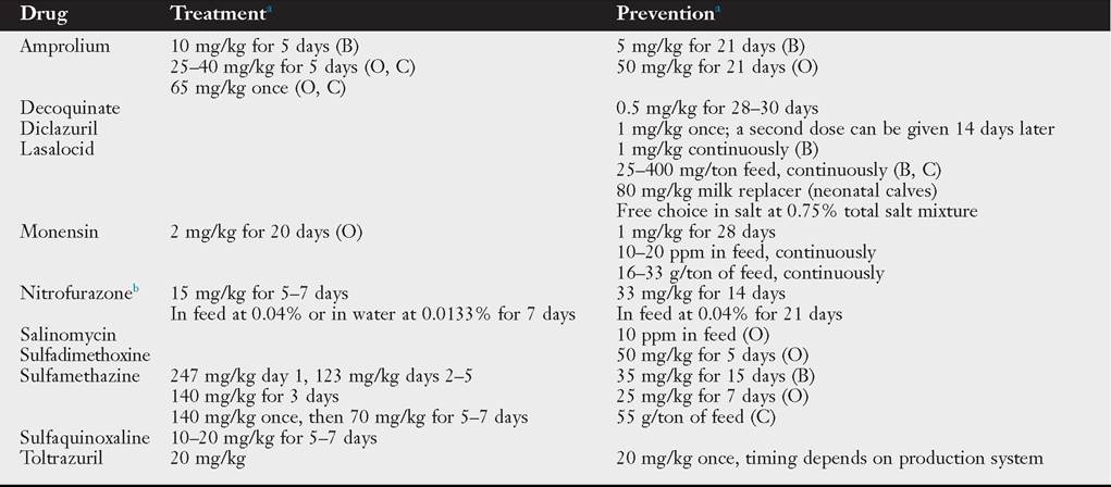
aOral administration; dose or regimen may vary among references.
bNo published data, and extralabel use prohibited in cattle in the United States.
B, Bovine; C, caprine; O, ovine.
Note: Coccidiostats used in poultry feeds may be toxic to ruminants.
contamination with and survival of oocysts should be addressed first; however, where these issues cannot be improved, it is best to use drugs prophylactically or metaphylactically rather than therapeutically to prevent losses. Administration of anticoccidials during this time does not completely prevent infection but prevents clinical disease without interfering with the production of protective immunity.4 Timing is also crucial, as improper metaphylactic use could result in wasted money or increased susceptibility to massive infections later in life.18 Preventive measures are best used during periods of highest risk, such as in calves after weaning or in crowded conditions. If coccidiosis occurs in nursing calves, addition of anticoccidials to creep feed may be necessary. In contrast, lambs and kids are most at risk while still nursing. Thus administration of anticoccidials for 30 days prior to lambing or kidding has been recommended to reduce environmental contamination of the lambing or kidding areas.2 Resistance to anticoccidials can occur and is a problem in the poultry industry; however, evidence for resistance in livestock is mostly circumstantial and unproven.2
OTHER STRATEGIES FOR MINIMIZING CLINICAL INFECTIONS. No vaccine for ruminant coccidiosis exists. Immunization of calves with a “trickle dose” of oocysts before turnout onto infected pasture attenuates the effects of natural challenge20 but is not commonly used. Use of growth implants containing estradiol and progesterone can attenuate the effects of high oocyst challenge in dairy calves but is not used for this purpose.21 It has been reported that oocysts of E. alabamensis may survive the hay-making process,22 and hay from infected pastures should not be used to feed susceptible calves. However, whether all species of Eimeria can survive the hay-making process or how common survival is with various types of hay production is unknown.
Concerns regarding the potential impact of anticoccidial drug use within the food chain and on the environment has sparked increasing interest and efforts to find novel agents characterized by low risks for consumer health and better environmental compatibility as well as sustainability.23 Evaluations of secondary plant metabolites, including polyphenols, flavonoids, tannins, and saponins, show promise in animal models (mice) or poultry,23 but few efforts have addressed livestock. Because much effort has focused on the anthelmintic properties of tannin-containing legumes and their use in controlling GINs of ruminants,24 particularly where anthelmintic-resistant populations of these parasites occur, some efforts evaluating their use as anticoccidials have occurred.25-31 The primary focus has been on small ruminants. Although all studies varied in their design (e.g., plus/minus concurrent GIN infections) and some lack certain information (e.g., species of Eimeria present), these data do indicate an overall reduction in oocyst excretion, which would aid in reducing environmental contamination.
■ Evaluation of Preventive Programs Evaluation of any coccidiosis prevention plan is based on the presence and extent of clinical disease. Herds or flocks that require preventive strategies do so because they usually have a history of prior disease. Program efficacy therefore is evaluated on the basis of a decrease in prevalence of clinically ill animals.
■ Clinical Management
DIAGNOSIS. Diagnosis is presumptive and based on history (which may include a stressful event), thorough knowledge of the herd or flock management, and clinical signs of severe diarrhea (usually young animals). Postmortem findings of inflammation, hyperemia, and thickening of the cecum with masses of gamonts and oocysts in mucosal scrapings will be confirmatory. Because pathogenicity varies by species, the value of fecal oocyst counts is debated. In general, fecal oocyst counts alone are not helpful if the species of coccidia present are not identified. The mere presence of oocysts does not itself indicate disease, as high numbers of nonpathogenic species can be present in animals with other causes of diarrhea. Clinical signs can also develop in the early stages of the life cycle before oocyst shedding. If fecal examinations are done during a suspected outbreak, multiple animals should be sampled. Due to the short life cycle, oocyst counts are highest in early patency and drop quickly after about 1 week. Thus fecal samples should be collected when diarrhea starts. Any standard fecal flotation method can be used to find oocysts.2-4
TREATMENT. Drugs used in the treatment of coccidiosis are listed in Table 49.5. Supportive therapy, when indicated, is primarily directed at replacing fluid losses and supporting the animal until the gut epithelium regenerates.
Anthelmintic Use1
SherriU A. Fleming
An anthelmintic is defined as an agent that acts to destroy or expel parasitic intestinal helminths. Anthelmintic drugs are used to treat animals that are suffering the adverse effects of a parasitic burden. They may also be used to prevent and minimize the economic losses associated with parasitic infection. Anthelmintics may be used on individual animals or a group of animals.
Drug Action
To use an anthelmintic properly, it is necessary to consider its mode of action, spectrum of activity, and duration of effect. The pharmacodynamics of an anthelmintic is specific for each species that is treated. Efficacy for a given drug may be defined as its ability to kill adult or larval parasites, suppress parasitic egg production, or promote the expulsion of worms from the gastrointestinal tract.
The emergence of drug-resistant strains of nematodes has necessitated the use of each class of anthelmintic to be customized to the premise where it will be used. Package inserts should be regarded as guidelines rather than gospel. Refer to the Anthelmintic Drugs and the Evaluation of Parasite Control Programs sections for particulars on anthelmintic performance. Readers should refer to the sections on horses, cattle, and small ruminants for more specific recommendations.
Anthelmintic Drugs (see Table 49.4)1,2
Avermectins and Milbemycins
The avermectins and milbemycins are macrocyclic lactones. They act by increasing the permeability of parasite cell membranes to chloride ions, which results in nonspastic paralysis and death of the parasite. These drugs may also act by potentiating presynaptic release of γ-aminobutyric acid (GABA), an inhibitory neurotransmitter, although this theory has been challenged.
These products have a high level and broad spectrum of activity against adult and larval nematodes. They are also effective against various ectoparasites such as mites, lice, ticks, bots, and cattle grubs; however, they are ineffective against flukes and tapeworms. These drugs suppress nematode egg production for longer than other anthelmintics. Because of its long duration of effect, moxidectin suppresses fecal egg counts and protects against reinfection for longer than ivermectin. Concern has been expressed about the environmental impact of these long-acting anthelmintics in grazing animals.
Several products have been developed for use in animals. The avermectins include ivermectin, abamectin, doramectin, and eprinomectin. The milbemycins include nemadectin and moxidectin. Oral (drench, sustained-release intraruminal device), topical (pour-on), and injectable formulations are available, depending on the drug. Ivermectin is safe to use during pregnancy, but currently it is not approved for use in lactating dairy animals or females of breeding age. Similarly, doramectin and moxidectin are not approved for use in female dairy cattle of breeding age. Eprinomectin is unique in that it has no withholding period for milk or meat, so there are no restrictions on its use in cattle. At labeled doses, moxidectin is a safe drug; however, accidental overdosage has caused neurologic signs in foals and miniature horses. Some of these products have extended efficacy periods but run the risk of having subthera- peutic periods, which increase the selection for resistant nematodes.
Benzimidazoles
The benzimidazoles (BZDs) comprise a large class of anthelmintics that interfere with parasitic carbohydrate metabolism by inhibiting the enzyme fumarate reductase. Many BZDs have been developed and marketed. They include albendazole, fenbendazole, mebendazole, oxfendazole, oxibendazole, parbendazole, ricobendazole, thiabendazole, triclabendazole, and the probenzimidazole drugs febantel and netobimin.
BZDs are widely used in horses and ruminants. In general, they exhibit a high degree of safety and a broad spectrum of activity against GINs and lungworms. Some members of this class (e.g., albendazole) are also active against liver flukes and certain cestodes in ruminants. Albendazole and netobimin can cause teratogenicity and embryo toxicity in sheep when given during early pregnancy. Fenbendazole, oxfendazole, and oxibendazole are considered safe for use in pregnant animals.
Resistance to BZDs has been documented in certain equine, ovine, and caprine parasites (see species-specific sections). In general, a strain of parasite resistant to one BZD drug quickly develops resistance to other BZDs or pro-BZDs, a phenomenon known as side resistance.
Levamisole (Imidazothiazoles)
Levamisole acts by causing neuromuscular depolarization and paralysis of the parasite. It has been widely used in ruminants to treat GINs and lungworm infections. However, levamisole resistance has become a problem in many areas. Levamisole is the only anthelmintic that has shown resistant parasites reverting to sensitivity if not used for several years. Recurrence of resistance will develop fairly rapidly with reuse of levamisole.
The dose of levamisole should be calculated carefully because toxic doses are only one to two times therapeutic doses. Signs of toxicity may mimic those of organophosphate (OP) toxicity, including muscle fasciculations around the lips and eyelids, hypersalivation, spastic movements, depression, and diarrhea. In ruminants, muzzle foam may develop after oral administration of the drug but usually disappears within a few hours after administration. In horses, transitory excitement has been seen after treatment. Levamisole is not recommended for use as an anthelmintic in horses. Concurrent administration of morantel, pyrantel, diethylcarbamazine, or OPs could enhance the toxic effects of levamisole.
Morantel and Pyrantel (Tetrahydropyrimidines)
The tetrahydropyrimidines morantel and pyrantel are cholinergic agonists that exert their anthelmintic effect by depolarizing neuromuscular junctions and causing irreversible paralysis of susceptible parasites, similar to the action of imidazothiazoles. Morantel is slower in its onset of action but much more potent than pyrantel. These products are effective against many species of adult nematodes but do not appear to be active against larval stages. Pyrantel is also effective against tapeworms in horses when given at twice the recommended dose. The margin of safety is relatively wide, and there is no contraindication to using morantel or pyrantel with other cholinergic drugs. However, it is recommended that morantel and pyrantel not be used concurrently and that neither be given with levamisole. Piperazine antagonizes the effects of morantel and pyrantel, so it should not be used with either of these drugs. Resistance to morantel and pyrantel has been documented in strains of H. contortus and in some Cyathostomes (equine small strongyles).
Organophosphates
OPs block neurotransmission by inhibiting acetylcholinesterase. Various formulations of OP drugs are available for treating gastrointestinal nematodiasis. Commonly used OPs include haloxon, coumaphos, trichlorfon, and dichlorvos. Toxicity occurs with these products in a dose-related manner, so dosages should be calculated with care. In addition, the potential danger to humans administering these products should not be overlooked. Atropine is recommended in cases of overdose in livestock.
Phenothiazine
The mode of action of phenothiazine (PTH) has not been clarified; it is thought to interfere with anaerobic metabolism of nematodes. The various formulations of PTH differ in purity and particle size. The purified product (99% PTH) with small particle size (2 μm) is the most effective.
Although PTH is effective against a wide spectrum of GINs, resistant strains of parasites have emerged in several species. The drug is synergistic with piperazine; combinations of these drugs have effective activity against PTH-resistant nematodes. PTH used in combination with piperazine can be administered at a much lower dose.
PTH toxicity has been reported. Toxic reactions include corneal inflammation, abortion, ataxia, hemolytic anemia, photosensitization, and nephrotoxicity. The drug should not be administered to debilitated or anemic animals or to animals in the last month of pregnancy.
Piperazine
Piperazine salts block neuromuscular transmission, resulting in paralysis of susceptible GINs. The worms are then passively removed from the gastrointestinal tract by intestinal peristalsis. Piperazines have low toxicity and are safe in young or pregnant animals. However, their spectrum of activity is limited, in practical terms, to ascarids. Piperazine must be used with caution in horses heavily infested with ascarids because the paralyzed ascarids can cause an impaction that may culminate in bowel rupture. Diethylcarbamazine is a piperazine derivative that has been used to control lungworm infection in sheep and cattle.
Monepantel
Monepantel is a new amino-acetonitrile derivative (ADD) compound that has a unique mode of paralyzing nematodes and is intended to be used as an oral drench. This product has been released for use in New Zealand but is not available in the United States at this time. The countries that do have this drug available are beginning to report resistance in nematode populations.3,4
Combination Dewormers
Combination dewormers have been shown to extend the effectiveness of anthelmintics. Many of the newer products reaching the market contain two or more classes of anthelmintics, but to date none of these products is available in the United States. One study showed that not only did combination anthelmintics not increase resistance development but there was some evidence of reversion to susceptibility in T. circumcincta populations.5
Other Anthelmintics
Praziquantel is a cesticidal drug that causes spastic paralysis, decreased glucose uptake, and disruption of the tapeworm’s tegument. Although not approved for this use, praziquantel is effective for treating tapeworm infestation in horses. It has also been used to control various cestodes in small ruminants. Nitroxinil is a halogenated phenol that has flukicide activity (Fasciola hepatica) and some nematodes and is available in some countries as an injection. Salicylanilides are a chemical class of antiparasitic active ingredients with efficacy against certain roundworms, tapeworms, and/or flukes. The class includes closantel (flukes and some nematodes), niclosamide (tapeworms and rumen flukes [Paramphistomum spp.]), oxyclozanide (some flukes), and rafoxanide (some flukes and nematodes).