ACCESSORY ORGANS
1. What are the names of the three pairs of salivary glands? Where are their openings? How are their secretions described?
2. Describe the location of the pancreas. What are its secretions?
3.
Which animal has clearly visible connective tissue septa surrounding each lobule of the liver?4. Study the triad of vessels and.ducts present in a liver lobule (Figure 12-21). What is the name of the large phagocytic cells that line the sinusoids of the liver lobules?
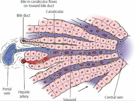
■ FIGURE 12-21 Portion of a liver lobule (highly magnified). Blood from the portal vein and hepatic artery flows into sinusoids (lined with Kupffer cells) and empties into the central vein. Bile travels in the opposite direction in canaliculi to empty into bile ducts in the triad areas. (From Ham AW. Histology. 7th edn. Philadelphia, PA: JB Lippincott, 1974.)
The salivary glands, pancreas, and liver supply secretions to the digestive tract and provide for digestion within the lumen. These secretions are in addition to those supplied by the many glands of the stomach and intestine and include electrolytes, water, digestive enzymes, and bile salts. This combination of secretions causes dietary substances to be degraded within the lumen so that the new substances can interact with the epithelial enzymes.
The salivary glands consist of three pairs of well-defined glands and some lesser-defined scattered salivary tissue. The larger glands are known as the parotid, mandibular, and sublingual salivary glands. These are connected to the oral cavity by one or more excretory ducts that have openings through the cheeks or ventral to the tongue. The general location of the salivary glands is shown in Figure 12-18 for the dog.
Salivary glands are serous, mucous, or mixed, depending on their secretion.
A serous secretion is a watery, clear fluid as compared with mucus, which is a viscid, tenacious material that acts as a protective covering throughout the digestive tract. A mixed gland secretes both serous and mucous fluids. Blood vessels and nerves enter each gland where the ducts exit. Innervation is provided by the sympathetic and parasympathetic divisions of the autonomic nervous system.The pancreatic gland has both endocrine and exocrine functions: it produces hormones (endocrine) and digestive (exocrine) secretions. The pancreas is always located near the first part of the duodenum and appears as an elongated gland of loosely connected aggregated nodules. The main pancreatic duct enters the first part of the duodenum close to the common bile duct, which comes from the liver (see Figure 12-8). In sheep and goats, a single pancreatic duct empties directly into the common bile duct so that a mixture of bile and pancreatic juice enters the duodenum. The accessory duct, if present, opens a short distance from the main duct. The endocrine portions of the pancreas, the pancreatic islets (formerly called islets of Langerhans), are isolated groups of cells scattered throughout the gland. The beta cells produce insulin and the alpha cells produce glucagon. Secretions from the alpha and beta cells are made directly into the blood (ductless secretions). Islet cells are clearly visible with a microscope (Figure 12-19).
The liver is a multipurpose organ; its production of bile and bile salts is only one of its many important functions. The epithelial cells of liver lobules are metabolically active in synthesis, storage, and metabolic conversions. The location of the liver varies among species, but it is always located immediately behind the diaphragm. In ruminants it tends to be on the right side. The lobules of the liver are clearly demarcated; in the pig they are surrounded by visible connective tissue septa. Other animals have fewer connective tissue divisions and accordingly cannot be seen.
The liver and its location in a pig are shown in Figure 12-20.The liver receives arterial blood for its many cells from the hepatic artery and venous blood through the portal vein from the stomach, spleen, pancreas, and intestines. Blood from both sources is circulated through the sinusoids, the second capillary bed of the hepatic portal system. Here it is detoxified and modified before reentering the central vein (second venous drainage of hepatic portal system) for return to the hepatic veins, and from there it proceeds to the heart through the caudal vena cava (see Chapter 9). The arrangement of a liver lobule with its triad of vessels and ducts (branches of portal vein, hepatic artery, and bile duct) is shown in Figure 12-21. Bile flows opposite to the direction of blood flow in the hepatic artery and portal vein branch.
The largest part of the macrophage system is present in the liver and is represented by the fixed macrophages, the Kupffer cells. Kupffer cells are highly phagocytic and remove foreign materials entering the blood from the stomach and intestines. They also remove tissue debris, such as old and fragile erythrocytes.
■