Atrial Cells Have Shorter Action Potentials Than Do Ventricular Cells
The previous description of cardiac ion channels, action potentials, and contractions is based on properties of normal ventricular cells. Atrial cells are basically similar, except that their action potentials are shorter than action potentials in ventricular cells.
As with ventricular cells, atrial cells have fast Na+ channels that open briefly at the beginning of an action potential and then become inactivated. Likewise, atrial slow Ca2+ channels open during the action potential, and K, channels close. The differences between atrial and ventricular cells are that atrial slow Ca2k channels typically stay open a shorter time than those in ventricular cells, and atrial K’ channels stay closed for a shorter time. As a result, the plateau of an atrial cell's action potential is shorter and not as “flat” as the plateau of a ventricular cell’s action potential (see Figure 19-3, bottom). As a consequence of having a shorter action potential, atrial cells have a shorter refractory period than do ventricular cells. Therefore the atrial cells are capable of forming more action potentials per minute than are ventricular cells; that is, the atria can “beat” faster than the ventricles. The implications of this difference are discussed later in this chapter.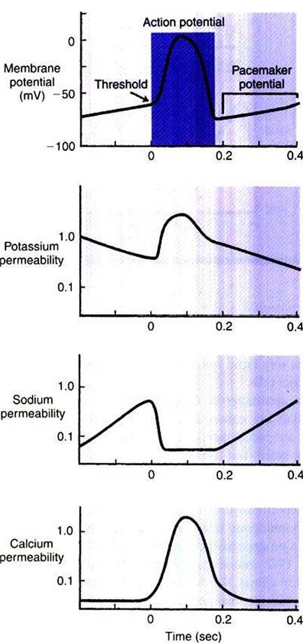
FIGURE 19-7 A cardiac pacemaker cell depolarizes spontaneously to threshold and initiates its own action potential (top).The spontaneous depolarization (called the pacemaker potential) is the result of a spontaneous, progressive decrease in K* permeability (second from top) and an increase in Nat permeability (second from bottom). An increase in Ca2* permeability makes a late contribution to the depolarization toward threshold (bottom). Once threshold level is reached, an action potential is produced. The action potential is driven primarily by a large, prolonged increase in Ca2* permeability.The absence of fast Na+ channels in pacemaker cells causes the upstroke of the pacemaker action potential to be much slower than that seen in nonpacemaker cells. (Compare with Figure 19-5.)