Specialized Ion Channels Cause Cardiac Pacemaker Cells to Depolarize to Threshold and Form Action Potentials
As mentioned, the cardiac pacemaker cells of the SA node spontaneously depolarize to threshold and then form action potentials. The spontaneous depolarization is called a pace-
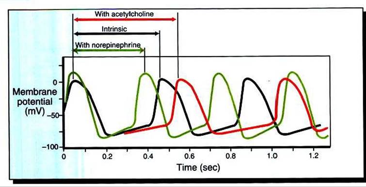
FIGURE 19-8 In the absence of ∩eurohumoral influences, a pacemaker cell of the SA node spontaneously depolarizes to threshold and initiates a series of action potentials, three of which are shown by the black line.The interval between action potentials under these conditions determines the intrinsic, or spontaneous, heart rate.
Acetylcholine decreases the rate of depolarization and therefore lengthens the interval between action potentials. Norepinephrine increases the rate of depolarization and therefore shortens the interval between action potentials.maker potential, and it is the distinguishing feature of a pacemaker cell (Figure 19-7, top). The action potentials of cardiac pacemaker cells typically have a rounded appearance; they lack the very rapid (phase 0) depolarization seen in ventricular and atrial cells.
The spontaneous depolarizations and rounded action potentials are consequences of the particular ion channels found in pacemaker cells. Pacemaker cells lack the usual voltage-gated fast Na4 channels. Instead, these cells have pacemaker Na" channels (also called “funny Na‘ channels**)» which close during an action potential and then begin to open again, spontaneously, once an action potential has finished. The spontaneous opening of the pacemaker Na* channels causes a progressive increase in the cell’s Na4 permeability (see Figure 19-7» second from bottom). The increase in Na4 permeability allows Na+ to enter the cell from the extracellular fluid, which depolarizes the cell toward threshold.
Pacemaker cells also have an unusual set of K4 channels, which participate in their spontaneous depolarization. At the end of one action potential, K* permeability in pacemaker cells is quite high, because most K+ channels are open. Then some K4 channels begin to close. As K1 permeability decreases, less K4 leaves the cells, which makes the cells progressively less negatively charged inside. Ca2* channels also make a small contribution to the pacemaker potential. Late in the pacemaker potential, just before a pacemaker cell reaches threshold, slow Ca24Channels begin to open, and Ca24 permeability begins to increase. The resulting entry of Ca2* into the ceil speeds its final approach to threshold. Thus the pacemaker potential is caused by the opening of pacemaker Na4 channels, the closing of K4 channels, and (late in the process) the opening of Ca24 channels. These spontaneous changes in Na4, K4, and Ca24 channels in pacemaker cells are in contrast to the stable status of the ion channels in normal, resting ventricular or atrial cells.Once threshold is reached in a pacemaker cell, an action potential occurs. The upstroke of the action potential is quite slow compared with the rapid, phase 0 depolarization in a normal ventricular or atrial cell, because there are no fast Na4 channels in pacemaker cells and therefore no sudden influx of Na+. The ion primarily responsible for the action potential in a pacemaker cell is Ca2+. Once threshold is reached, many of the cell’s slow Ca2' channels open. The permeability to Ca24 increases, and extracellular Ca24 flows into the cell. The action potentials in pacemaker cells are often called slow action potentials, because they lack a rapid, phase 0 depolarization and because they are caused primarily by the opening of slow Ca24 channels. In contrast, normal ventricular or atrial action potentials are called fast action potentials. Note, however, that all cardiac action potentials (even the “fast” ones) have a very long duration compared with action potentials in nerve or skeletal muscle cells.