BASIC PLAN AND DEVELOPMENT
Even a cursory examination of the head, intact or in sagittal section, shows that it consists of two principal parts. One, the neural part, comprises the brain together with the encasing structures; the other, the facial part, is much larger in most adult mammals and is formed by the jaws and the initial parts of the respiratory and digestive systems.
The distinction between neural and facial parts is already plain in embryos at the somite stage (Figure 2-28).At this stage of development the dorsal structures predominate, and the size and form of the head are largely determined by the brain.
The neural part (cranium) of the skull has its primordium in a series of cartilages that form ventral to the brain and are supplemented by cartilaginous capsules enclosing the primitive olfactory organs, eyeballs, and labyrinths of the ears. Later, “dermal bones” appear by ossification within the membrane that covers the brain to the sides and above; ultimately, all of these elements fuse with each other and with the bones of the face.
The ventral part of the head—the future face—is much smaller and at this stage blends smoothly with the neck, largely occupied by the heart. It exhibits a quite different pattern of segmentation imposed by the pharyngeal arches, serial thickenings of the unsplit mesoderm lateral and ventral to the rostral part of the foregut that becomes the pharynx.
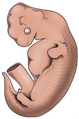
Figure 2-28 Pig embryo (1.5 cm) to show dominance of the neural over the facial part of the head at this stage.
| Table 2-1 Derivatives of the Pharyngeal Arches | |||
| Pharyngeal Arch | Skeleton | Muscles | Motor Innervation |
| First (mandibular) | Mandible (in part); certain ear ossicles (malleus and incus) | Muscles of mastication; mylohyoideus; digastricus (in part); tensor veli palatini; tensor tympani | Mandibular division of trigeminal nerve (V3) |
| Second (hyoid) | Hyoid apparatus (in part); ear ossicles (stapes) | Muscles of facial expression; digastricus (in part); stapedius | Facial nerve (VII) |
| Third | Hyoid apparatus (remaining part) | Stylopharyngeus caudalis; possibly other pharyngeal muscles | Glossopharyngeal nerve (IX) |
| Fourth (and subsequent arches) | Most laryngeal cartilages | Pharyngeal and laryngeal muscles; muscles of accessory nerve field | Vagus nerve (X); (medullary) part of accessory nerve (XI) |
The formation, significance, and detailed fate of these arches is not described here; at present it is sufficient to recall that a cartilaginous skeleton with associated musculature innervated by a specific cranial nerve develops within the core of each arch. Each arch is also supplied by an arterial loop connecting the ventral to the dorsal aorta.
The structures formed within the various pharyngeal arches are listed in Table 2-1; from this it can be seen that the cartilaginous parts ultimately make only a small contribution to the skeleton of the face. The definitive facial skeleton is mainly provided by dermal bones formed in the connective tissue of the jaws, although certain elements for a time obtain support from cartilaginous precursors such as the cartilage of the first arch and the nasal capsule.In most mammals the facial part enlarges disproportionately and comes to lie as much before as below the brain. Despite many qualitative and quantitative differences the basic arrangement is the same in all species. The relationships and topography of the major organs and cavities of the head should be studied before passing on to more detailed matters. Figures 4-2 and 4-3 provide the necessary information.