THE SKULL
The complete skeleton of the head comprises the skull, the mandible or lower jawbone, the hyoid apparatus, the ossicles of the middle ear, and the cartilages of the external ear, nose, and larynx.
The skull (in the narrower sense) is a mosaic of many bones, mostly paired but some median and unpaired, that fit closely together to form a single rigid construction. The separate elements, which are named individually, develop from independent centers of ossification and have, for the most part, well established homologies. In the young animal they are separated from each other by narrow strips of fibrous tissue—cartilage in a few situations—and this pattern of joints or sutures provides for growth. Once growth has ceased, sutures are no longer necessary and ossification extends into the connective tissue, finally welding the bones together. This process is drawn out, and it may never be completed; the outlines of most bones are therefore discernible, even in skulls of old animals. Acquaintance with the names, positions, and approximate extents of the individual bones (Figure 2-29) is essential as it provides a useful system of reference to regions of the head, but a detailed knowledge of the disarticulated units has little practical value; most readers are better served by an appreciation of the skull as a whole.
Conventional descriptions are based on the views obtained from various directions with the skull resting on a flat surface, even though this may not be its habitual orientation in life. In most views the two distinct portions of the skull are immediately apparent: the caudal part encasing the brain and the rostral part supporting the face. The orbits, the fossae containing the eyeballs, are part of the face but lie at the boundary. In most domestic animals the facial part of the skull is larger than the neural part and is situated mainly in front of this.
However, the ratio varies among species and also with breed, age, and individual conformation. The many particular differences make it impossible to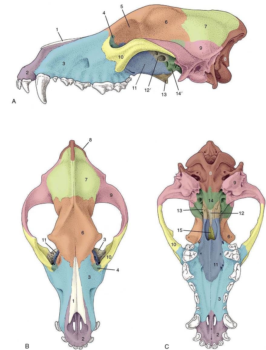
Figure 2-29 Lateral (A), dorsal (B), and ventral (C) views of the canine skull to show the extents of the cranial bones. 1, Nasal bone; 2, incisive bone; 3, maxilla; 4, lacrimal bone; 5, orbit; 6, frontal bone; 7, parietal bone; 8, occipital bone; 9, temporal bone; 10, zygomatic bone; 11, palatine bone; 12, presphenoid; 12', wing of presphenoid; 13, pterygoid bone; 14, basisphenoid; 14', pterygoid process of basisphenoid; 15, vomer.
provide even a general description of the skull that is valid for all species.
The Skull of the Dog
This initial account is of the skull of an adult dog of average (mesaticephalic) conformation, neither shortheaded (brachycephalic) like a Pekingese nor longheaded (dolichocephalic) like a Borzoi. Some salient breed differences are mentioned later (p. 374).
In the dorsal view (Figure 2-30), the ovoid cranium meets the bones of the face where the zygomatic processes (Figure 2-30Z√') of the frontal bones project laterally to form the dorsocaudal parts of the orbital walls. The caudal extremity of the cranium is marked by the external occipital protuberance in the midline; its demarcation from the caudal (nuchal) surface is completed by the nuchal crests that extend laterally to each side. The median sagittal crest that extends forward from the occipital protuberance is most prominent in robust, well-muscled animals. All these features are easily palpated in life. The dorsal and lateral surfaces of each half of the cranium blend in a continuous and slightly roughened surface from which the temporalis muscle arises. Rostral to the zygomatic processes of the frontal bones the dorsal surface of the skull dips, sometimes quite markedly, before continuing as the straight and narrow dorsum of the nose.
This ends at the wide nasal aperture beyond which the bony skull is prolonged by pliant nasal cartilages.The orbit is the most prominent feature of the lateral view (Figure 2-31). Behind the orbit, the dorsolateral part of the braincase forms the wall of the temporal fossa (Figure 2-31Z16). The ventrolateral part is more complicated and presents the zygomatic arch and ear regions. The zygomatic arch (Figure 2-31Z15) springs free from the braincase and, bowing laterally, passes below the orbit to rejoin the facial part of the skull. It is formed by two bones, the squamous temporal and zygomatic, which meet at an overlapping suture. The ventral surface of the caudal part of this arch carries the articular surface for the mandible, shaped as a transverse gutter in this species; the articular area continues caudal to this onto the rostral surface of a ventral projection, the retroarticular process (Figure 2-31Z6). The large, smooth dome of the tympanic bulla (Figure 2-31Z9) (enclosing part of the cavity of the middle ear) and the rough mastoid process lie behind the retroar- ticular process. Three openings are present in this region of the skull: the retroarticular foramen emits a major vein draining the cranial cavity, the stylomastoid foramen gives passage to the facial nerve, and the external acoustic meatus is, in the fresh state, closed by a membrane (eardrum) that separates the canal of the external ear from the cavity of the middle ear. The paracondylar process (Figure 2-31Z11) is conspicuous at the caudal limit of the skull.
The orbit is funnel shaped, and in the macerated state its walls are very incomplete. In life the orbital rim
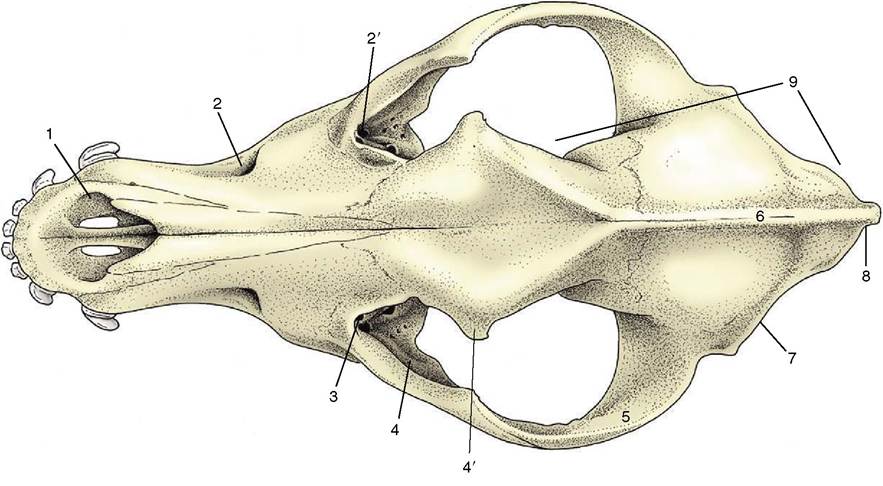
Figure 2-30 Dorsal view of canine skull. 1, Nasal aperture; 2, infraorbital foramen; 2', maxillary foramen; 3, fossa for lacrimal sac; 4, orbit; 4', zygomatic process of frontal bone; 5, zygomatic arch; 6, external sagittal crest; 7, nuchal crest; 8, external occipital protuberance, 9, cranium.
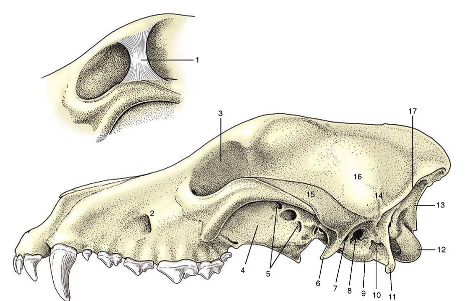
Figure 2-31 Lateral view of canine skull. 1, Orbital ligament (inset); 2, infraorbital foramen; 3, orbit; 4, pterygopalatine fossa; 5, optic canal, orbital fissure, and rostral alar foramen; 6, retroarticular process; 7, retroarticular foramen; 8, external acoustic meatus; 9, tympanic bulla; 10, stylomastoid foramen; 11, paracondylar process; 12, occipital condyle; 13, nuchal surface; 14, mastoid process; 15, zygomatic arch; 16, temporal fossa; 17, nuchal crest.
is completed by a ligament (Figure 2-31/7) that connects the zygomatic process of the frontal bone to the zygomatic arch. Ventrally the orbital cavity is continuous with the pterygopalatine fossa (Figure 2-31/4), but in the fresh state these regions are separated by the periorbita, a dense fascial sheet that completes the definition of the orbit. Two groups of foramina are visible in this region. The caudal group (Figure 2-31/5) comprises the optic canal, orbital fissure, and rostral alar foramen. The optic opening, placed at the apex of the conical orbital cavity, is the portal of entry of the optic nerve. The more ventral orbital fissure transmits the nerves (ophthalmic, oculomotor, trochlear, and abducent) that supply ancillary structures of the eye and the external ophthalmic vein. Most ventrally the rostral alar foramen provides a common opening for the maxillary nerve, passing from the cranial cavity, and the maxillary artery, which transverses a canal (alar canal) in the sphenoid bone.
The rostral group of foramina comprises the maxillary, sphenopalatine, and caudal palatine foramina. The maxillary foramen (Figure 2-30/2') leads to the infraorbital canal, the sphenopalatine foramen to the nasal cavity, and the caudal palatine to the palatine canal, which emerges on the hard palate; each opening conveys like-named branches of the maxillary artery and nerve. More dorsally the rostral orbital wall contains the lacrimal fossa for the lacrimal sac (Figure 2-30/5).
An opening in the depth of the fossa leads to a passage that conveys the nasolacrimal (tear) duct to the nose.The infraorbital foramen (Figure 2-30/2) is the most prominent feature of the lateral aspect of the face and is easily palpable in the live animal; it is the site of emergence of the infraorbital nerve, which continues from the maxillary nerve through the infraorbital canal. Toward the alveolar margin the facial skeleton is molded over the roots of the teeth, most especially over the large root of the canine tooth.
In the ventral view (Figure 2-32), three regions of the skull are distinct: the base of the cranium, the choanal region where the nasal cavities open into the pharynx, and the hard palate. The first shows at its caudal limit the ovoid, obliquely oriented occipital condyles that flank the foramen magnum (Figure 2-32/72) through which the spinal cord connects with the brain. Rostral to this the median area is generally flat, although
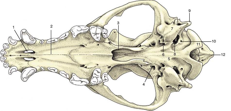
Figure 2-32 Ventral view of canine skull. 1, Palatine fissure; 2, hard palate; 3, choanal region; 4, oval foramen; 5, base of cranium; 6, foramen lacerum; 7, tympanic bulla; 8, jugular foramen; 9, paracondylar process; 10, hypoglossal canal; 11, occipital condyle; 12, foramen magnum.
midway along its length, tubercles are present for the attachment of muscles that flex the head on the neck. The tympanic bulla and paracondylar process occupy much space to each side. The medial aspect of the bulla (Figure 2—32/7) meets the occipital bone, and this fusion separates two openings that are confluent in some other species (e.g., horse; see Figure 2-37), namely, the more caudal jugular foramen and the more rostral foramen lacerum (Figure 2-32/8,6). The glossopharyngeal, vagus, and accessory nerves emerge through the jugular foramen together with a large vein draining the interior of the cranium. Between the jugular foramen and the condyle is the hypoglossal canal, which transmits the hypoglossal nerve.
Lateral to the foramen lacerum, small fissures exist for the exit of the chorda tympani (a branch of the facial nerve) and for the communication of the cartilaginous auditory tube with the cavity of the middle ear. Rostral to these is the prominent oval foramen (Figure 2-32/4), through which the mandibular nerve emerges.
The openings (choanae) that lead from the nasal cavities to the nasopharynx are the main features of the middle part of the ventral aspect. The choanal region is bounded dorsally by the floor of the cranium and laterally by the thin plates of bone whose outer surfaces were earlier noted as forming the medial walls of the pterygopalatine fossae. The soft palate, which arises from the free margin of the hard palate, in life provides the floor of the space—essentially the first part of the nasopharynx—enclosed by these formations. The palate, which lies rostral to this, is broad behind and narrower in front. It is margined by the alveoli or sockets in which the upper teeth are implanted. Toward its rostral extremity, it is perforated by the large bilateral palatine fissures. Several smaller foramina toward the caudal extremity of the palate are rostral openings of the palatine canal.
The nuchal surface (Figure 2-31//5), broadly triangular, is limited dorsally by the external occipital protuberance and the nuchal crests. Its lower part presents the foramen magnum, the occipital condyles, and the paracondylar processes. The remainder of the surface is roughened for the attachment of dorsal muscles of the neck.
The apex of the skull is formed by the nasal aperture situated dorsal to the rostral extremities of the jaws that carry the incisor teeth.
The cavities of the skull are described with the respiratory system (Chapter 4), central nervous system (Chapter 8), and ear (Chapter 9).
The lower jaw or mandible comprises two parts (Figure 2-33). In the dog these are firmly but not rigidly united by the connective tissues of the mandibular symphysis. Each half is divided between a body, or horizontal part, and a ramus, or vertical part. The body carries the alveoli of the lower teeth and is laterally compressed. Except at its rostral extremity, it diverges from its fellow to bound an intermandibular space. Toward its rostral extremity the lateral surface presents several mental foramina, one generally much larger than the rest;
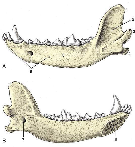
Figure 2-33 Lateral (A) and medial (B) views of the left half of the canine mandible. 1, Coronoid process; 2, vertical part (ramus); 3, condylar process; 4, angular process; 5, horizontal part (body); 6, mental foramina; 7, mandibular foramen; 8, symphysial surface.
through these emerge the mental branches of the inferior alveolar nerve and vessels. The ramus (Figure 2-33/2) is wider but less robust. Its dorsal extremity ends in the high recurved coronoid process, which projects into the temporal fossa and gives attachment to the temporalis muscle, and the lower and more caudal condylar process (Figure 2-33/3), which carries an articular head shaped like a portion of a truncated cone. The lower part of the caudal margin of the ramus carries the projecting angular process that enlarges the areas of attachment of the masseter and medial pterygoid muscles. The lateral surface is scooped out to provide a roughened depression where the masseter inserts. The medial surface gives insertion to the pterygoid muscles and also presents the large mandibular foramen (Figure 2-33/7), where the inferior alveolar vessels and nerve enter the bone.
The hyoid apparatus consists of a series of bony rods, jointed together and forming a means of suspending the tongue and larynx from the skull. The names given to the several parts are shown in Figure 2-34, which illustrates their arrangement and the attachment of the apparatus as a whole to the temporal region of the skull. The transversely placed basihyoid may be palpated within the intermandibular space; other parts are palpable—indeed their positions are visible—when the walls of the pharynx are inspected through the mouth.
Some Comparative Features of the Skull
When equipped with the mandible the skull of the cat (Figure 2-35) appears globular. Several features combine to create this conformation: the rounded cranial capsule, surmounted by a short, often weak sagittal crest, and corresponding closely to the contours of the brain; the very salient convex zygomatic arches; and the relative shortness of the face, which may account for as little as 20% of the total length. Breed differences are more pronounced than sometimes supposed. The skulls of Siamese and similar cats have much longer faces, which often blend smoothly with the cranium without any break (stop) in the dorsal contour. In contrasting types, for example, the Persian, the face is short and shallow and the stop is prominent.
The orbital region is distinctive. The orbits are large, face more directly forward than in the dog, and have more complete bony margins. The frontal process of the zygomatic bone and the zygomatic process of the frontal bone leave only a small gap in the ovoid margin to be
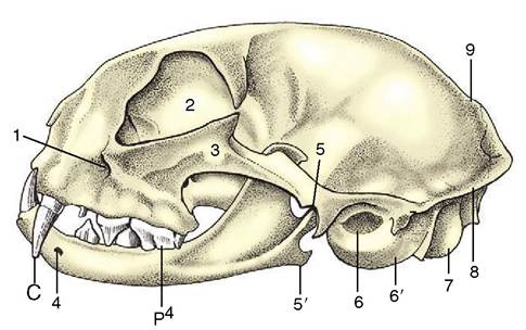
Figure 2-35 Feline skull with mandible. 1, Infraorbital foramen; 2, orbit; 3, zygomatic arch; 4, mental foramen; 5, temporomandibular joint; 5', angular process of mandible; 6, external acoustic meatus; 6', tympanic bulla; 7, occipital condyle; 8, nuchal crest; 9, sagittal crest; C, canine tooth; P4, upper fourth premolar.
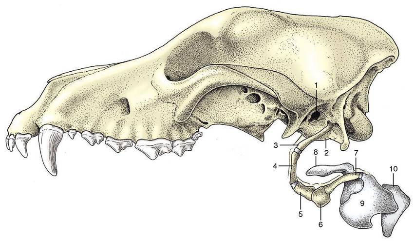
Figure 2-34 Hyoid apparatus and larynx suspended from the temporal region of a canine skull. 1, External acoustic meatus; 2, tympanic bulla; 3, stylohyoid; 4, epihyoid; 5, ceratohyoid; 6, basihyoid; 7, thyrohyoid; 8, epiglottic cartilage; 9, thyroid cartilage; 10, cricoid cartilage.
closed by the orbital ligament. The zygomatic arch is surprisingly strong where it contributes to the orbital rim. The infraorbital foramen is placed close to the rostroventral part of the orbit, where it may be palpated.
On the ventral aspect, the hard palate is short, wide, and carries alveoli for only four cheek teeth. That for the largest (P4) of these teeth is located dangerously close to the orbit, which may become involved in a spreading alveolar abscess. Caudally, the deep gutter of the temporomandibular articulation is bounded by a prominent retroarticular process. The very large tympanic bulla is so salient that it may be palpated between the caudal part of the zygomatic arch and the wing of the atlas.
As in the dog, the halves of the mandible do not fuse, even in old age, and a small degree of movement is allowed at the mandibular symphysis. Each half carries sockets for only three cheek teeth.
The equine skull (Figure 2-36) is characterized by a relatively long face, a feature that develops further with increasing size; it is therefore more pronounced in mature than in juvenile animals and in large than in small breeds. The cranium is relatively narrow and generally not unlike that of the dog. The external sagittal crest is weaker. The forehead is wide between the origins of the zygomatic processes of the frontal bones, which bend ventrally to join the zygomatic arches.
The zygomatic arch (Figure 2-36/7) is conspicuously strong, even without taking into account the extra support it obtains from the zygomatic process connecting it with the frontal bone. It is not bowed laterally to any extent and carries a rather complicated articular surface on its caudoventral aspect; this comprises a rostral tuber, an intermediate fossa, and a salient retro-
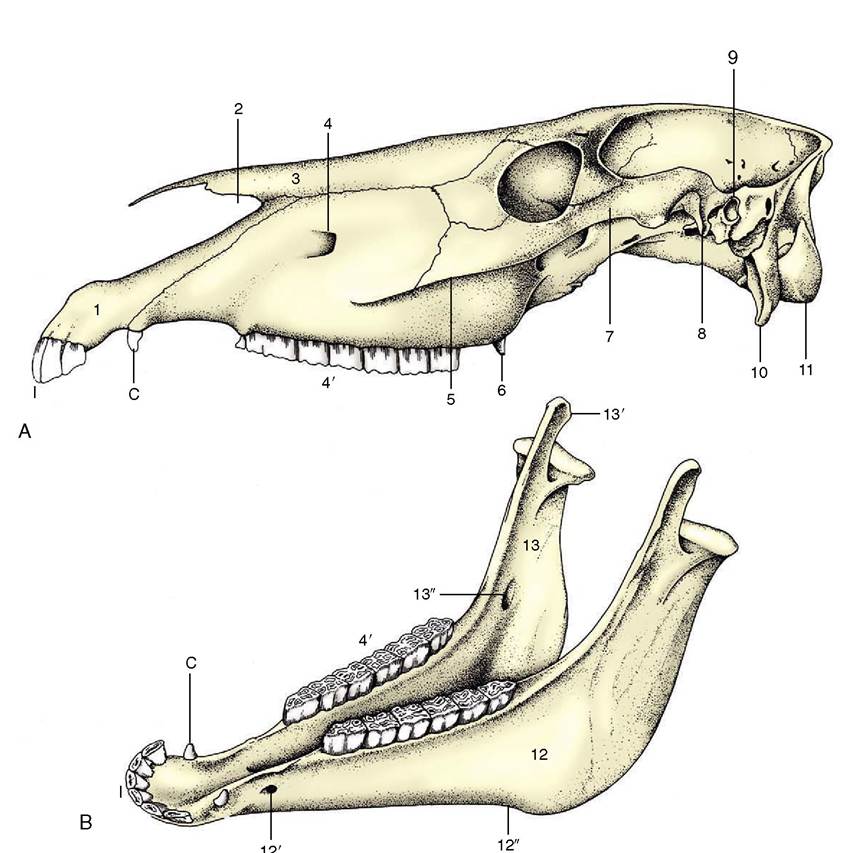
Figure 2-36 A, Equine skull, and B, equine mandible. 1, Incisive bone; 2, nasoincisive notch; 3, nasal bone; 4, infraorbital foramen; 4', cheek teeth; 5, facial crest; 6, hamulus of pterygoid bone; 7, zygomatic arch; 8, retroarticular process; 9, external acoustic meatus; 10, paracondylar process; 11, occipital condyle; 12, horizontal part (body) of mandible; 12', mental foramen; 12", vascular notch; 13, vertical part (ramus) of mandible; 13', coronoid process; 13", mandibular foramen; I, incisors; C, canine tooth (present only in the male).
articular process (Figure 2-36/8). The orbit faces almost laterally and has a complete bony rim. A large maxillary tuberosity appears to continue the alveolar process directly. The zygomatic arch is continued rostrally, beyond the orbit, as a prominent ridge on the lateral surface of the face. This ridge, the facial crest (Figure 2-36/5), runs parallel to the dorsal contour of the nose and ends above a septum between the alveoli of the third and fourth cheek teeth in the adult.
A deep (nasoincisive) notch separates the pointed nasal bone from the incisive bone (Figure 2-36/1,2,3). This notch and the rostral end of the facial crest are both very easily identified landmarks; they are used as guides to the position of the infraorbital foramen, which lies a little caudal to the middle of the connecting line (Figure 2-36/4).
The features visible on the ventral view lie more or less on one level. The caudal part of this surface is distinguished by the large and very salient paracondylar processes (Figure 2-36/10) and the jagged outlines of the large openings to each side of the occipital bone. Each opening results from the failure of the temporal bone to reach the lateral margin of the occipital bone, which permits the confluence of several foramina that are distinct in the dog. The caudal part is the equivalent of the jugular foramen; the cranial part (foramen lacerum) combines the oval and carotid foramina (Figure 2-37/7,6). In life the greater part of the large opening is occluded by membrane that leaves barely sufficient passage for the various nerves and vessels. The tympanic bulla is not prominent, but styloid (for the hyoid apparatus) and muscular processes of the temporal bone are well developed.
The choanae lie almost in the plane of the hard palate. The vertical plate of bone that separates the choanal from the pterygopalatine region carries a prominent hamular process (Figure 2-36/6). The palate is flat and unremarkable. The greater part of its margin is occupied by the alveoli of the incisor and cheek teeth.
A well-marked external occipital protuberance is present on the nuchal surface, midway between the nuchal crest and the dorsal margin of the foramen magnum.
The mandible is massive, and its right and left halves diverge at a relatively small angle (Figure 2-36, B). The symphysis becomes obliterated quite early, usually about 2 years after birth. The lower margin carries a prominent vascular notch where the facial vessels wind onto the face (Figure 2-36/72"). The ramus is high, the coronoid process projects far into the temporal fossa, and the articular process carries the ovoid articular surface well above the occlusal plane of the cheek teeth.
The parts of the hyoid apparatus (see Figure 4-8) are of different proportions to their counterparts in the dog and are laterally compressed. A substantial lingual process projects from the basihyoid into the root of the tongue.
Figure 2-37 Left caudolateral parts of the base of the equine (A) and canine (B) cranium, showing portions of the occipital (O), sphenoid (S), and temporal (T) bones; ventral view (schematic). 1, Foramen magnum; 2, occipital condyle; 3, hypoglossal canal; 4, jugular foramen; 5, foramen lacerum; 5', petrooccipital suture; 6, carotid canal; 6', carotid notches; 7, oval foramen; 7’, oval notch.
The bovine skull (Figure 2-38) is relatively short and wide: its general form is pyramidal. Cornual (horn) processes project from the frontal bones of horned breeds where the dorsal, lateral, and nuchal surfaces meet; their size and direction vary greatly with breed, age, and sex. The very wide and flat frontal region is bounded by a prominent temporal line that overhangs the deep temporal fossa and confines this to the lateral aspect of the skull. The forehead continues smoothly into the dorsal contour of the nose.
The principal features of the lateral aspect are the confinement of the temporal fossa and the elevation of the orbital rim above its surroundings. The rim is complete and is formed by the meeting of processes from the zygomatic and frontal bones in its caudal part. There is no facial crest, only a discrete facial tuberosity from which the rostral part of the masseter arises. The infraorbital foramen is directly above the first cheek tooth, rather low toward the palate.
The ventral surface is very uneven, and the cranial base is located in a considerably more dorsal plane than the palate. The temporal and occipital bones are separated by a narrow fissure, which is an arrangement intermediate between the suture of the dog and the wide opening of the horse and pig. The tympanic bulla is prominent and laterally compressed. The choanae are separated by the caudal prolongation of the ventral part of the nasal septum and are enclosed laterally by very extensive plates of bone. The palate, long and narrow, is bounded by high alveolar processes. Of course, no alveoli are present for incisor or canine teeth, which are lacking in the upper jaws of ruminants.
The mandibular symphysis ossifies late, if at all, in ruminants. In general, the mandible is weaker than that of the horse, which is a feature very apparent in the body of the bone with its gently convex ventral border. The coronoid process is high and caudally inflected. The articular surface is concave and widened laterally.
The few remarks necessary regarding the skulls of the small ruminants and pig are found on pages 646 and 752, respectively.
The Joints of the Head
The articulations between the skull and mandible (temporomandibular joints) and that between the halves of the mandible (mandibular symphysis) are appropriately considered in the following chapter (p. 112) because the teeth, the muscles of mastication, and the joints form a single functional complex.