Biological Membranes Are a Mosaic of Proteins Embedded in a Phospholipid Bilayer
Before continuing the discussion of the cellular basis of physiological control, an additional basic structure must be introduced. This is the phospholipid bilayer of the biomembranes of cells.
Phospholipids are molecules that have two long tails of hydrophobic fatty acid and a head containing a charged, hydrophilic phosphate group. Under appropriate aqueous conditions, these molecules spontaneously form an organized membrane structure, similar to the film of a soap bubble. This filmy layer is composed of two layers (a bilayer) of phospholipid molecules. In both layers the hydrophilic heads point outward to hydrogen bond with water, and the oily, fatty-acid tails point inward, toward one another and away from the water. Proteins embedded in this lipid bilayer, called intrinsic membrane proteins or just membrane proteins, produce the fluid mosaic structure of biomembranes shown in Figure 1-6. All biological membranes share this fluid mosaic structure, whether the membrane is the outer plasma membrane separating cytoplasm from extracellular fluid or the membrane surrounding intracellular membranous organelles such as endoplasmic reticulum or lysosomes. It is called a “fluid mosaic” because of the mosaic of proteins among phospholipids, and because the phospholipid layer is fluid; proteins can move around and diffuse within the plane of the bilayer “like icebergs floating in a phospholipid sea” (the apt phrase of S.J. Singer, one of the originators of the model).Biological membranes are another crucial molecular structure underlying physiological control. The basic fluid mosaic structure serves four broad functions: (1) compartmentalization, (2) selective transport, (3) information processing and transmission, and (4) organizing biochemical reactions in space.
Compartmentalization is the ability to separate and segregate different regions by composition and function.
For example, the lysosome is a membranous organelle within cells that contains hydrolytic (digestive) enzymes that can potentially digest the cell. The lysosomal membrane compartmentalizes these potentially harmful enzymes, segregating them from the bulk cytoplasm. The rigor mortis, mentioned earlier, that begins shortly after death is transitory because on death the lysosomes begin to break open, releasing their enzymes, and the actomyosin cross-bridges are eventually digested apart.Clearly, the membrane cannot keep a compartment perfectly sealed; material must enter and leave the cell and its internal compartments. Selective transport results partly from the properties of the phospholipid bilayer but mostly from transport proteins embedded in the membrane. These proteins are characteristically selective in their transport functions; for example, the protein that is the specialized ion channel underlying neuronal signaling is 15 times more permeable to sodium ions (Na’) than to potassium ions (IC). Transport is a major topic of cell physiology and is discussed in more detail later.
If the cells of an organism are to respond to external changes, they must receive information about the state of the outside world. Just as we higher animals have our sensory organs—eyes, ears, nose, and so forth—arrayed on our outside surface, so too cells have most of their environmental information processing and transmission apparatus on their external surfaces. These are intrinsic membrane proteins of the plasma membrane, called receptors, that serve a purely informational function, as discussed earlier.
At first glance it might seem odd that a fluid membrane could provide spatial organization for biochemical reactions.
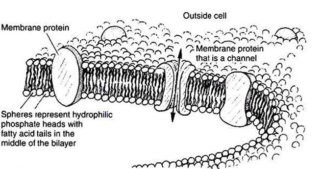
FIGURE 1-6 Fluid mosaic model for biomembranes. Biomembranes consist of a lipid bilayer in which membrane proteins are embedded.
However» returning to the “icebergs in a phospholipid sea” analogy» random collisions are much more likely for material in the two-dimensional membrane surface than for material moving through the three-dimensional volume of the cytoplasm. (If the Titanic had been able to dive or fly» it would have had additional ways to avoid the iceberg!) This much larger collision probability is exploited by the cell in a number of physiological processes. Membranes can also be fenced off into distinct regions» across which there is limited diffusion of membrane proteins. For example» certain cells in the kidney have two membrane regions that are quite distinct with respect to transport proteins, which is important in the regulation of salt and water balance by the animal.