Blood Pressure Represents a Potential Energy That Propels Blood Through the Circulation
The systemic circulation has the aorta as its inlet point and the venae cavae as its outlet. The remainder of the circulation (i.e., right heart, pulmonary circuit, and left heart) is, by definition, the central circulation.
Blood enters the central circulation from the venae cavae and leaves the central circulation through the aorta.Figure 22-1 shows the normal pressure profile in the systemic circulation. This figure portrays the pressures that would be measured if a miniature pressure gauge were inserted into the various vessels that blood encounters in its passage through the systemic circulation. The blood pressure is highest in the aorta (typically, mean aortic pressure is 98 mm Hg) and lowest in the venae cavae (typically, 3 mm Hg). It is this pressure difference that forces blood to move (via bulk flow) through the systemic vessels; that is, 98 minus 3 mm Hg is the perfusion pressure (difference) that drives systemic blood flow (the concept of perfusion pressure is described in Chapter 18).
Aortic blood pressure can be thought of as the potential energy available to move blood; the decrease in pressure in the sequential segments of the systemic circuit represents the amount of this potential energy that is “used up” in moving blood through each segment. Pressure energy is used up through/γ∕i7∕oh, which is generated as the molecules and cells of blood rub against each other and against the walls of the
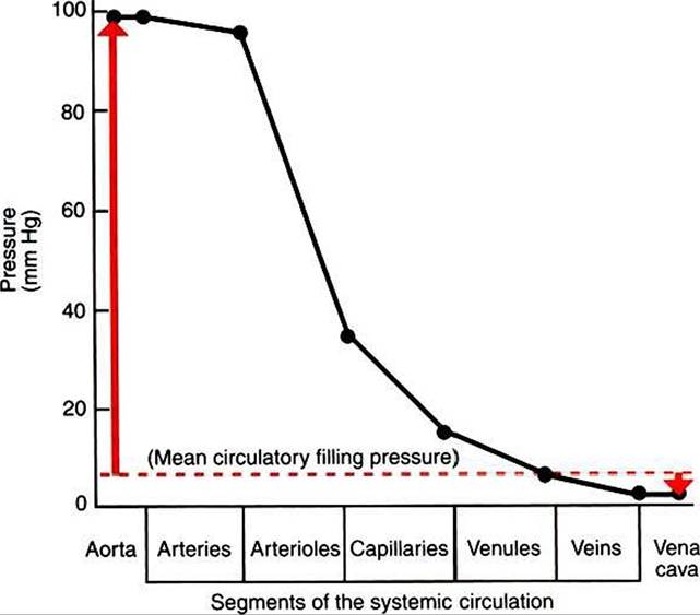
FIGURE 22-1 Graph of the blood pressures (hydrostatic pressures) that typically exist in the systemic circulation of a dog at rest (solid black line).The actual blood pressure in the aorta and arteries is pulsatile, increasing with each cardiac ejection and falling between ejections.The values plotted here are the average (mean) values of those pulsatile pressures.
Mean circulatory filling pressure (dashed red line) is the pressure that would exist if the heart were stopped. Red arrows show the contrasting directions and magnitudes of the pressure changes that would occur in the aorta and vena cavae if a stopped heart were restarted and cardiac output returned to normal (see text). All pressures are measured at heart level, with reference to atmospheric pressure (taken as zero).blood vessels. The energy used up through friction is actually converted to heat, although the increase in the temperature of the blood and blood vessels as a result of friction is very small.
The amount of the blood pressure energy used up in each of the sequential segments of the systemic circulation depends on the amount of friction or resistance that the blood encounters. The aorta and large arteries offer very little resistance to blood flow (very little friction), so the blood pressure decreases only a little in these vessels (from 98 to about 95 mm Hg). The greatest pressure decrease (greatest loss of pressure energy through friction) occurs as blood flows through arterioles; that is, the resistance to blood flow is greater in the arterioles than in any other segment of the systemic circulation. The capillaries and the venules offer a substantial resistance to blood flow, but the resistance (and therefore the pressure decrease) is not as great in these vessels as it is in the arterioles. The large veins and the venae cavae are low-resistance vessels, so little pressure energy is expended in driving the blood flow through these vessels.
The pumping of blood by the heart maintains the pressure difference between the aorta and the venae cavae. If the heart stops, blood continues to flow for a few moments from the aorta toward the venae cavae. As this blood leaves the aorta, the aortic walls become less distended, and the blood pressure inside the aorta decreases. As extra blood accumulates in the venae cavae, they become more distended than before, and the blood pressure inside the venae cavae increases.
Soon, there is no pressure difference between the aorta and the venae cavae. Blood flow in the systemic circuit ceases, and the pressure everywhere in the systemic circulation is the same. It has been demonstrated experimentally that this eventual pressure is about 7 mm Hg. This pressure, in a static circulation, is called the mean circulatory filling pressure. The mean circulatory filling pressure is above zero, because there is a “fullness” to the circulation; that is, even if the heart stops, blood still distends the vessels that contain it. The vessel walls, being elastic, recoil (“push back”) against this distention, which accounts for the persistence of pressure in the circulation even if the heart stops. If a transfusion of blood is administered to an animal with the heart stopped, the vessels become more distended, and the mean circulating filling pressure rises above 7 mm Hg. Conversely, if blood is removed from an animal with the heart stopped, the pressure everywhere falls to a level below 7 mm Hg.Consider what happens if the heart is restarted in an animal after the pressure has equalized everywhere at 7 mm Hg. With each heartbeat, the heart takes some blood out of the venae cavae and moves it into the aorta. The volume of blood in the venae cavae decreases, so the venae cavae become less distended and vena caval pressure drops below 7 mm Hg. The volume of blood in the aorta increases, so the aorta becomes more distended and aortic pressure rises above 7 mm Hg. The vena caval pressure drops about 4 mm Hg (from 7 to 3 mm Hg), and the aortic pressure rises about 91 mm Hg (from 7 to 98 mm Hg). It is important to understand why the pressure decreases only a little in the venae cavae and why it increases so much in the aorta, even though the volume of blood removed from the venae cavae with each heartbeat is the same as the volume of blood added to the aorta. The reason is that the veins are much more compliant (distensible) than the arteries; one can add or remove blood from veins without changing the venous pressure very much, whereas the addition or removal of blood from arteries changes the arterial pressure a great deal.
A compliant vessel readily distends when pressure or volume is added. It yields to pressure. By definition, compliance is the change in the volume within a vessel or a chamber divided by the associated change in distending (transmural) pressure, as follows:

Compliance corresponds to the slope of a volume-versus- pressure graph. Veins are about 20 times more compliant than arteries (as illustrated in Figure 22-2). Therefore, veins can accept or give up a large volume of blood without incurring much of a change in pressure. Veins readily expand or contract to accommodate the changes in blood volume that occur with fluid intake (c.g., drinking) or fluid loss (e.g., sweating). Veins thus function as the major blood volume reservoirs of the body. In contrast, arteries function as pressure reservoirs, providing the temporary storage site for the surge of pressure energy that is created with each cardiac ejection. Arteries are tough vessels, with low compliance. Therefore, arteries can accept a large increase in pressure during a cardiac ejection and then sustain the pressure high enough between cardiac ejections to provide a continuous flow of blood through the systemic circulation.