BLOOD VESSELS
1. What is the relationship of the blood vessel linings to the heart?
2. What is the order of blood vessels from the ventricles back to the atria? Which one of the vessel divisions permits exchange with interstitial fluid?
3.
What is the function of elastic fibers in arteries?4. What composes a capillary? Do they have muscle fibers and elastic fibers in their walls?
5. Is backflow of blood possible in veins? Do veins have muscle fibers in their walls?
6. Which blood vessels have the lowest pressure within them?
7. Differentiate between the pulmonary and systemic circulations (origin and distribution).
The inner aspect of the pericardium is described as the outer cell layer of the heart (because of fusion) and is known as the epicardium (visceral pericardium). The cardiac muscle cells of the heart occupy the middle layer of the heart, and the innermost cell layer is known as the endocardium. The endocardium is described here because it continues as the lining (endothelium) for all of the blood vessels. Endothelial cells are classified as simple (single-layered), squamous (plate-like) epithelium (a primary type of tissue that also covers the body surfaces and forms active parts of glands). Simple squamous epithelium is found wherever a smooth surface is required to reduce friction. In this regard, it is ideal for lining the inner aspects of the heart, its valves, and the inner coat of the blood vessels to minimize the resistance (and hence the energy requirement) for blood flow. Inflammation of the endothelial lining in the heart is called endocarditis; if it involves the lining of valves, it is called valvular endocarditis.
The blood vessels provide a continuous route for blood leaving the heart to return to the heart. From the ventricles back to the atria they are, in order, the arteries, arterioles, capillaries, venules, and veins.
An overview of the functional circulatory system is shown in Figure 9-8. The large arteries have a greater proportion of their mass composed of elastic tissue than do the small arteries. This elastic tissue provides for expansion as blood is pumped into them, and the expanded fibers serve as a source of energy for continuing the circulation of blood when the ventricles relax. The small arteries have some portion of their elastic fibers replaced by smooth muscle. Contraction of the smooth muscle constricts these vessels and permits reduced blood flow to a particular part and diversion of blood flow to other parts. The arterioles are muscular just before emptying into the capillaries. Changes in their muscle tone (degree of contraction) regulate blood flow to capillary beds (Figure 9-9).
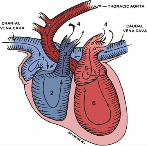
■ FIGURE 9 -7 Schematic of the blood pathway through the heart. Venous blood from the entire body (except for the lungs and some of the heart) enters the right atrium (1) and then flows sequentially through the right ventricle (2), pulmonary trunk (3), pulmonary arteries/capillary bed/pulmonary veins (4), left atrium (5), left ventricle (6), and ascending aorta (7), and into all of the body (except for the alveoli). (From Adams DR. Canine Anatomy: A Systemic Study. Ames, IA: Iowa State Press, 2004.)
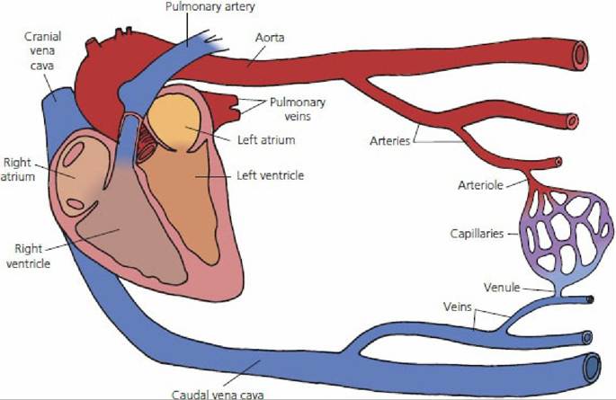
■ FIGURE 9-8 Schematic representation of the functional circulatory system. A network of arteries, arterioles, capillaries, venules, and veins exists between the aorta and cranial and caudal venae cavae.
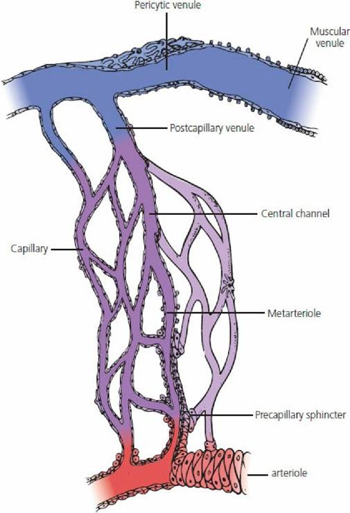
■ FIGURE 9-9 Schematic drawing of the microvasculature. Capillaries arise from both an arteriole and a metarteriole; precapillary sphincters are present. The metarteriole continues into a central channel, followed by a postcapillary venule, and as the venules increase in diameter they are called pericytic venules in which pericytes form a continuous layer.
(From Eurell JA, Frappier BL. Dellmann’s Textbook of Veterinary Histology. 6th edn. Ames, IA: Blackwell Publishing, 2006.)The volume of the capillary bed is small (4% of the total blood volume), but the vast number of capillaries provides for a large total cross-sectional area that leads to a slow rate of blood flow that favors transcapillary exchange. Capillaries are endothelial tubes with a diameter ranging from 5 to 10 μm. The walls are composed of endothelial cells, associated basal lamina (basement membrane), and pericytes. The basal lamina encloses both the endothelial cells and the pericytes (Figure 9-ιo). Pericytes are undifferentiated mesenchymal cells with potential to transform into other cell types (e.g., fibroblasts, smooth-muscle cells). Also, by this means, capillaries can transform into other types of vascular tubes (i.e., arteries, veins) if the internal flow characteristics change. Capillary pericytes synthesize and release constituents of the basement membrane.
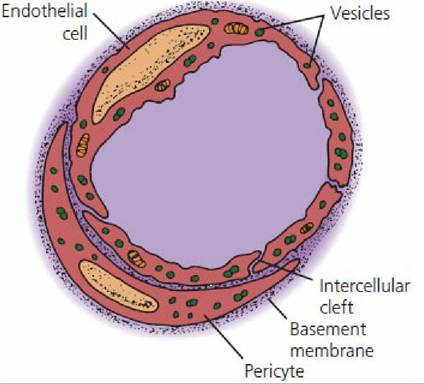
■ FIGURE 9-10 Schematic representation of a cross-,section through the endothelial wall of a muscle (continuous) capillary. Portions of endothelial cells are shown; these are separated from each other by intercellular clefts. Pericytes are outside of endothelial cells and are enclosed by a common basement membrane. Many pinocytotic vesicles are also shown.
Where the endothelial cells border each other, a thin slit (slit pore) or intercellular cleft exists and allows for the diffusion of dissolved substances in plasma. The limited size of the slit pores inhibits the passage of large molecules (e.g., protein molecules). Pinocytotic vesicles are also present in the endothelial cells. These are formed at one surface of the cell and migrate to the opposite surface, where they discharge their contents. Many of the protein molecules are probably transported through the endothelial cells in this manner.
The capillaries unite with one another to form larger vessels known as venules and the venules unite with other venules to form the veins. The largest veins are the venae cavae, which return the blood to the right atrium of the heart.The veins are thin-walled tubes reinforced by connective tissue and they also contain smooth muscle fibers. Contraction of the muscle fibers increases resistance to blood flow and helps regulate the circulation. Venous constriction increases the blood pressure in all vessels that precede the veins. Valves are present in veins at irregular intervals that are directed (or opened) toward the heart (Figure 9-11). External pressure on veins causes blood to advance in only one direction because backflow is prevented by valve closure. Similarly, backflow does not occur when external pressure is released.
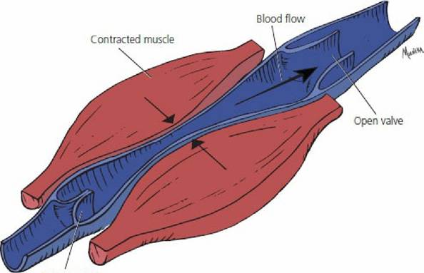
Closed valve
■ FIGURE 9-11 Valves of a vein showing the pumping action of adjacent muscles. External pressure on veins causes blood to advance in only one direction because backflow is prevented by venous valve closure.
The pressures within the veins are the lowest of the vessel pressures (Figure 9-12). This follows from the physical law of pressure dissipation as distance from the source (heart) increases. The pressure noted for capillaries might seem to be greater than what could be tolerated for a single-celled tube but, because of the extremely small diameter, the tension exerted on the capillary wall is extremely low. For a given pressure within the vascular system, the wall tension increases with the radius of the vessel according to Laplace’s law:

where T = wall tension, P = pressure in the vessel, and r = radius of the vessel.
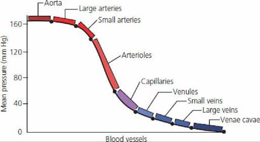
■ FIGURE 9-12 Graphic illustration of decreasing pressures from major arteries to major veins.
Note the sharp decrease in pressure in the arterioles and the more gentle slope in the much wider vascular bed made up of capillaries. (Drawing made from The Dukes Physiology Film Series (DKS- 15), Ames, IA: Iowa State University, 1969.)Blood Circulatory Systems
The blood vessels that have been described serve two separate circulatory systems (Figure Q-13). The pulmonary system circulates blood through the lungs (Figure 9-14). The pressure providing this circulation.originates from the right ventricle. The capillaries of the pulmonary system are associated intimately with the smallest terminations of the air passages, the pulmonary alveoli. Blood from this system is returned to the left atrium.
■ FIGURE 9-13 General scheme of mammalian circulation showing the pulmonary system, which serves the lungs, and the systemic system, which serves the remainder of the body. The pulmonary circulation is shown in black.
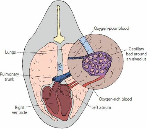
■ FIGURE 9-14 Schematic representation of the lungs and the pulmonary circulation. The circled inset represents a functional unit of the lung, the alveolus. Mixed venous blood leaves the right ventricle through the pulmonary trunk and is oxygenated at the level of the alveoli. Oxygenated blood returns to the left atrium through the pulmonary veins.
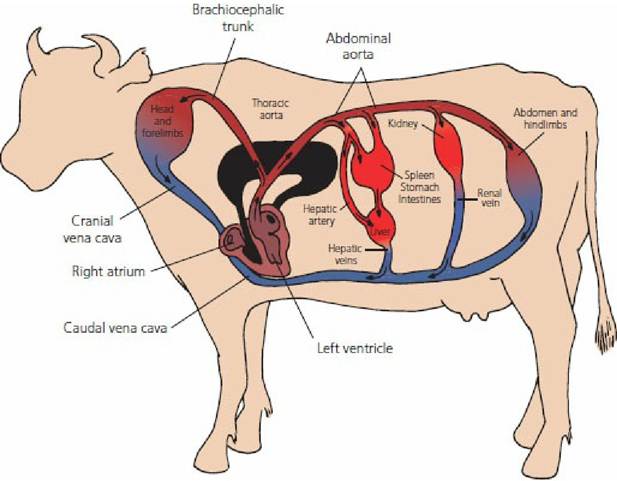
The systemic circulation carries blood that has returned from the lungs to all areas of the body. The pressure necessary for this circulation originates from the left ventricle. Blood that traverses this system leaves the left ventricle through the aorta and is returned to the right atrium through the venae cavae. The first branches of the aorta supply the heart muscle through the coronary arteries (Figure Q-15). Within the systemic circulation are a few portal systems. A portal system departs from the usual pattern of circulation in that a vein returning blood to the heart branches to reform capillaries, which reunite to form veins.
The primary example of a portal system is the hepatic portal system in the liver (Figure 9-16). The reformed capillaries are the sinusoids of the liver, which are lined by cells involved in many liver functions and by those that assist in the cleansing of blood or the removal of harmful substances by macrophages (Kupffer cells).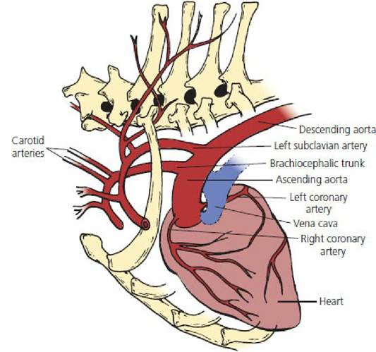
■ FIGURE 9-15 Cranial aspects of the systemic circulation (dog). The first branches of the aorta supply the heart muscles through the coronary arteries. The descending aorta is composed of the thoracic and abdominal aorta. The main arteries to the forelimbs arise from the left subclavian artery on the left side and from the brachiocephalic trunk on the right side. The carotid arteries ascend to the head.
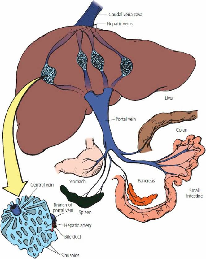
■ FIGURE 9-16 The mammalian hepatic portal system. Blood in the portal vein from the stomach, spleen, pancreas, and intestines goes to the liver, where it flows through the sinusoids and is reformed by the central vein of each lobule. It finally enters the caudal vena cava through the hepatic veins.
■