HEART AND PERICARDIUM
1. Know the orientation of the heart within the thorax with regard to its base and apex.
2. What is the pericardial sac and where is it attached to the heart? What is its function?
3.
Know the chambers of the heart. Which chamber normally has the greatest thickness?4. Know the location of atrioventricular and semilunar valves. What prevents eversion of atrioventricular valves when ventricles contract?
5. Follow a drop of blood from its entrance to the heart from the venae cavae until its ejection from the heart into the aorta.
The heart is a cone-shaped, hollow, muscular structure located in the thorax (Figure 9-1). The large arteries and veins are continuous with the heart at its base. Its base is directed upward (dorsal) and forward (cranial). The opposite end of the cone is known as the apex. During early embryonic development, the heart is pushed into a serous sac known as the pericardium. The part of the sac next to the heart becomes fused to the muscle of the heart and is known as the visceral pericardium, or epicardium (Figure 9-2). The outer part of the sac is continuous with the epicardium and extends outward from its fusion at the base to envelop the heart completely. The apex of the heart is free (unattached) within the pericardium. This outer layer is known as the parietal pericardium. The pericardial cavity is a potential space and contains a small amount of fluid to provide lubrication for the outer surface of the heart during its near-continuous motion. It is referred to as a potential space because it can increase its fluid volume during times of inflammation. A major cause of inflammation in cattle is traumatic pericarditis, such as when a foreign object (e.g., nail, wire) penetrates from the forward compartment (reticulum) of the bovine stomach (Figure 9-3). A splashing sound, similar to water in a washing machine, can sometimes be heard with each beat of the heart because of the increased fluid.
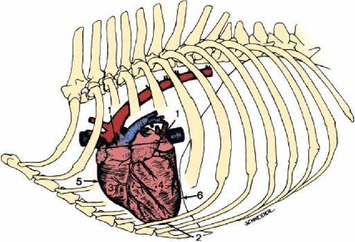
■ FIGURE 9-1 The canine heart and its major vessels in the thorax (left lateral view). 1, Flattened base;.2, apex; 3, right ventricle; 4, left ventricle; 5, right ventricular margin; 6, left ventricular margin. (From Adams DR. Canine Anatomy: A Systemic Study. Ames, IA: Iowa State Press, 2004.)
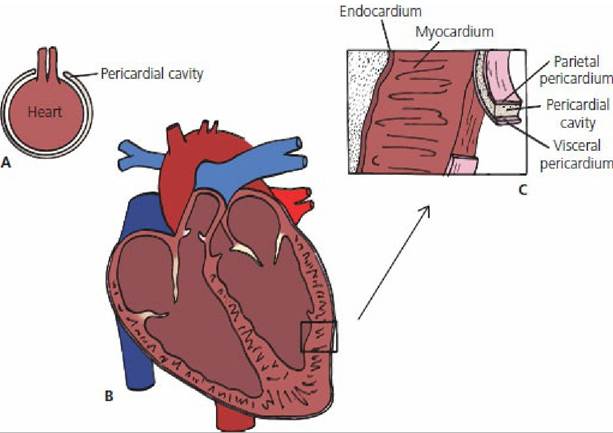
■ FIGURE 9-2 Cross-sectional schematic representation of a mammalian heart. A. Embryologic invagination of the heart into the pericardial coelom (becomes the pericardial sac). B. Sagittal section of the heart with the pericardial sac. C. Details of the heart wall and pericardium.
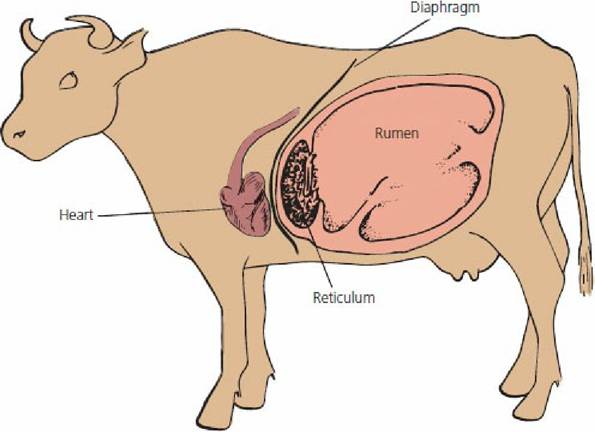
■ FIGURE 9-3 Left view of the bovine thorax and abdomen showing location of the heart relative to the reticulum. Foreign objects (nails, wire), sometimes ingested by cattle, accumulate in the reticulum (one of the bovine forestomachs). Contraction of the reticulum can force pointed objects through the reticulum wall and the diaphragm, causing final penetration of the pericardium and subsequent inflammation (pericarditis).
Myocardium
The muscular part of the heart is known as the myocardium, which forms the walls.for the compartments (chambers) of the heart. The muscle fibers are arranged so that, when they contract, the blood is ejected from the chambers (Figure 9-4). The heart chambers are divided into those on the right side of the heart and those on the left side (Figure 9-5); each side has an atrium and a ventricle. To conserve space, each atrium has an extension known as an auricle, with a shape that conforms to that of adjacent parts. The atria receive blood from veins and the ventricles receive blood from the atria. The right and left ventricles pump blood from the heart through the pulmonary trunk and aorta, respectively.
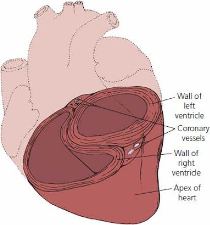
■ FIGURE 9-4 Cross-sectional view of horse heart at the ventricular level showing the relative thickness of the myocardium and the orientation of the muscle fibers.
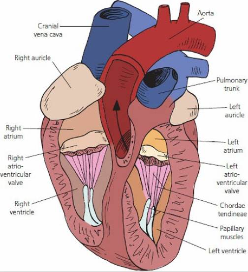
■ FIGURE 9-5 A sagittal section of the canine heart. The right and left chambers are shown with separation of the atria and ventricles by atrioventricular valves. The auricles are extensions of the atria. The aorta is seen to be arising from the left ventricle. The pulmonary trunk arises from the right ventricle (origin not visible) and divides into right and left pulmonary arteries beyond the pulmonary semilunar valve. The cranial vena cava and caudal vena cava (not visible) deliver venous blood (low oxygenated) into the right atrium.
Heart Valves
The valves located between the atria and ventricles are known as the atrioventricular (A-V) valves (Figure 9-6A). The valve on the right side has three cusps (flaps) and is called the tricuspid valve; the left A-V valve has two cusps and is called the bicuspid, also known as the mitral valve. The A-V valves prevent expulsion of ventricular blood into the atria when the ventricles contract. Because of the pressure associated with the expulsion of blood from the ventricles, the A-V valves could be everted into the atria. This is prevented by cords (chordae tendineae) attached to the free margin of the cusps at one end and to small muscles (papillary muscles) at the other end that extend from the myocardium. Papillary muscle contraction is synchronized with the myocardial contraction so that tension to the chordae tendineae is appropriately timed. Backflow of blood that has just been ejected from the ventricles is prevented by valves located at the exits of the arteries from the ventricles (Figure 9-6B). The valves on both the right and left sides have three cusps and are known as the semilunar valves.
The valve on the right side is known as the pulmonary semilunar valve because of its location relative to the pulmonary trunk and the valve on the left side is known as the aortic semilunar valve because of its location relative to the aorta.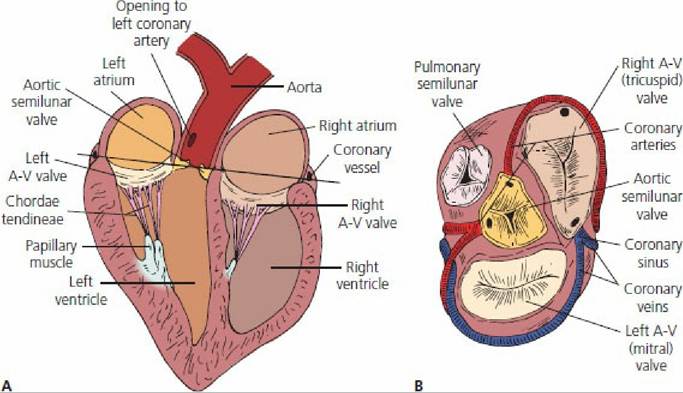
■ FIGURE 9-6 Heart valves. A. Location relative to the chambers and the aorta. The pulmonary trunk and its semilunar valve are not shown. B. A view of the heart from above the ventricles with the atria removed (at the level of the straight line shown in A to show the semilunar and atrioventricular valves). The first branches from the aorta are the coronary arteries. The coronary sinus opens into the right atrium and receives blood from the heart wall through the coronary veins.
Blood Flow through the Heart
Blood that originally enters the heart and is finally ejected follows a specific route (Figure 9-7). Blood that circulates to the tissues returns to the heart through the cranial vena cava (blood from forward parts of the body) and the caudal vena cava (blood from rear parts of the body). This is the venous blood. It has lost oxygen to the tissues, gained carbon dioxide, and must now be directed to the lungs, where it becomes arterial blood by gaining oxygen and losing carbon dioxide. The venous blood enters the right atrium during the atrial relaxation phase of the cardiac cycle. At the appropriate time in the cardiac cycle, the blood is directed through the right A-V valve into the right ventricle. The ventricles contract and the blood goes through the pulmonary semilunar valves to the lungs through the pulmonary arteries. These are called arteries, even though they transport venous blood, because they transport blood away from the heart. After the blood has circulated through the lungs, it reenters the heart through the pulmonary veins (contain arterial blood). It enters the left atrium; from here the blood is directed to the left ventricle, from which it is pumped to the systemic (whole body) circulation through the aorta. The left ventricle has the greatest muscle mass of the heart chambers because of the greater work required to pump blood throughout the entire body.
■