LYMPHATIC SYSTEM
1. What is meant by the lymphatic system? What is the fluid of its vessels known as?
2. Does protein ever leak from capillaries? What is its turnover rate?
3. What is the route for return of protein to the blood after it has leaked?
4.
What is one of the most important functions of the lymphatics?5. What is the location and what are the functions of lymph nodes?
An important adjunct to the circulatory system is the lymphatic system. The,lymphatic vessels have blind beginnings (lymph capillaries) in the interstitial spaces (the spaces between cells and outside of the blood vessels) and the continuation vessels tend to parallel the veins (Figure 9-17). Lymph vessels join with each other and eventually form a few large lymph vessels that empty directly into the large veins. The fluid of the lymph vessels is called lymph. There is little difference between the composition of lymph and that of interstitial fluid. Although blood capillaries permit most plasma constituents to diffuse through their endothelium, protein molecules are somewhat restrained because of their size. It is essential for proteins to enter the interstitial fluid, however, because they act.as carriers for cell products or for substances needed by cells. In addition, antibodies (protein substances) are needed in the interstitial space for more intimate association with antigen. There is a complete turnover (from capillaries and return to blood) of plasma protein once every 12 to 24 hours. Because the concentration of protein is higher in the plasma than in the interstitial space, the gradient for diffusion is to the interstitial space. Protein in the interstitial space does not diffuse back to the plasma; it can only return to the plasma through the lymphatic vessels. The blind beginnings of the lymph vessels are adapted for the intake of large molecules, and concentration and pressure gradients favor this route (Figure 9-18).
Anchoring filaments prevent collapse of the vessels when the tissue swells with excess fluid (edema). Also, the overlap of endothelial cells with each other permits easy access of interstitial fluid, but because of their valve-like arrangement, backflow is prevented. The return of protein that has leaked or that is otherwise transported from the blood capillaries back to the systemic circulation is one of the most important functions of the lymphatics.
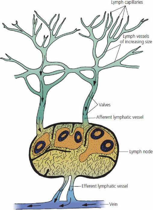
■ FIGURE 9-17 Schematic representation of lymph drainage. Interstitial fluid gains access to the blind beginnings of lymph capillaries and proceeds centrally through lymph vessels of increasing size. Lymph nodes are located along the course of lymph vessels. Lymph is returned to blood by drainage into veins.
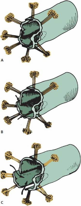
■ FIGURE 9-18 Special structure of the lymphatic capillaries that permits passage of high- molecular-weight substances into the lymph. The structures radiating from the capillaries are anchoring filaments that give support to portions of endothelial cells where the capillaries begin. The unsupported portion of the endothelium allows fluid to flow into the capillary (arrows), as shown in B and C. Raised pressure in the capillary closes the flap against the overlapping supported endothelium, as shown in A. (From Leak LV. The fine structure and function of the lymphatic vascular system. In: Meessen H, ed. Handbuch der Allgemeinen Pathologie. New York: SpringerVerlag, 1972.)
Lymph nodes are nodular structures of varying sizes located along the course of lymph vessels. They contain clusters of germinal cells that reproduce to form lymphocytes (Figure 9-19). The lymphocytes in turn can be of a type that produce antibodies or they can be sensitized lymphocytes. In both cases they are highly specific against substances (antigens) that are foreign to the body.
The antibodies and sensitized lymphocytes leave the lymph nodes with the lymph in the vessels and enter the blood, where they can be circulated throughout the body.Lymph nodes also contain fixed macrophages that are attached to the reticulum (inner framework) of the lymph nodes. Lymph circulating through the nodes thus is in intimate contact with the macrophages and, because the macrophages are highly phagocytic, foreign materials in lymph (e.g., bacteria, cellular debris) are engulfed and prevented from progressing further. Infection or inflammation of a body part often results in enlarged lymph nodes serving that part because of the entrapment and because of lymphocyte proliferation stimulated by the presence of these antigenic materials. Cancer cells might be routed from their origin and be entrapped by lymph nodes, where they can proliferate and continue into the next lymph node in the chain. Inspection of lymph nodes for enlargement is an important part of the postmortem carcass examination procedure for animals that are slaughtered for food consumption.
The lymph vessels are one-way channels that contain valves similar to those in veins, which prevent backflow of lymph once it has progressed toward the veins. Lymph progresses through the channels by contractions of the lymph vessels and by a massaging action of muscles that overlie lymph vessels. Forward movement of lymph lowers the pressure in the part of the vessel evacuated and, because there is no backflow of lymph, entry of lymph from the backward parts is favored. There is no central pump, such as the heart, to facilitate lymph circulation, and disturbances in lymph flow can cause accumulation of interstitial fluid in low-lying body parts. The return of lymph is assisted by elevation of these parts, such as the limbs, to a level higher than the centrally located veins and by muscle movement from exercise.
■