BLOOD VESSELS AND LYMPHATIC STRUCTURES
The axillary artery, the main supply to the limb, is used occasionally as a source of arterial blood; it may be located on deep palpation where it winds around the first rib. The courses and branches of the arteries in the proximal segments of the limb follow the general pattern closely enough to make description unnecessary.
The account may commence where the median artery accompanies the deep digital flexor tendon through the carpal canal. It runs with a satellite vein and the median nerve where it enters the metacarpus to continue medial to the flexor tendons under cover of a thick deep fascia (Figure 30-13) but becomes superficial and vulnerable at the fetlock joint. Its course now takes it over the palmar surface of the medial branches of the flexor tendons before diving into the interdigital space. The
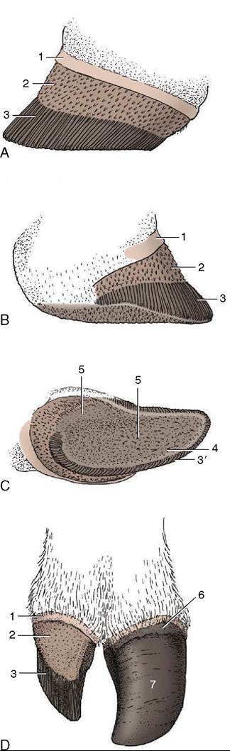
Figure 30-12 Dermis over which the horn of the hoof is produced. A to C, Abaxial, axial, and ground surface. D, Dorsal surface of dermis and hoof. 1, Perioplic dermis; 2, coronary dermis; 3, laminar dermis; 3', terminal papillae at the distal ends of the laminae; 4, sole dermis; 5, dermis of the bulb; 6, periople; 7, wall of hoof.
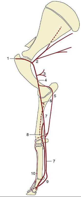
Figure 30-13 The principal arteries on the bovine right forelimb; medial view. 1, Axillary a.; 2, subscapular a.; 3, deep brachial a.; 4, brachial a.; 5, collateral ulnar a.; 6, common interosseous a.; 7, median a.; 8, radial a.; 9, palmar common digital artery III; 10, dorsal common digital artery III.
artery and accompanying vein bulge visibly at this level in thin-skinned animals, but though the artery may be palpated, a pulse cannot usually be perceived. It now bears a new title, palmar common digital artery III, and within the space it gives off a number of branches of minor importance before dividing into the two axial palmar digital arteries.
Each of these passes distally to reach and enter the distal phalanx through the large foramen located by the extensor process. Lesser palmar abaxial digital arteries, derived from arteries of the forearm, enter the distal phalanges at the palmar ends of their abaxial surfaces. Within the bone the axial and abaxial arteries anastomose to form a terminal arch from which numerous branches are released to thedermis. Other small arteries on the dorsal aspect of the digits are of little importance. All the arteries are severed when a digit is amputated; the stump of the axial palmar artery bleeds most profusely, and it, at least, must be ligated.
The limb veins are divided between a deep system, satellite to the arteries, and a quasi-independent superficial system. The two systems are connected by prominent anastomoses at the elbow, above the carpus, and in the foot and eventually join into one when the cephalic vein opens into the external jugular at the base of the neck. The superficial system comprises the cephalic and accessory cephalic veins and the tributaries of the latter in the foot (Figure 30-14, A). Most can be palpated, and especially in young, thin-skinned subjects, they may provide visible surface landmarks: their positions are more certainly revealed when raised by a tourniquet. They are now much used for obtaining surgical anesthesia of the digits by retrograde intravenous injection. Those that lend themselves to the procedure are shown in Figure 30-14, B-C. The technique is simpler and more reliable than the alternative method, which requires the deposit of anesthetic solution over several nerves.
The lymph nodes of the forelimb comprise the large proper axillary node, which lies against the thoracic wall caudal to the shoulder joint, and a few small accessory nodes (Inn. axillares primae costae) placed over the first rib and adjoining intercostals space. The axillary node receives lymph from the deeper structures of the upper
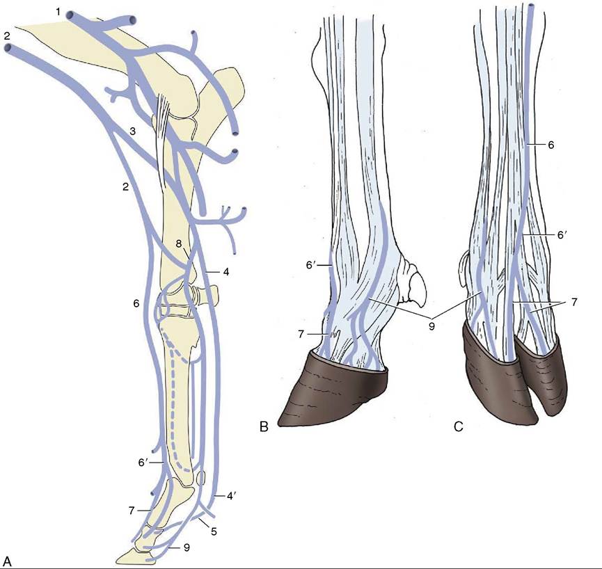
Figure 30-14 The principal veins of the bovine forelimb.
A, Right limb; medial view. B, Left foot; lateral view. C, Right foot, dorsal view. 1, Brachial v.; 2, cephalic v.; 3, median cubital v.; 4, median v.; 4’, palmar common digital v. III; 5, axial palmardigital vv.; 6, accessory cephalic v.; 6', dorsal common digital v. III; 7, dorsal digital vv.; 8, radial v.; 9, abaxial palmar digital vv.
segments of the limb, including the ventral girdle muscles, and forwards it first to the accessory nodes and thence either to the caudal deep cervical nodes or directly to one or other of the veins at the thoracic inlet. This node may be inspected through an incision of the first intercostal space of the split carcass. The dorsal girdle muscles, the skin and subcutaneous fascia of the shoulder, arm, and forearm, and all structures distal to the carpus drain directly to the superficial cervical node, which may be palpated in front of the shoulder.