BONE FORMATION
1. What method of bone formation is represented by the os penis of some animals and the os cordis of the bovine heart?
2. What is endochondral ossification?
3. What method of bone formation forms the flat bones of the skull and face?
4.
What is the name of the oldest zone within the epiphyseal plate?5. Visualize the elongation of a bone by virtue of cells dividing, secreting matrix, and thus pushing the zone of reserve cartilage away from the diaphysis.
6. Does cartilage have a blood supply?
7. Are cartilage lacunae connected by canaliculi?
8. What causes chondrocytes to die?
9. What previously occupied the tunnels that exist in the zone of calcified matrix?
0. Note the invasion of the tunnels by capillaries as a prerequisite for new bone formation.
11. Visualize the development of lamellae (layers) around the capillary such that the tunnel is reduced to a narrow canal (a Haversian system).
2. As a bone grows in width, why do the walls not become unduly thick? What is the appositional mechanism of bone growth?
3. What osteogenic layer accounts for the outer circumferential lamellae? The inner circumferential lamellae?
4. What cell provides for the erosion needed to form new channels during the process of bone remodeling?
15. After the erosion, what sequence of events forms new Haversian systems?
6. How is bone mass correlated with increased muscle mass and exercise?
Bone formation (ossification) is identified according to the environment in which it is formed as either heteroplastic, endochondral, or intramembranous. Ossification is heteroplastic if it is formed in tissue other than the skeleton. This type occurs with the os penis of some animals and the os cordis of the bovine heart, but mostly it is pathologic. Endochondral ossification is that which develops from cartilage and is mostly preformed in the fetus but continues after birth from cartilage plates located between the metaphysis and epiphysis, and from the periosteum that surrounds the cortex.
Most long bones are developed by this method. Intramembranous bone formation is that which is formed without the intervention of cartilage. These bones are preformed in a fibroid membrane that is then infiltrated with osteoid tissue that later becomes calcified. Bones formed by this method are the flat bones of the skull and face, the mandible, and the clavicle (cat). The previously mentioned mechanisms refer only to the manner in which existing bone was originally formed. Remodeling of bone is established on the preexisting bone and the mechanism of remodeling is identical whether the original bone was formed by endochondral or intramembranous,ossification. The sequence of actual bone formation during remodeling consists of osteoblasts laying down osteoid tissue that is subsequently calcified.Growth of Long Bones
Increase in length of a bone depends on the presence of a cartilage plate (epiphyseal plate), wherein four zones are recognized that extend from the epiphysis to the diaphysis (Figure 7-15). These are termed zones of reserve cartilage (the youngest), proliferation, hypertrophy, and calcified matrix (the oldest). Beyond the zone of calcified matrix are the developing trabeculae that make up the spongy bone of the metaphyses.
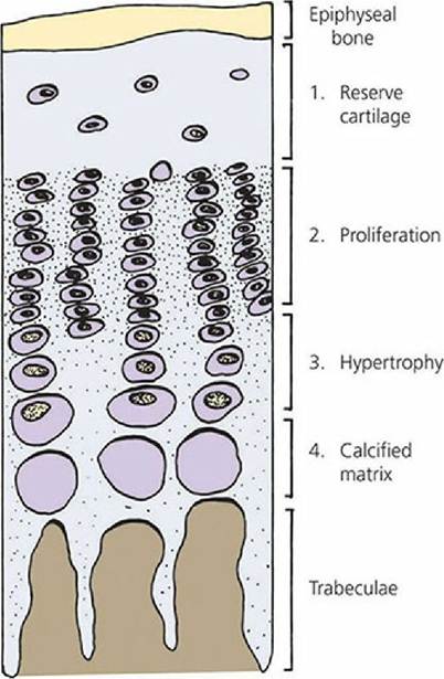
■ FIGURE 7-15 The four zones of a cartilage (epiphyseal) plate.
Cartilage does not have a blood supply and nutrition of the cartilage cells (chondrocytes) depends on diffusion of extracellular fluid from its source to the chondrocytes that lie within their lacunae. Also, unlike osteocytes, chondrocytes are still able to divide after they have become embedded in cartilage matrix. When the chondrocytes from the zone of reserve cartilage undergo division, the chondrocytes become organized into distinct columns and a zone of proliferation that is directed toward the diaphysis is recognized. The columns are formed because of chondrocyte capture within lacunae.
Each daughter cell within a lacunae produces a matrix and this causes the cartilage matrix to expand from within. This has the effect of pushing the epiphysis away from the diaphysis and, thus, elongation of the bone.Each division of chondrocytes brings about larger cells, and hence the zone of hypertrophy (see Figure 7-15). This has the effect of compressing the matrix into linear bands between the columns of hypertrophied cells. After several divisions, the hypertrophied cells become further removed from the epiphyseal plate and become active in bringing about calcification of the cartilage matrix. Calcification, coupled with increasing distance from the nutritional source, causes the chondrocytes to die and the matrix becomes the zone of calcified matrix. The appearance of longitudinal and cross sections of different areas of the epiphyseal plate and metaphysis at the periphery of a growing shaft are presented in Figure 7-16. A cross section at the level of the calcified matrix (Figure 7-16C2) would show that tunnels exist where nests of hypertrophied cells previously occupied the space between the linear bands of compressed cartilage matrix (now calcified). What are seen as trabeculae (columns) in longitudinal sections actually constitute a honeycombed structure in cross sections and the spaces seen between the trabeculae in longitudinal sections are seen as tunnels in cross sections.
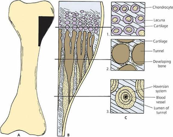
■ FIGURE 7-16 The appearance presented by both longitudinal and cross sections of different areas of the epiphyseal plate and metaphysis at the periphery of a growing shaft. A. The blackened area is the location on the long bone for parts B and C. B. Horizontal lines extend to their respective cross sections. Brown areas are tunnels or openings to tunnels. The oblique lines represent cartilage and the stippled structures represent calcified matrix. C1. Chondrocytes in their lacunae in the zone of hypertrophy.
C2. Tunnels formed in the zone of calcified matrix. Trabeculae are composed of both cartilage and bone. C3. Haversian system transforming tunnels into compact bone.The tunnels are now invaded from the diaphysis by capillaries and osteoblasts line up along the sides of the tunnels and deposit bone on their inner surfaces. The osteoblasts continue to divide and each division of osteoblasts pushes the original osteoblast layer closer to the capillary in the center. Concentric lamellae of bone substance are thus established, with osteocytes occupying lacunae and canaliculi. After several layers of bone (concentric lamellae) have been deposited, the tunnel is reduced to a narrow canal, which contains a blood vessel, some osteoblasts or osteogenic cells, and perhaps a lymphatic vessel (Figure 7-16C3). This arrangement is known as a Haversian system, the unit of structure of compact bone.
While a long bone is growing in length, it is also growing in width. New layers of bone are being added to the outside of the shaft at the same time that bone is dissolved away from the inside of the shaft. Although the shaft of the bone becomes wider, its walls do not become unduly thick, and the width of the marrow cavity gradually increases. The shaft of a bone grows in width by the appositional mechanism (Figure 7-17). The periosteum provides the osteogenic layer and, by repeated proliferation, new bone is formed to fill in the grooves between the longitudinal ridges of Haversian systems that were formed while the bone was elongating. The same process of appositional growth occurs on the inner aspect of the bone shaft from endosteum. The bone formed from the periosteum and endosteum accounts for the outer and inner circumferential lamellae, respectively (see Figure 7-13).
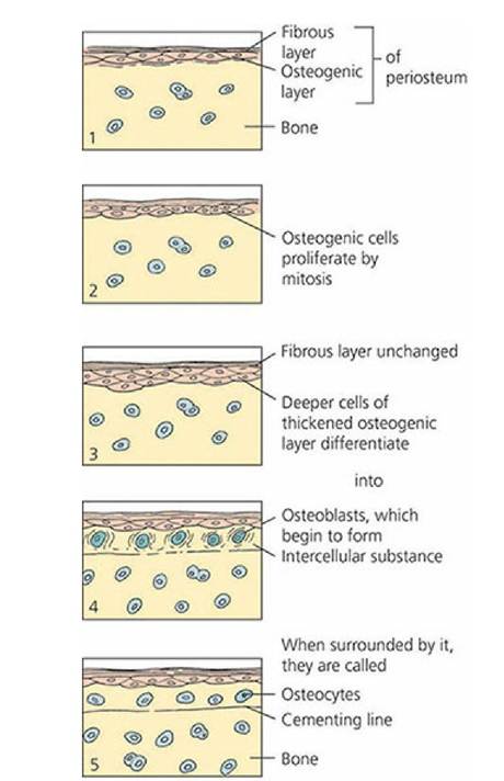
■ FIGURE 7-17 Bone growth by apposition. (Adapted from Ham AW. Histology. 1st edn. Philadelphia, PA: J.B.
Lippincott, 1950.)Bone Remodeling
As described previously, the growth of bones does not simply involve an increase in their thickness. Rather, there is a coordinated formation of new bone at the outer surfaces and resorption of bone at the inner surfaces (Figure 7-18). This also occurs to the bones of the neurocranium to accommodate the growing brain during its maturation. In each instance, the two processes of appositional growth and bone resorption are the only ways that the shape and size of a bone can change during prenatal and postnatal life. Because this applies to long bones of the body, the shape of the bone does not grossly change during growth, and its marrow cavity is enlarged to ensure a sufficient area for blood cell requirements. During growth, Haversian systems are.being formed, resorbed, and remodeled. The general process for new Haversian systems is generally initiated by osteoclasts concurrent with the invasion of blood vessels (Figure 7-19). The osteoclasts are on the leading edge of the invading blood vessels. New tunnels are thus formed by erosion through the endosteal surface that are oriented with the long axis of the shaft. A layer of osteoblasts forms on the surface of the eroded tunnel (that has a central blood vessel), and concentric lamellae are formed as previously described for Haversian systems. The blood vessels grow and branch, with accompanying osteoclast and osteoblast activity, whereby new channels are made and new Haversian systems form to fill them. In addition to the remodeling that occurs to accommodate growth, remodeling also occurs in response to stress placed on bones. Reduction in bone mass accompanies loss of muscle mass and decreased mobility, whereas an increase in muscle mass and exercise is accompanied by an increase in bone mass. Therefore, the organization of bone changes to meet mechanical and other stresses placed on the skeleton, and represents a balance between bone formation and bone resorption.
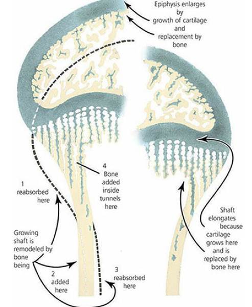
■ FIGURE 7-18 Sites of bone deposition and resorption in the process of lengthening and remodeling of long bones.
Bone is shown in tan, cartilage in blue-green. (From Ham AW and Cormack DH. Histology. 8th edn. Philadelphia, PA: JB Lippincott Company, 1979.)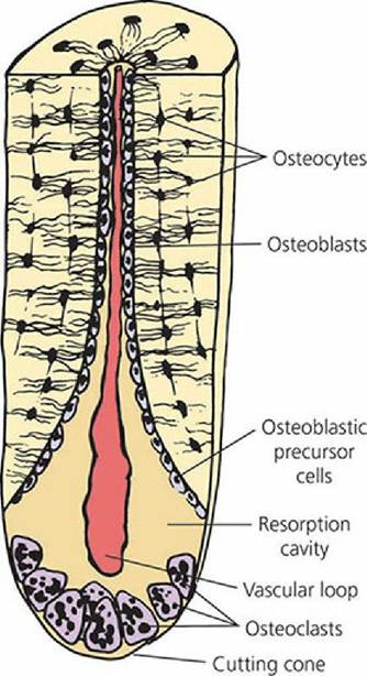
■ FIGURE 7-19 Osteoclastic activity that precedes bone remodeling. Osteoclasts advance a resorption cavity into the bone and are immediately followed by a vascular loop accompanied by precursor cells that multiply and differentiate into osteoblasts. Osteoblasts lay down new layers of osteoid. Canaliculi are formed and osteoblasts become osteocytes. Successive layers of new bone are deposited to give the concentric lamellar rings of Haversian bone. (From Whittick WG. Canine Orthopedics. 2nd edn. Philadelphia, PA: Lea & Febiger, 1990.)
■