Capillaries, the Smallest Blood Vessels, Are the Sites for the Exchange of Water and Solutes Between the Bloodstream and the Interstitial Fluid
Because of their small size, the capillaries are sometimes called the microcirculation. They are also called the exchange vessels, because the exchange of water and solutes between the bloodstream and the interstitial fluid takes place across the walls of the capillaries.
Each type of blood vessel in the body is structurally suited for its primary function, and the walls of the capillaries are especially well adapted for their exchange function.Figure 23-1 shows the contrasting features of the walls of the various types of blood vessels. The distinguishing feature of the walls of the aorta and large arteries is the presence of a large amount of elastic material along with smooth muscle. These vessels are called the elastic vessels; elasticity is necessary because the aorta and large arteries must distend with each pulsatile ejection of blood from the heart. The arterial walls are also strong and quite stiff (low compliance). There is no contradiction in saying that the arteries are elastic and have low compliance. Elasticity denotes distensibility and an ability to return to the original shape after the distending force or pressure is removed. Compliance is a measure of how much force or pressure is required to achieve distention. The arteries are elastic, but a high pressure (systolic pressure) is required to distend them.
Small arteries, and particularly arterioles, have relatively thick walls with less elastic tissue and a predominance of smooth muscle, so they are called the muscular vessels. The muscle enables these vessels to constrict or dilate, which varies their resistance to blood flow. The muscular vessels vary the total
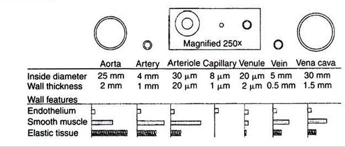
FIGURE 23-1 Each type of blood vessel in the systemic circulation is specifically suited to its particular function by its size, wall thickness, and wall composition.
In this drawing, each type of vessel is shown in cross section. The drawings are to scale (the arteriole, capillary, and venule are magnified 250 times to make them visible). Also shown are the relative proportions of the three most important types of tissue found in blood vessel walls.peripheral resistance and direct blood flow toward or away from particular organs or particular regions within an organ.
Capillaries are the smallest vessels, being about 8μm in diameter and about 0.5 mm long. Capillaries are so small that red blood cells (7.5 μm in diameter) must squeeze through in single file. Capillary walls consist of a single layer of endothelial cells. The small diameter of the capillaries and the thinness of their walls facilitate the exchange of water and solutes between the blood within capillaries and the interstitial fluid immediately outside the capillaries.
Venules and veins are larger than capillaries, and they have thicker walls. Venules and veins have both elastic tissue and smooth muscle in their walls. However, the walls of veins are not as thick or as muscular as the walls of arteries or arterioles. The primary role of veins is to serve as reservoir vessels. Veins are very compliant. Furthermore, it is normal for many veins in the body to be in a state of partial collapse. Therefore, substantial changes in venous blood volume can occur without much change in venous pressure.
Capillaries form a network (see Figure 18-4). In most tissues the capillary network is so dense that each cell of the tissue is within 100 μm of a capillary. However, not all the capillaries of a tissue carry blood at all times. In most tissues the arterioles alternate between constriction and dilation, so blood flow is periodically reduced or even stopped in most capillaries. Also, in some tissues (e.g., intestinal circulation), tiny cuffs of smooth muscle encircle capillaries at the points where they branch off from arterioles. Contraction of these precapillary sphincters can reduce or stop the flow of blood in individual capillaries. When the metabolic rate of a tissue increases (and therefore its need for blood flow increases), the arterioles and precapillary sphincters still constrict periodically, but they spend more time in the dilated (relaxed) state. I his increases the fraction of capillaries in which blood is flowing at any one time. At maximal metabolic rate (e.g., maximal exercise in a skeletal muscle), blood flows through all the capillaries all the time. Sending blood flow to all the capillaries not only increases the total blood flow through a tissue but also minimizes the distance between each cell of the tissue and the nearest capillary carrying blood by bulk flow. Both these effects speed up diffusional exchange between the capillary blood and the tissue cells.