Lipid-Soluble Substances Diffuse Readily Through CapilIaryWaIIsrWhereas Lipid- Insoluble Substances Must PassThrough Capillary Pores
The rate of diffusional exchange between capillary blood and the surrounding interstitial fluid depends both on the features of the capillary wall and on the properties of the substance
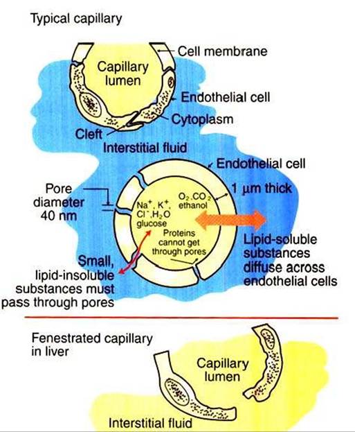
FIGURE 23-2 Capillaries in cross section.
Most capillaries have pores, or clefts, between endothelial cells (top}. Water and lipid-insoluble compounds move between the capillary plasma (yellow) and the interstitial fluid (b∕ue∏hrough these water-filled channels (center). Lipid-soluble substances diffuse directly through the capillary endothelial cells.The size of the capillary pores varies greatly from tissue to tissue, with the largest capillary pores found in the liver capillaries (liver sinusoids) (bottom).being exchanged. In most tissues there are water-filledpores, or clefts, between the endothelial cells that form capillary walls (Figure 23-2). These pores provide channels through which water and lipid-insoluble substances can move from the capillary lumen to the interstitial space» or vice versa. Plasma electrolytes, glucose, and amino acids are among the lipid- insoluble substances that must pass through these water-filled channels. By contrast, lipid-soluble substances in blood, such as dissolved oxygen and carbon dioxide, fatty acids, ethanol, and some hormones, can dissolve in and diffuse through the endothelial cells that form the capillary wall. For a lipid- soluble substance, this process takes only a fraction of a second. In fact, the diffusional exchange of lipid-soluble substances is much more rapid than that of lipid-insoluble substances, because the lipid-insoluble substances are restricted to passage through the capillary pores, which constitute only about 1% of the total wall surface area of a typical capillary.
The characteristics of the capillary pores vary from tissue to tissue.
Two extremes are found in the liver and the brain. The pores or clefts in the liver capillaries are so large that even plasma proteins such as albumin and globulin can pass through them. This is an appropriate feature for the liver capillaries (hepatic sinusoids) because the plasma proteins are produced in the liver. The large clefts permit the newly synthesized protein molecules to enter the bloodstream. The large pores in liver capillaries are also appropriate for the role of the liver in detoxification. Some toxins that become bound to plasma proteins are removed from the bloodstream by the liver and chemically changed into less toxic substances. Because of their large pores, capillaries (sinusoids) in the liver are called fenestrated capillaries (“capillaries with windows”) (Figure 23-2, bottom).The brain capillaries represent the other extreme in pore size. The pores of brain capillaries are so small that only water and electrolyte molecules can pass through them; not even glucose and amino acid molecules can pass through these tiny pores. The tight barrier provided between the bloodstream and the brain tissue by the small pores is called the blood-brain barrier (see Chapter 15). One function of the blood-brain barrier is to protect brain neurons from exposure to toxic substances. Glucose is moved across the brain capillary endothelial cells by means of specialized protein carrier molecules that are embedded in the cell membranes of the endothelial cells. The energy to drive this facilitated diffusion comes from the glucose concentration difference between the blood and the brain interstitial fluid. Brain neurons require glucose to carry out their normal metabolism.