CARDIAC CONTRACTILITY
1. What is the sinoatrial node? What is its pacemaker function?
2. What are the two syncytia of the heart and how are they separated?
3. What is the contraction sequence of the two syncytia and what function is thereby served?
4.
Describe conduction of the impulse throughout the heart. What purpose is served by fast conduction?5. Do both atria contract at the same time? Do both ventricles contract at the same time?
6. Define diastole and systole.
7. Describe the events of the.cardiac cycle.
All muscles seem to have an inherent rhythmicity of contraction. If the three muscle types (cardiac, skeletal, smooth) are removed from the nerve and blood supply and placed into physiologic fluids, contraction begins in a rhythmic manner. The frequency of contraction is greatest in cardiac muscle, followed by skeletal muscle, and finally by smooth muscle.
Origin of the Heartbeat
In cardiac muscle, the atria have a higher frequency of contraction than the ventricles. In addition, a small area of specialized cardiac muscle fibers near the junction of the cranial vena cava with the right atrium has a contraction frequency higher than that of the atria. These specialized muscle fibers constitute what is known as the sinoatrial (S-A) node. Impulses originating in the S-A node spread throughout the musculature of the atria, and the impulse is conducted to the ventricles by way of internodal pathways. Because the contraction frequency for the S-A node exceeds that of the atria and ventricles, the S-A node impulse becomes the stimulus for contraction of the atria and ventricles, whereby the contraction frequency of the S-A node becomes the contraction frequency of the atria and ventricles. The S-A node therefore serves a pacemaker function.
Conduction of the Impulse
The muscle fibers of the atria and those of the ventricles are arranged to form an atrial and a ventricular syncytium.
A syncytium is an arrangement of muscle fibers in which the fibers form an interconnected mass of fibers. The atrial syncytium is separated from the ventricular syncytium by a fibrous ring that surrounds the A-V valves. The fibrous ring acts as an insulator between the two syncytia. An impulse that spreads throughout the atria does not spread to the ventricles, and an impulse from the ventricles does not spread to the atria. This permits independent contraction and provides an opportunity for the atria and ventricles to coordinate their function of emptying, so that the ventricles are filled during their relaxation by the contraction and emptying of the atria.It is desirable for the muscle fibers in each syncytium to contract as simultaneously as possible. All fibers contribute to the pressure increase needed for evacuation of the blood from the chambers of the syncytium. Fibers contracting at different times could not attain sufficient pressure for efficient evacuation. Because the function of the atria is to fill the ventricles before they contract, impulse conduction is completed first throughout the atria. After a slight delay, the impulse is then conducted throughout the ventricles.
To facilitate rapid conduction (and coordinated contraction), the heart has a specialized conduction system composed of specialized conduction tracts and fibers called Purkinje fibers (Figure Q-22). The S-A node conducts the impulse throughout the atria through several small tracts of fibers called internodal pathways. Depolarization of these pathways provides the stimulus for depolarization of adjacent muscle fibers; the transmission of impulses and subsequent depolarization of other muscle fibers is facilitated by the intercalated disks interposed between muscle fibers. Impulse conduction by the internodal pathways is received by the A-V node, which is located at a point between the atria and ventricles. The A-V node is continued through the fibrous ring by the A-V bundle. A-V bundle fibers are smaller in diameter than the other Purkinje fibers and impulse conduction is slowed to about 10% of the velocity of cardiac muscle fibers.
This permits a delay of an impulse to facilitate complete emptying of the atria before the ventricles contract. Conduction fibers are continued from the A-V bundle in the wall dividing the right from the left ventricles as Purkinje fibers distribute to the right ventricle (right bundle branch) and as Purkinje fibers distribute to the left ventricle (left bundle branch). These large fibers transmit impulses about two to three times faster than cardiac muscle fibers. The muscle in the walls of the ventricles is thicker than the muscle in the walls of the atria and the distance of conduction is greater. Therefore, to achieve coordinated contraction of muscle fibers of the ventricles, it is essential to have a greater velocity of conduction that is provided by the Purkinje fibers.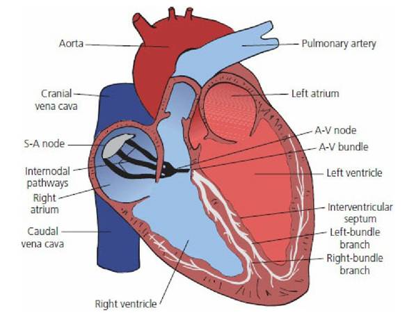
■ FIGURE 9-22 Conduction system of the mammalian heart. Impulse originates in the S-A node located near the junction of the venae cavae with the right atrium. The internodal pathways conduct the impulse throughout the atria and the left- and right-bundle branches of Purkinje fibers conduct the impulse throughout the ventricles. The A-V node and bundle conduct the impulse from the atria to the ventricles.
Not only does cardiac muscle contract more slowly than skeletal muscle but it also has a longer refractory period. A refractory period is the period during repolarization when a stimulus cannot evoke another depolarization. This is advantageous for the heart because, when the impulse completes its travel through each syncytium, the impulse is stopped because all the previously stimulated fibers are refractory to further stimulation. When the impulse is stopped, the muscle fibers are allowed to relax and the chambers fill with blood in preparation for the next cycle.
During this discussion about impulse conduction, it should be noted that both atria contract at the same time, which completes the filling of the ventricles, and that both ventricles contract at the same time, thus pumping blood to the pulmonary circulation and systemic circulation simultaneously.
Contraction and relaxation of the muscle fibers within a syncytium are synchronized. When contraction of muscle fibers and relaxation of other muscle fibers occur at the same time in the same syncytium, the condition is referred to as fibrillation. Electrical current conducted through the heart during fibrillation causes simultaneous depolarization of all fibers, called defibrillation, and the heart can then start a new cycle with impulses that begin in the S-A node.Cardiac Cycle
The cardiac cycle refers to the sequence of events that occurs during one complete heartbeat. These events are continuous and the assigned periods are arbitrary for descriptive purposes. Diastole refers to relaxation of a heart chamber before and during filling of the chamber. Systole refers to contraction of a heart chamber in the process of emptying. During atrial diastole the atria are filled with blood. After ventricular systole, and during ventricular diastole, the following sequence of events occurs (Figure 9-23):
1. Volume and pressure increase in the atria as they fill by receiving blood from the venae cavae and pulmonary veins (occurs during ventricular systole); A-V valves open when atrial pressure exceeds the ventricular pressure (occurs at the beginning of ventricular diastole).
2. Blood flows into relaxed ventricles (accounts for up to 70% of ventricular filling).
3. Atria contract (accomplishes complete filling or priming of ventricles).
4. Atria relax and begin refilling.
5. Ventricles begin contraction, and A-V valves are closed because ventricular pressures exceed atrial pressures.
6. Continued contraction of ventricles creates sufficient pressure to exceed arterial pressures.
7. Semilunar valves are opened.
8. Blood is ejected from ventricles.
9. Ventricles begin to relax.
0. Arterial pressures begin to exceed the ventricular pressures and the semilunar valves close.
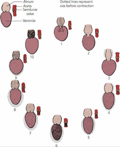
■ FIGURE 9-23 The cardiac cycle of the mammalian heart.
As shown in the key for the cycle sequence, the single chambers represent both right and left atria and ventricles. The single semilunar valve represents both the pulmonary and aortic semilunar valves that separate the ventricles from their respective pulmonary trunk and aorta. The dotted lines surrounding the ventricles correspond to the would-be size related to expansion and contraction. 1, A-V valves open;2, ventricles receive blood; 3, atria contract and empty; 4, ventricles begin contraction and close A-V valves; 5, atria relax and begin to fill; 6, ventricular pressure increases; 7, semilunar valves open; 8, blood ejection begins from ventricles through semilunar valves; 9, ventricles begin relaxation; and 10, semilunar valves close, while A-V valves are still closed.
The cycle ends and is repeated at a frequency consistent with the heart rate for each species. The process of recording these changes is called electrocardiography and the record obtained is known as the electrocardiogram (ECG).
■